What is discoid lupus erythematosus
Discoid lupus erythematosus is a chronic inflammatory autoimmune cutaneous disease that can lead to significant disfiguration and scarring 1. Discoid lupus erythematosus is the most common chronic form of cutaneous lupus erythematosus. Discoid lupus erythematosus is characterized by persistent scaly, disc-like plaques on scalp, face and ears that may cause pigmentary changes, scarring and hair loss.
Discoid lupus erythematosus is the most common form of chronic cutaneous erythematosus and can occur as localized form (80%) with lesions on the face, ears, and scalp or as disseminated discoid lupus erythematosus (20%) with lesions above and below the neck 2. The disseminated form of discoid lupus erythematosus, especially when involving the trunk, is associated with an increased risk of progression to systemic lupus erythematosus (SLE). Patients who present with discoid lesions may have associated arthralgias, but, over time, only approximately 10% to 20% of these patients eventually meet the classification criteria for systemic lupus erythematosus (SLE).
It is unusual for discoid lesions to be present below the neck without lesions also being present above the neck. Occasionally, discoid lesions develop on mucosal surfaces, including the lips, nasal mucosa, conjunctivae, and genital mucosa. Some patients with discoid lesions exhibit a photodistribution. Sun exposure seems to play a role in the development of lesions. However, patients can have discoid lesions on the sun-protected skin, and there is no clear association between sun exposure and their development.
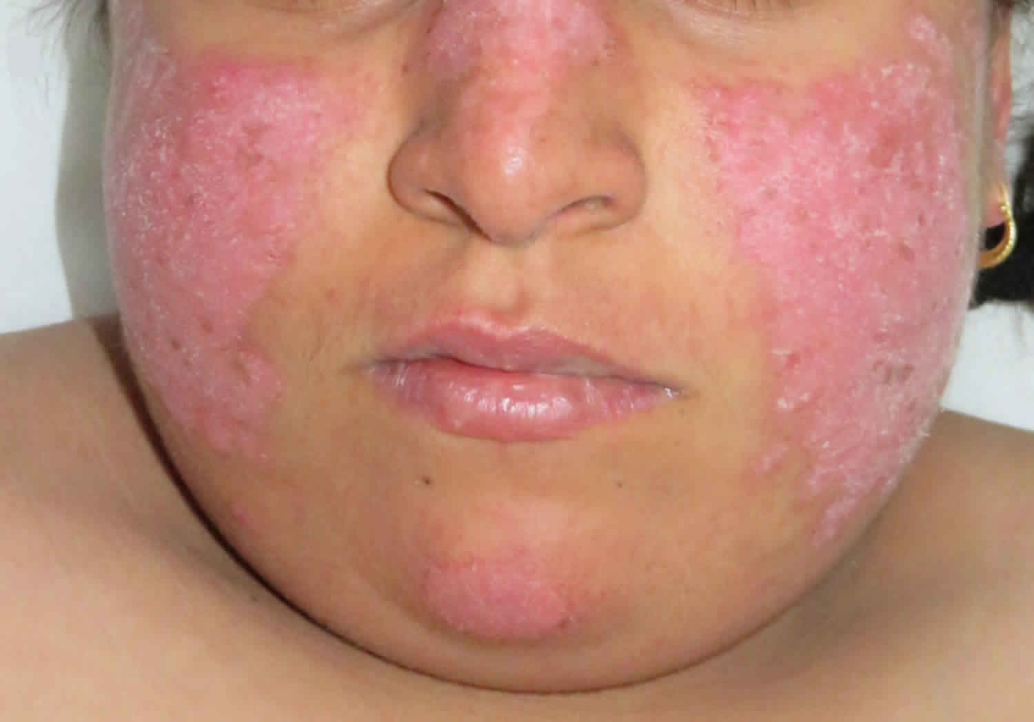
The first morphological sign of discoid lupus erythematosus is a well-defined, annular erythematous patch or plaque of varying size followed by follicular hyperkeratosis, which is adherent to the skin. By removing the adherent scale, follicle-sized keratotic spikes similar to carpet tacks can be seen (“carpet tack sign”). The lesions slowly expand with active inflammation and hyperpigmentation at the periphery leaving depressed central atrophy and scarring, telangiectasia, and hypopigmentation. discoid lupus erythematosus can progress to irreversible scarring alopecia on the scalp. Although uncommon, a squamous cell carcinoma can develop in a longstanding discoid lesion.
Lupus erythematosus is a group of diverse, persistent inflammatory autoimmune diseases that predominately affects the skin. There are several types of cutaneous lupus erythematosus. The most common types are acute cutaneous lupus erythematosus (ACLE), subacute cutaneous lupus erythematosus (SCLE) and discoid lupus erythematosus (DLE) 2.
Discoid lupus erythematosus can affect males and females of any age. Discoid lupus erythematosus is five times more common in females than males, and onset is most often between the ages of 20 and 40 years. Discoid lupus erythematosus is more common than systemic lupus erythematosus (SLE). The estimated prevalence is around 20–40 people in every 100,000.
Discoid lupus erythematosus may be more common in patients with darker colored skin than in fair Caucasians.
Discoid lupus erythematosus is more common and more severe in smokers compared to non-smokers. Smoking also reduces the effectiveness of antimalarials and other therapies.
Early treatment of discoid lupus erythematosus lesions may lead to the total clearing of skin lesions, but treatment failure results in permanent scarring. Hair loss, depressed scars, and pigmentary changes are often disfiguring, particularly in darker-skinned people. Some general measures, such as sun avoidance and liberal application of sunscreen, are encouraged because cutaneous lesions are known to be exacerbated by sunlight. Smoking cessation is encouraged, as this can increase discoid lupus erythematosus disease activity. Studies demonstrate a statistically significant decrease in efficacy of antimalarial medication in individuals who have currently or ever smoked 3.
Current first-line treatment for discoid lupus erythematosus consists of photoprotection in conjunction with topical or oral corticosteroids, topical calcineurin inhibitors, and systemic antimalarial therapy. Chronic discoid lupus erythematosus lesions that are not responsive to topical corticosteroids or topical calcineurin inhibitors may be responsive to intralesional corticosteroid injections. When discoid lupus erythematosus is refractory to these measures, other agents with varying degrees of proven efficacy are used. Currently, no medications have been approved specifically, and many of the drugs described in the literature were developed for use in other autoimmune disorders.
Antimalarials are immunotherapeutic and are considered first-line systemic therapy in cutaneous lupus erythematosus 2. Hydroxychloroquine and chloroquine with or without quinacrine are currently utilized in the treatment of discoid lupus erythematosus. Hydroxychloroquine is preferred over chloroquine due to the lower risk of side effects, specifically retinal toxicity.
Thalidomide, a potent teratogen, has been used in the treatment of discoid lupus erythematosus. An early report of its use in the treatment of discoid lupus erythematosus of 60 individuals treated with 50 to 100 mg per day found complete or marked regression in 54 individuals (90%) with disease relapse in 71% of individuals with medication discontinuation. Side effects included drowsiness, constipation, rash, edema, xerostomia, and 25% of individuals complained of slight to moderate polyneuritis symptoms. Lenalidomide is a thalidomide analog that may also prove useful in the treatment of discoid lupus erythematosus. Evidence suggests lenalidomide is effective in treating discoid lupus erythematosus and has a less severe side effect profile than thalidomide but may be similarly limited by a tendency to relapse once discontinued.
Other treatment modalities such as retinoids are vitamin A analogs with anti-keratinizing and anti-inflammatory effects are sometimes used in cutaneous lupus erythematosus, but documentation in the literature is limited. Immunosuppressive agents such as mycophenolate mofetil, azathioprine, intravenous immune globulin (IVIG), cyclophosphamide, and cyclosporine have all been trialed in the treatment of discoid lupus erythematosus but thought to be second-line when refractory to other treatments.
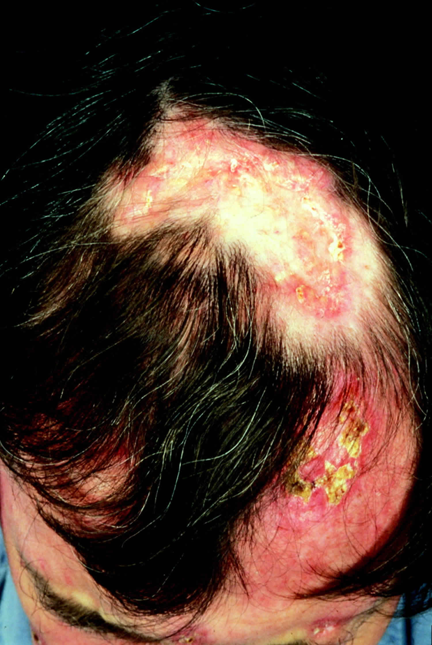
Figure 1. Discoid lupus erythematosus
Footnote: Critical alopecia on scalp caused by discoid lupus erythematosus.
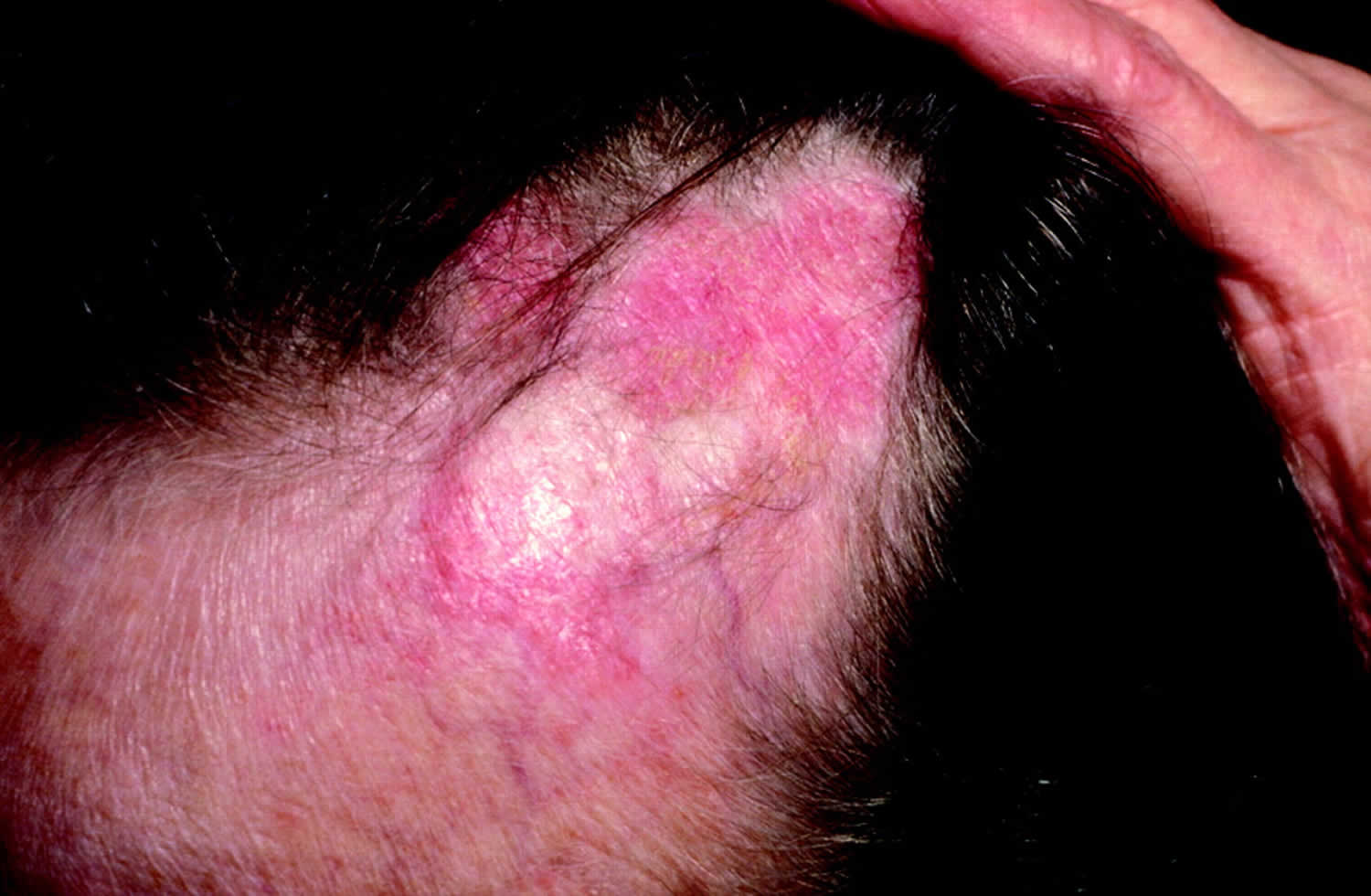
[Source 4 ]Figure 2. Discoid lupus erythematosus
Footnote: Extensive loss of scalp hair in a patient with discoid lupus.
[Source 4 ]Discoid lupus erythematosus causes
Factors leading to discoid lupus erythematosus include:
- Genetic predisposition
- Exposure to sunlight (often several weeks before presentation)
- Toxins such as cigarette smoke
- Hormones.
The manifestations of discoid lupus erythematosus are due to loss of regulation of the immune system in the skin.
Discoid lupus erythematosus pathophysiology
The cause of cutaneous lupus erythematosus is multifactorial with an interplay between genetic and environmental factors. Some contributing environmental factors include ultraviolet radiation (UVR), medications, cigarette smoking, and possibly, viruses 2. The interaction between these multiple factors triggers an inflammatory cascade of cytokine, chemokine, and inflammatory cell responses. Genes previously associated with SLE are TYK2, IRF5, and CTLA4 and also confer an increased risk for developing discoid lupus erythematosus.
An analysis of 405 patients by Bockle et al. 5 found that smoking is highly associated with discoid lupus erythematosus. Bockle et al. 5 hypothesized smoking may play a pathogenic role in cutaneous lupus erythematosus variants (discoid lupus erythematosus, tumid lupus) by the induction of apoptosis, its ability to stimulate T-cell proliferation, and increase photosensitivity. Another explanation might be that smoking provokes DNA damage, resulting in the formation of DNA adducts and the production of ds-DNA antibodies. Keratinocytes may also participate in lupus skin damage by increasing the apoptotic rate and the production of proinflammatory cytokines such as IFN-alpha and IL-6 for SLE, and IFN-lambda for discoid lupus erythematosus 6.
Discoid lupus erythematosus symptoms
Most patients with discoid lupus erythematosus just have skin involvement (cutaneous lupus erythematosus). Between 5% and 25% of patients with discoid lupus erythematosus develop systemic lupus erythematosus (SLE), in which there may be other forms of cutaneous lupus, and other organs may develop the disease. Typically, systemic symptoms are mild in these patients.
Discoid lupus erythematosus may be localized (above neck in 80%) or generalized (above and below the neck in 20%).
Signs of localized discoid lupus erythematosus include:
- Initial lesions are dry red patches
- These evolve to indurated red or hyperpigmented plaques with adherent scale
- Follicular keratosis, or plugs of keratin within hair follicles, is noted when the surface scale is removed, for example with tape (carpet-tack sign)
- Older lesions are hyperpigmented, especially on the edge of the plaques
- Scarring results in central loss of pigment (white patches) and skin atrophy (tissue loss)
- Discoid lupus erythematosus is typically located on the nose, cheeks, ear lobe and concha
- It may involve lips, oral mucosa, nose or eyelids
- Scalp lesions cause temporary or permanent patches of hair loss
- Hypertrophic (warty) lupus erythematous describes red, very thickened plaques.
Signs of generalized discoid lupus erythematosus include:
- Plaques on anterior chest, upper back, backs of hands
- Sometimes, plaques on upper and lower limbs
- Can affect palms and soles
- Can affect anogenital mucosa.
The patient’s main concern is the unsightly appearance of the plaques, but they may also be itchy or sore.
Cutaneous Lupus Erythematosus Disease Area and Severity Index
The Cutaneous Lupus Erythematosus Disease Area and Severity Index (CLASI) was developed in an attempt to classify the severity of cutaneous lupus erythematosus 7. A score of activity and damage due to the disease is calculated in each of 12 anatomical locations.
Total activity score is made up of:
- A degree of redness (0–3) and scale (0–2)
- Mucous membrane involvement (0–1)
- Recent hair loss (0–1), nonscarring alopecia (0–3).
Total damage score is made up of:
- The degree of dyspigmentation (0–2), and scarring (0–2)
- Persistence of dyspigmentation more than 12 months doubles the dyspigmentation score
- Scalp scarring (0, 3, 4, 5, 6).
Discoid lupus erythematosus complications
Discoid lupus erythematosus possible complications:
- Arthritis
- Myositis
- Hypertension
- Renal failure
- Neuropsychiatric symptoms
- Pleuropericarditis
- Pancreatitis, mesenteric vasculitis
- Optic neuritis
About 25% of patients with discoid lupus erythematosus also develop systemic lupus erythematosus (SLE) within months to decades of the diagnosis of skin disease.
Discoid lupus erythematosus may leave permanent scars, even when the active disease has responded to treatment.
Discoid lupus erythematosus diagnosis
Discoid lupus erythematosus is often diagnosed from its distribution in sun-exposed sites and the clinical appearance of the plaques. After a careful history, the patient with discoid lupus erythematosus should undergo a thorough general examination, to find out if other forms of lupus may be present.
The diagnosis is usually confirmed by skin biopsy, in which typical features of lupus are noted: interface and periadnexal dermatitis, follicular plugging, atrophy and scarring. Direct immunofluorescence is often positive in lesional skin in discoid lupus erythematosus (positive lupus band test).
Blood tests
Patients with discoid lupus erythematosus will usually have blood tests at the time of diagnosis and from time to time afterwards.
- Complete blood count (CBC)
- Renal function test
- Inflammatory markers such as C-reactive protein (CRP)
- Antinuclear antibody (ANA, ANF; if present, they are usually in low titre)
- Extractable nuclear antibody (ENA)
- Anti-annexin 1 antibodies—these may be a diagnostic marker for discoid cutaneous lupus erythematosus
Circulating autoantibodies are found in about 50% of patients with discoid lupus erythematosus.
Discoid lupus erythematosus treatment
Preventative measures
The following measures are important to reduce the chance of flares of discoid lupus erythematosus.
- Careful year-round protection from sun exposure using clothing, accessories and thickly applied SPF 50+ broad-spectrum sunscreens. Sunscreens alone are not adequate.
- Indoors, some patients may also need to stay away from glass windows, or these can be treated with UV-blocking films.
- Vitamin D supplements should be recommended for those who strictly avoid the sun.
- Smoking cessation.
Topical therapy
Intermittent courses of potent topical corticosteroids are the main treatment for discoid lupus erythematosus. They should be applied accurately to the skin lesions for several weeks. Potency should be selected to suit the body site and thickness of the plaque. Very potent topical steroids may cause thinning of the surrounding skin and increase blood vessel formation (telangiectasia). Intralesional injections of corticosteroids are sometimes used, especially for hypertrophic discoid lupus erythematosus.
The calcineurin inhibitors tacrolimus ointment and pimecrolimus cream can also be used.
Camouflage makeup is useful to improve appearance.
Systemic therapy
Typically, any of the following drugs may be used to treat discoid lupus erythematosus alone or in combination. Treatment is less effective in smokers than in non-smokers.
- Hydroxychloroquine and other antimalarials—response rates are about 80% in cutaneous lupus erythematosus.
- Systemic corticosteroids such as prednisone or prednisolone. These are rarely required for discoid lupus erythematosus.
- Methotrexate—best response in subacute cutaneous lupus erythematosus and discoid cutaneous lupus erythematosus
- Retinoids isotretinoin and acitretin
- Mycophenolate
- Azathioprine
- Dapsone
- Thalidomide
- Belimumab
Discoid lupus erythematosus prognosis
Discoid lupus erythematosus is an unpredictable and a highly variable disorder. Discoid lupus erythematosus tends to persist for years or decades. In some patients, all signs of active disease resolve in time. The outcome is much improved for patients with only skin and musculoskeletal involvement. The outcomes are worst for patients with central nervous system (CNS) and renal involvement.
Squamous cell carcinoma (SCC) can rarely arise within a longstanding discoid lupus erythematosus plaque in the skin or mucous membrane. It presents as an enlarging warty growth or ulcer. It is usually treated surgically.
Today, with treatment there is an 80% survival at ten years, but failure to comply with treatment can lead to early death 2. At some point in time, the majority of lupus patients will develop hypertension, lipid disorders, diabetes, infections, osteoporosis and malignancies like lymphomas and liver cancer 8.
References- Idoudi S., Litaiem N., Ferjani M., Zeglaoui F. 2018. Dermoscopy of discoid lupus erythematosus. Eur. J. Pediat. Dermatol. 28 (3): 135-138. DOI: 10.26326/2281-9649.28.3.1866
- McDaniel B, Tanner LS. Discoid Lupus Erythematosus. [Updated 2019 Feb 28]. In: StatPearls [Internet]. Treasure Island (FL): StatPearls Publishing; 2019 Jan-. Available from: https://www.ncbi.nlm.nih.gov/books/NBK493145
- Muangchan C, van Vollenhoven RF, Bernatsky SR, Smith CD, Hudson M, Inanç M, Rothfield NF, Nash PT, Furie RA, Senécal JL, Chandran V, Burgos-Vargas R, Ramsey-Goldman R, Pope JE. Treatment Algorithms in Systemic Lupus Erythematosus. Arthritis Care Res (Hoboken). 2015 Sep;67(9):1237-45.
- Early Diagnosis and Treatment of Discoid Lupus Erythematosus. Suresh Panjwani. The Journal of the American Board of Family Medicine Mar 2009, 22 (2) 206-213; DOI: 10.3122/jabfm.2009.02.080075 https://www.jabfm.org/content/22/2/206
- Böckle, B. C., & Sepp, N. T. (2015). Smoking is highly associated with discoid lupus erythematosus and lupus erythematosus tumidus: analysis of 405 patients. Lupus, 24(7), 669–674. https://doi.org/10.1177/0961203314559630
- Kahn JS, Deverapalli SC, Rosmarin DM. JAK-STAT signaling pathway inhibition: a role for treatment of discoid lupus erythematosus and dermatomyositis. Int. J. Dermatol. 2018 Aug;57(8):1007-1014.
- Walling HW, Sontheimer RD. Cutaneous lupus erythematosus: issues in diagnosis and treatment. Am J Clin Dermatol. 2009;10(6):365-81. doi: 10.2165/11310780-000000000-00000
- Jessop S, Whitelaw DA, Grainge MJ, Jayasekera P. Drugs for discoid lupus erythematosus. Cochrane Database Syst Rev. 2017 May 05;5:CD002954


