Splenic abscess
Splenic abscess is an uncommon disease, and it has been noted to occur at a rate of 0.14% to 0.7% in autopsy studies 1. Splenic abscesses generally occur in patients with neoplasia, immunodeficiency, trauma, metastatic infection, splenic infarct or diabetes 2. The incidence of splenic abscess is thought to be growing, due to the increasing number of immunocompromised patients who are particularly at risk for this disease, and also due to the widespread use of diagnostic imaging modalities such as computated tomography (CT) and ultrasonography 3.
Splenic abscess has a bimodal age distribution with peaks in the third and sixth decade of life. Approximately two-thirds of splenic abscesses in adults are solitary, and one-third are multiple 4. Mortality rates are high and fluctuate with immune status and the type of abscess. There is up to an 80% mortality in immunocompromised patients with multilocular abscesses and 15% mortality in immunocompetent patients with unilocular abscesses 5.
Splenic abscess has very high mortality reaching more than 70% when the diagnosis is missed, however, with appropriate treatment, the mortality can be reduced to less than 1% 5. Today with the availability of a CT scan, splenic abscess is not only rapidly diagnosed, but it also helps with treatment by aspirating the collection 6.
The management of splenic abscess is based on medical therapy with antibiotics and splenectomy or percutaneous drainage, with good results 7. However, the optimal treatment remains unclear. While it is recognized that percutaneous drainage may be appropriate for some patients, a high failure rate (14.3-75%) has been observed for this procedure, and surgery remains the gold standard treatment and must be considered if the available less-invasive treatment methods fail 8.
Splenic abscess causes
Splenic abscesses usually result from bacteremia, particularly in the setting of abnormalities caused by trauma, embolization, or hemoglobinopathy. Immunocompromised states that from human immunodeficiency virus (HIV) infection, may also be a risk factor. Some reports indicate that splenic abscesses have occurred from a contiguous focus of infection. Other recognized risk factors are neoplasms, metastatic infection, splenic infarction, and diabetes. Splenic abscesses have also been found to be associated with parasitic infection of the spleen 9.
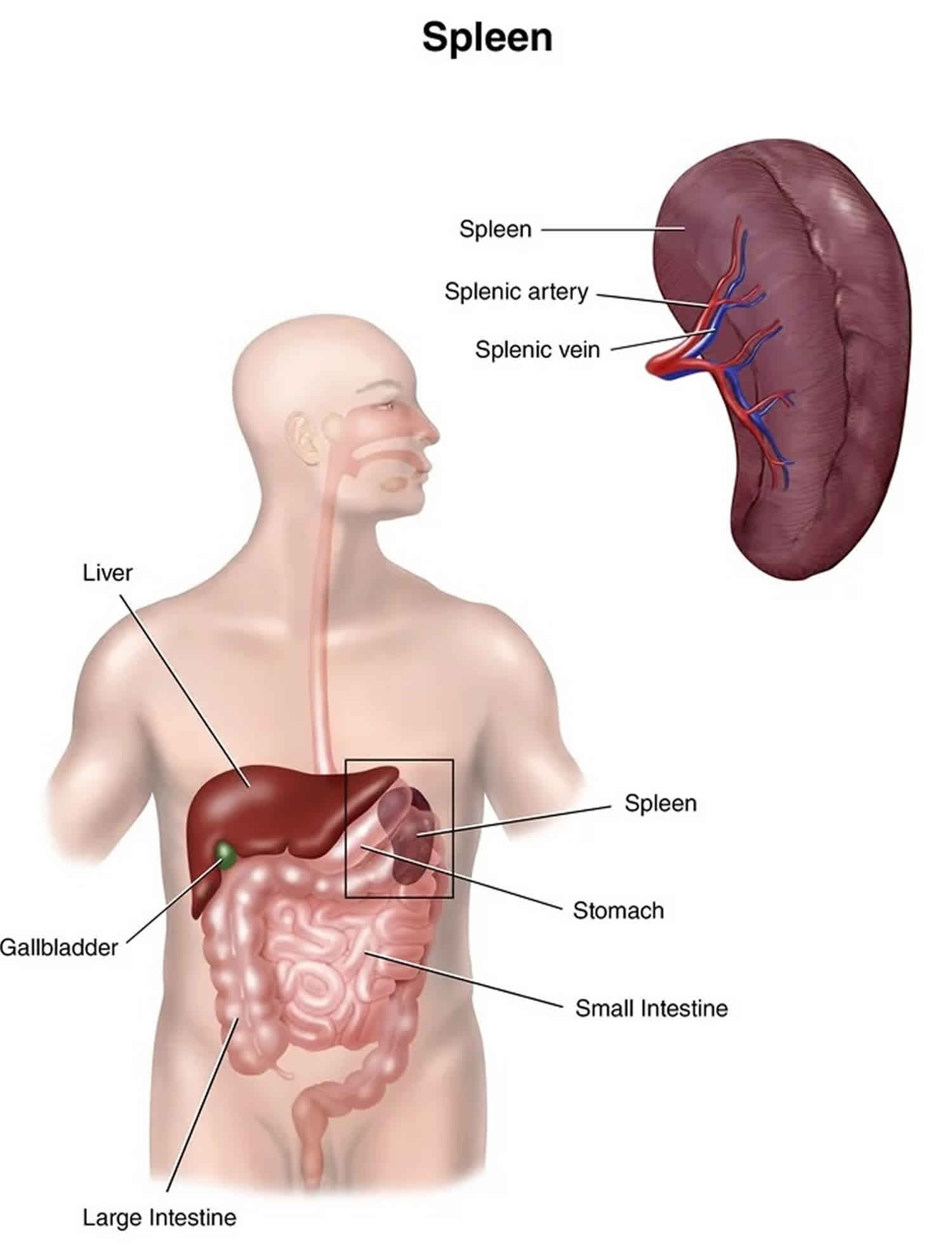
In some cases, an abscess elsewhere in the abdomen may communicate and involve the spleen. It is known as pancreatic abscesses, and diverticulitis may sometimes extend and involve the spleen.
Organisms commonly associated with a splenic abscess include the following:
- Aerobes
- Anaerobes
- Fungi usually Candida
- Polymicrobial in more than 50% of cases
- Rare organisms like Burkholderia, Mycobacterium, and Actinomycetes
Splenic abscesses are most regularly seen as a complication of infective endocarditis, which occurs in about 5% of patients. Frequently, isolated pathogens include Streptococcus, Staphylococcus, (due to endocarditis being the most common cause of splenic abscess), Mycobacterium, fungi, and parasites. Burkholderia pseudomallei is a cause of splenic abscesses in predisposed individuals in some parts of the world.
Splenic abscess symptoms
Fever is the most common symptom, followed by abdominal pain and a tender mass on palpation of the left upper quadrant of the abdomen. The common signs and symptoms described of a splenic abscess include the triad of fever, left upper quadrant tenderness, and leukocytosis is present only in one-third of the cases.
The physical exam will reveal the following:
- Muscle guarding in the upper left quadrant
- Edema of the overlying soft tissues
- Costovertebral tenderness
- Splenomegaly
- Left basilar rales
- Dullness at the left lung base.
Splenic abscess complications
Complications of a splenic abscess include the following:
- Pneumothorax
- Atelectasis
- Life-threatening hemorrhage
- Left-sided pleural effusion
- Subphrenic abscess
- Perforation of the stomach, small bowel or colon
- Pancreatic fistula
- Postsplenectomy thrombocytosis
- Pneumonia
The respiratory complications can be minimized by advocating incentive spirometry, pain control, and aggressive chest physical therapy.
If a subphrenic abscess develops, it usually requires prompt drainage.
Post-splenectomy sepsis is always a risk, especially in young people who have had the spleen removed. These patients should undergo immunization against Meningococcus, Streptococcus pneumoniae and Haemophilus influenzae.
Splenic abscess diagnosis
Diagnosis of a splenic abscess is a clinical challenge 10.
Blood work will reveal leucocytosis with a left shift, and the blood cultures may be positive.
Plain radiographs of the chest can reveal many findings indicative of splenic abscesses such as an elevated left hemidiaphragm and left-sided pleural effusion with or without left basal atelectasis. An ultrasonogram typically demonstrates an area of decreased or absent echogenicity and splenomegaly. An ultrasonogram is quick and can be done at the bedside. A CT scan is the gold standard for diagnosis. The scan also helps doctors to plan treatment by delineating the details of the abscess and the topography of the surrounding structures.
In many cases, a diagnostic aspiration guided by ultrasound or CT scan can help confirm the diagnosis.
Splenic abscess treatment
Hospital admission is recommended for all patients with a splenic abscess 11.
High-dose parenteral broad-spectrum antibiotics are of paramount importance while further diagnostic and therapeutic arrangements are made. The culture results guide the choice of antibiotics.
The gold standard for treatment of splenic abscess is splenectomy; however, recent studies have shown success using different approaches based on abscess characteristics. Percutaneous aspiration may be a less invasive option in patients who are at high risk for surgery, or a temporary solution used as a bridge to surgery, avoiding the risk of a fulminant and potentially life-threatening infection. A percutaneous aspiration is a successful approach when the abscess collection is unilocular or bilocular, with a complete and thick wall and no internal septations. Aspiration is easier to achieve when the content is liquid enough to be drained. If there are multiple collections, or there is associated coagulopathy, either laparoscopic or open surgical treatment is preferred 12.
Percutaneous drainage is less likely to be successful in patients with multilocular abscess, ill-defined cavities, necrotic debris, and thick, viscous fluid. Contraindications for percutaneous drainage include the following:
- Multiple small abscesses
- Debris filled cavities
- Coagulopathy
- Poorly defined cavities
- Diffuse ascites
- Difficult access
Medical treatment alone is not recommended and remains a controversial subject. Mortality rates of more than 50% have been reported in patients only managed with antibiotics. In patients who do not respond, one should consider fungi, actinomycetes or Mycobacterium as a cause.
Fungi are known to respond well to antifungal treatment alone. One study also noted that corticosteroid therapy in these patients could be beneficial.
Open drainage is sometimes required when percutaneous drainage fails. The routes for open drainage include:
- Abdominal extraperitoneal
- Tranapleural
- Retroperitoneal
Splenic abscess prognosis
Unlike the past, the prognosis of a splenic abscess today is markedly improved. The availability of percutaneous CT guided drainage is not only safe and less invasive, but it also avoids the morbidity of open surgery in suitable patients 13. Furthermore, laparoscopic splenectomy has been a promising alternative to the open method, with faster recovery and short hospital stays 14. Surgical splenectomy is the treatment of last choice since most cases can be managed with percutaneous guided drainage and antibiotics.
References- Lee WS, Choi ST, Kim KK. Splenic abscess: a single institution study and review of the literature. Yonsei Med J. 2011;52(2):288–292. doi:10.3349/ymj.2011.52.2.288 https://www.ncbi.nlm.nih.gov/pmc/articles/PMC3051211
- Ng KK, Lee TY, Wan YL, Tan CF, Lui KW, Cheung YC, et al. Splenic abscess: diagnosis and management. Hepatogastroenterology. 2002;49:567–571.
- Chang KC, Chuah SK, Changchien CS, Tsai TL, Lu SN, Chiu YC, et al. Clinical characteristics and prognostic factors of splenic abscess: a review of 67 cases in a single medical center of Taiwan. World J Gastroenterol. 2006;12:460–464.
- Liu YH, Liu CP, Lee CM. Splenic abscesses at a tertiary medical center in Northern Taiwan. J Microbiol Immunol Infect. 2014 Apr;47(2):104-8.
- Waheed A, Mathew G, Zemaitis MR. Splenic Abscess. [Updated 2019 Jul 4]. In: StatPearls [Internet]. Treasure Island (FL): StatPearls Publishing; 2019 Jan-. Available from: https://www.ncbi.nlm.nih.gov/books/NBK519546
- Chen H, Hu ZQ, Fang Y, Lu XX, Li LD, Li YL, Mao XH, Li Q. A case report: Splenic abscess caused by Burkholderia pseudomallei. Medicine (Baltimore). 2018 Jun;97(26):e11208.
- Chou YH, Hsu CC, Tiu CM, Chang T. Splenic abscess: sonographic diagnosis and percutaneous drainage or aspiration. Gastrointest Radiol. 1992;17:262–266.
- Tung CC, Chen FC, Lo CJ. Splenic abscess: an easily overlooked disease? Am Surg. 2006;72:322–325.
- Liverani E, Colecchia A, Mazzella G. Persistent Fever and Abdominal Pain in a Young Woman With Budd-Chiari Syndrome. Gastroenterology. 2018 Feb;154(3):495-497.
- Lee MC, Lee CM. Splenic Abscess: An Uncommon Entity with Potentially Life-Threatening Evolution. Can J Infect Dis Med Microbiol. 2018;2018:8610657
- De Pastena M, Nijkamp MW, van Gulik TG, Busch OR, Hermanides HS, Besselink MG. Laparoscopic hemi-splenectomy. Surg. Today. 2018 Jul;48(7):735-738.
- Divyashree S, Gupta N. Splenic Abscess in Immunocompetent Patients Managed Primarily without Splenectomy: A Series of 7 Cases. Perm J. 2017;21:16-139.
- Moll R, Sailer M, Reith HB, Schindler G. CT-gesteuerte Drainagenbehandlung der Milz bei Abscessen und Hamatomen. Klinikarzt. 2004 Jun. 33:183-8.
- Carbonell AM, Kercher KW, Matthews BD, Joels CS, Sing RF, Heniford BT. Laparoscopic splenectomy for splenic abscess. Surg Laparosc Endosc Percutan Tech. 2004 Oct. 14 (5):289-91.


