Straight leg raise test
Straight leg raise also called Lasegue test, is a neurodynamic test done during a physical examination to determine whether a patient with low back pain has an underlying herniated disc to assess the sciatic nerve compromise due to lumbosacral nerve root irritation 1. Straight leg raise test, along with relevant history and decreased range of motion, are considered by some to be the most important physical signs of disc herniation, regardless of the degree of disc injury. Straight leg raise is a neural tension test that can be used to rule in or out neural tissue involvement as a result of a space occupying lesion, often a lumbar disc herniation 2. It is one of the most common neurological tests of the lower limb 3. Straight leg raise test is an important physical examination finding during primary care to assess the need for imaging studies such as X-rays and MRI, and the potential need for a referral from primary care to a spine specialist. Straight leg raise test has a sensitivity of 28%-29% and a specificity of 88%-90% for nerve root impingement 4.
Straight leg raise test which was first described by Dr. Lazar Lazarevic 5 and wrongly attributed to Dr. Charles Lasegue, a French clinician can be positive in a variety of conditions, being lumbar disc herniation the most common. Nonetheless, there are multiple causes of a positive straight leg test such as facet joint cyst or hypertrophy 6. Overall, straight leg raise test is one of the most commonly performed maneuvers across clinical practice and provides important information when making the clinical decision to refer a patient to a specialist as well as among spinal surgeons to guide therapeutic decision-making 7.
Straight leg raise test or Lasegue test is basically a provocation test that evidences radicular irritation in the lumbosacral region by lower limb flexion and can be due to multiple causes. Radicular symptoms are primarily produced by nerve root inflammation by surrounded structures 8. The foramina are formed by the pedicle superiorly and inferiorly, ligamentum flavum posteriorly, disc and vertebral body anteriorly, and this small space normally allows the nerve root excursion of 4 mm, however during the straight leg raise test this root excursion can be compromised by several factors. Mechanical compression solely does not always generate radicular symptoms as many patients have asymptomatic foraminal stenosis in MRI 9, therefore, positive leg raise test may undergo influence by nerve root irritation secondary to inflammation as well as mechanical compression.
Previous analysis of the sensitivity and specificity of the straight leg raise test shows high sensitivity and low specificity of lumbar disc protrusion 10. However, most of the literature is limited by poor quality and were performed in surgical case-series at non primary care level, limiting the external validity of these findings. Also, some studies have shown restricted diagnosis accuracy of neurological examination in detecting disc herniation with radiculopathy 11. As straight leg raise test demonstrates high sensitivity, it could be useful to rule out lumbar disc protrusion; however, the utility is limited due to low specificity as it can be positive in ischialgia secondary to other causes.
Low back pain is one of the most common complaints among active workers and a significant cause of absenteeism from work. Sciatic pain is radiating pain from the buttocks to the leg and is frequently associated with low back pain 12. To this regard, neurological examination is fundamental in discriminating patients with isolated lower back pain from those with associated radiculopathy. Consequently, early recognition of radiculopathy allows a targeted treatment and diminishes disability 13. The specificity of the straight leg raise test has been reported to be low 14, making the diagnosis accuracy limited. However, the clinical usefulness of straight leg raise test remains important both for general practitioners as for spine surgeons and should still be considered a relevant component of the physical examination that, associated with proper imaging studies can lead to an accurate diagnosis and treatment.
Figure 1. Straight leg raise test
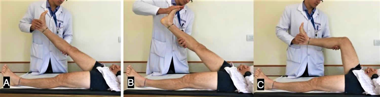
Footnote: A) Straight leg raise test. B) Bragaad’s Test to increase the test sensitivity. C) When flexing the knee the patient usually experience pain relief.
Straight leg raise test technique
The straight leg raise test is performed with the patient in a supine position. The examiner gently raises the patient’s leg by flexing the hip with the knee in extension, and the straight leg raise test is considered positive when the patient experiences pain along the lower limb in the same distribution of the lower radicular nerve roots (usually L5 or S1). A positive straight leg test is demonstrated when reproduction of symptoms radiating down the leg is produced at 30-70° of leg elevation 15. The straight leg raise test has a sensitivity of 91% and specificity of 26% 16. If pain radiates below the knee, L4-S1 nerve root impingement has been identified 17.
Furthermore, a positive straight leg raise test is determined when pain is elicited by lower limb flexion in an angle lower than 45 degrees. During the straight leg raise test, if the pain is reproduced during the leg straightening, patients usually request that the examiner aborts the maneuver and by flexing the patient’s knee, the buttock pain is usually relieved (Figure 1).
Sensitizing maneuvers
After the elicitation of symptoms, the examiner can slowly and carefully lower the leg until the patient no longer feels pain or tightness. Next, either the patient is asked to bring his or her chin to the chest, or the examiner may dorsiflex the patient’s foot (Bragard’s sign) or both actions may be done simultaneously; however, foot dorsiflexion is most commonly performed first. Both maneuvers are considered to be provocative or sensitizing tests for neurological tissue.
Pain that increases with neck flexion or foot dorsiflexion (Bragard’s sign) or both indicates stretching of the dura mater of the spinal cord or a lesion within the spinal cord (e.g. disc herniation, tumor, or meningitis) 18.
Pain that does not increase with neck flexion may indicate a lesion in the hamstring area (tight hamstrings) or in the lumbosacral or sacro-iliac joint.
- Inclusion of neck flexion in the straight leg raise is documented as Hyndman’s sign, Brudzinski’s Sign, Linder’s Sign, or the Soto-Hall test.
- Inclusion of ankle dorsiflexion in the straight leg raise is documented as Lasegue’s test or Bragard’s test.
- Inclusion of great toe extension in the straight leg raise (instead of ankle dorsiflexion) is documented as Sicard’s Test.
An additional maneuver is the crossed straight leg test (crossed over Lasegue), in which the examiner passively flexes the patient’s uninvolved limb while maintaining the knee in extension. A positive test is when the patient reports pain in the involved limb at 40 degrees of hip flexion with the uninvolved limb. A crossed straight test is positive in central disc herniation in cases of severe nerve root irritation 19.
Seated straight leg raise test
To perform a seated straight leg raise test, the patient is seated on the examination table with the hips and knees bent to 90° and legs hanging freely over the edge of the table. The physician slowly extends one knee from the 90° starting position. Extension of the leg continues until pain or reproduction of symptoms is appreciated down the tested leg. A positive test result is defined as reproduction of symptoms prior to reaching full extension.
While performing the straight leg raise test, the physician may produce symptoms in the contralateral leg being tested. Reproduction of symptoms in the opposite leg being tested is termed crossed straight leg raise test result (see video below) and indicates a large central lumbar disc herniation. This test has a sensitivity of 28%-29% and a specificity of 88%-90% for nerve root impingement 4.
Straight leg raise test interpretation
- If symptoms are primarily back pain, it is most likely the result of a disc herniation applying pressure on the anterior theca of the spinal cord, or the pathology causing the pressure is more central. “Back pain only” patients who have a disc prolapse have smaller, more central prolapses 18.
- If pain is primarily in the leg, it is more likely that the pathology causing the pressure on neurological tissue(s) is more lateral 18.
- Disc herniations or pathology causing pressure between the two extremes are more likely to cause pain in both areas 18.
Reasoning
- Neurologic pain which is reproduced in the leg and low back between 30-70 degrees of hip flexion is suggestive of lumbar disc herniation at the L4-S1 nerve roots.
- Pain at less than 30 degrees of hip flexion might indicate acute spondyloithesis, gluteal abscess, disc protrusion or extrusion, tumor of the buttock, acute dural inflammation, a malingering patient, or the sign of the buttock.
- Pain at greater than 70 degrees of hip flexion might indicate tightness of the hamstrings, gluteus maximus, or hip capsule, or pathology of the hip or sacroiliac joints.
- Camino Willhuber GO, Piuzzi NS. Straight Leg Raise Test. [Updated 2019 Nov 15]. In: StatPearls [Internet]. Treasure Island (FL): StatPearls Publishing; 2019 Jan-. Available from: https://www.ncbi.nlm.nih.gov/books/NBK539717
- Boyd BS, Villa PS. Normal inter-limb differences during the straight leg raise neurodynamic test: a cross sectional study. BMC Musculoskeletal Disorders. 2012;13:245. doi:10.1186/1471-2474-13-245.
- Butler DA: Mobilisation of the nervous system, Melbourne,1991,Churchill Livingstone.
- van der Windt DA, Simons E, Riphagen II, et al. Physical examination for lumbar radiculopathy due to disc herniation in patients with low-back pain. Cochrane Database Syst Rev. 2010 Feb 17. CD007431
- Drača S. Lazar K. Lazarević, the author who first described the straight leg raising test. Neurology. 2015 Sep 22;85(12):1074-7.
- Tawa N, Rhoda A, Diener I. Accuracy of clinical neurological examination in diagnosing lumbo-sacral radiculopathy: a systematic literature review. BMC Musculoskelet Disord. 2017 Feb 23;18(1):93.
- van den Hoogen HJ, Koes BW, Devillé W, van Eijk JT, Bouter LM. The inter-observer reproducibility of Lasègue’s sign in patients with low back pain in general practice. Br J Gen Pract. 1996 Dec;46(413):727-30.
- Stafford MA, Peng P, Hill DA. Sciatica: a review of history, epidemiology, pathogenesis, and the role of epidural steroid injection in management. Br J Anaesth. 2007 Oct;99(4):461-73.
- Tachihara H, Kikuchi S, Konno S, Sekiguchi M. Does facet joint inflammation induce radiculopathy?: an investigation using a rat model of lumbar facet joint inflammation. Spine. 2007 Feb 15;32(4):406-12.
- Rabin A, Gerszten PC, Karausky P, Bunker CH, Potter DM, Welch WC. The sensitivity of the seated straight-leg raise test compared with the supine straight-leg raise test in patients presenting with magnetic resonance imaging evidence of lumbar nerve root compression. Arch Phys Med Rehabil. 2007 Jul;88(7):840-3.
- Majlesi J, Togay H, Unalan H, Toprak S. The sensitivity and specificity of the Slump and the Straight Leg Raising tests in patients with lumbar disc herniation. J Clin Rheumatol. 2008 Apr;14(2):87-91.
- Hill JC, Konstantinou K, Egbewale BE, Dunn KM, Lewis M, van der Windt D. Clinical outcomes among low back pain consulters with referred leg pain in primary care. Spine. 2011 Dec 01;36(25):2168-75.
- Bertilson BC, Brosjö E, Billing H, Strender LE. Assessment of nerve involvement in the lumbar spine: agreement between magnetic resonance imaging, physical examination and pain drawing findings. BMC Musculoskelet Disord. 2010 Sep 10;11:202.
- Devillé WL, van der Windt DA, Dzaferagić A, Bezemer PD, Bouter LM. The test of Lasègue: systematic review of the accuracy in diagnosing herniated discs. Spine. 2000 May 01;25(9):1140-7.
- Bruno PA, Millar DP, Goertzen DA. Inter-rater agreement, sensitivity, and specificity of the prone hip extension test and active straight leg raise test. Chiropr Man Therap. 2014. 22:23.
- Chou R, Qaseem A, Snow V, et al. Diagnosis and treatment of low back pain: a joint clinical practice guideline from the American College of Physicians and the American Pain Society. Ann Intern Med. 2007 Oct 2. 147(7):478-91.
- Casazza BA. Diagnosis and treatment of acute low back pain. Am Fam Physician. 2012 Feb 15. 85(4):343-50.
- David J. Magee;Orthopaedic Physical Assessment; Chapter 9-Lumbar Spine;Fifth Edition: Pg 558-564.
- Hudgins WR. The cross-straight-leg-raising test. N. Engl. J. Med. 1977 Nov 17;297(20):1127.


