Tattoo ink allergy
A tattoo is the result of the deposition of exogenous pigment into the skin. This may be purposeful or accidental. The introduction of foreign substances into the skin can result in a number of adverse effects, including toxic or immunologic reactions to the tattoo pigments, transmission of infectious diseases, and the localization of skin disease within the tattoo. There is a significant body of literature on allergic reactions to tattoos and specifically to red dye tattoos. Red pigment is the most common cause of reactions in tattoos and this can present in various clinical and histological variants with dermatitis and lichenoid being the most common 1. Such allergic reactions can be due to contact dermatitis and photoallergic dermatitis. The ink or pigment allergy causes an acute inflammatory reaction and sometimes can produce granulomatous, lichenoid, and pseudolymphomatous types of responses.
Specifically, red tattoo dye may contain the following:
- Ferric hydrate (sienna)
- Mercury sulfide (cinnabar)
- Sandalwood
- Brazilwood
The high incidence of reactions in red tattoos is attributed to the toxic metals often found within the pigment, which predispose the skin to a higher incidence of adverse reactions 2. Furthermore, there is no quality control or legislation regarding the inks contained within red tattoos 3, especially inorganic cinnabar tattoos, highlighting that a safer outcome may be found with synthetic tattoos whereby the dye within them is 4. It has been reported that some inks have been obtained from the clothing industry (red dyes for clothes) and that there may have been batches contaminated in some way. One study analysed the decomposition of tattoo pigments using liquid chromatography and mass spectrometry; they found lasers broke down the pigments to produce 2-methyl-5-nitroaniline, 2-5-dichloraniline and 4-nitro-toluene. These materials are not only toxic but postulated to be carcinogenic 5, which may contribute to the high incidence of red tattoo reactions.
As noted, reactions to red tattoo pigments account for the majority of the reactions. However, the evaluation of a patient with a possible reaction to tattoo dye is complex because many times the dyes are mixed by the person applying the tattoo, and tattoo inks change their composition over time.
Usually reds are from mercury compounds, blue from cobalt, green from chromium and yellow from cadmium. Blue pigments may also include copper carbonate, sodium aluminum silicate, calcium copper silicate, other cobalt aluminum oxides and chromium oxides 6.
Cobalt compounds are associated with a nonimmunologic contact urticaria which results in a transient urticaria after creation of the tattoo 7. Generalized urticaria, presumably immunologic, has also been described and was attributed to blue pigment, with resolution after removal of the tattoo 8. The pathophysiologic mechanisms for tattoo reactions include nonimmunologic histamine release, T cell mediated delayed reactions, granulomatous reactions, lichenoid reactions (resemble a graft versus host reaction), mast cell degranulation and pseudolymphomatous response.
Table 1. Composition of tattoo pigment colors
| Color | Composition |
|---|---|
| Red |
|
| Black |
|
| Brown |
|
| Blue |
|
| Green |
|
| Yellow |
|
| Purple |
|
| White |
|
Tattoo artists use many compounds to create tattoo pigment and several allergic reactions can occur as a result of these additives. The compositions of many inks have been identified; however, as new mixtures are created it becomes difficult to identify the specific ingredients in a particular ink. Allergic reactions to a particular pigment can manifest in several ways including allergic contact dermatitis and photoallergic dermatitis. Subsequently, tattoo ink or pigment allergy reactions can be classified as acute inflammatory reactions, allergic hypersensitivities, and granulomatous, lichenoid, and pseudolymphomatous types of reactions.
Temporary tattoos made with henna have resulted in sensitivity to para-substituted amino-compounds leading to hair coloring sensitivity. Contact dermatitis has also been reported in people with henna tattoos. Henna tattoos are non-permanent tattoos where henna dye is painted onto the skin with an artist’s brush resulting in a brownish stain. Henna itself should be safe, but the dye is often mixed with paraphenylenediamine (PPD), a chemical substance that is well known for causing allergic reactions in people sensitive to it. In this case the dye is black in color, so-called black henna.
Tattoo ink allergy symptoms
The ink or pigment allergy causes an acute inflammatory reaction and sometimes can produce granulomatous, lichenoid, and pseudolymphomatous types of responses. The pathophysiologic mechanisms for tattoo reactions include nonimmunologic histamine release, T cell mediated delayed reactions, granulomatous reactions, lichenoid reactions (resemble a graft versus host reaction), mast cell degranulation and pseudolymphomatous response.
Although dermatitis and lichenoid reactions are by far the most common, there have been reports of some rarer forms of reactions to red tattoo pigment. Litvinov and Sassevile reported a case of pyoderma gangrenosum 9 in a red tattoo consistent with the Koebner phenomenon. One group reported a red tattoo granulomatous dermatitis that was histologically similar to granuloma annulare presented post acquisition of a red pigment tattoo 10.
A 2003 paper 11 found that the azo pigments and quinacridones found in red tattoo pigment led to skin reactions with differing histology found on biopsy: some lichenoid, some eczematous and others pseudolymphomatous (Table 2).
Table 2. Clinico-pathological patterns of red tattoo reactions
| Skin reaction | Histology |
|---|---|
| Allergic contact dermatitis | Acanthosis, spongiosis, perivascular lymphocytic inflammatory infiltrate |
| Lichenoid dermatoses | Acanthosis and thickening of the stratum corneum |
| Granulomatous | Giant cell positive or negative |
| Pseudolmphomatous | T- or B-cell lymphoma equivalent |
| Pseudoepitheliomatous hyperplasia | Reactive changes: similar to SCC and keratoacanthoma |
| Irregular acanthosis | |
| Low mitotic activity |
Allergic reaction to a tattoo ink causing rash
You can develop an allergic reaction to tattoo ink at any time. It can happen:
- Immediately
- Weeks or years later
- Decades afterward
Some people develop an allergic reaction after having a medical treatment. This reaction is most likely to occur if you:
- Start antiretroviral treatment for HIV
- Have joint-replacement surgery
Signs of an ink allergy: Most people develop an allergy to a specific color of ink. Red ink is often the culprit, but any color can cause an allergic reaction. When this happens, you may notice one or more of the following in only one color of ink:
- Redness and swelling
- Itch
- Small pimple-like bumps
- Raised, scaly patches
- Deep lumps
- Blisters
- Skin crusts or flakes off
- A watery fluid leaking from the area
Take action: If you suspect that you’re having an allergic reaction, dermatologists recommend the following:
Serious reaction: Seek immediate medical care.
- Signs of a serious reaction: You see a reaction in your tattoo and have one or more of the following: Trouble breathing, a racing heart, tightness in your chest, dizziness or lightheadedness, a stomachache, intense swelling, serious pain, flushing, or hives.
- Mild or moderate reaction: Tell your tattoo artist about the reaction, and ask if there’s anything you should do. If the reaction lasts longer than 1 or 2 weeks, see a board-certified dermatologist.
- Signs of a mild or moderate reaction: You have a noticeable change within your inked skin, but you don’t have any symptoms that affect another part of your body, such as trouble breathing or a stomachache.
Temporary tattoo rash
Temporary tattoo rash can occur at any time between getting the tattoo and 3 weeks later.
The allergic reaction is usually caused by a black dye. Many people have an allergic reaction to black dye that contains a chemical called PPD (paraphenylenediamine).
Signs of a reaction: If you or your child has an allergic reaction, you may notice one or more of the following in the temporary tattoo:
- Redness and swelling
- Intense itch
- Pain
- Tiny bumps
- Scaly, raised skin
- Blisters, which may open and leak
- Loss of skin color
- Scarring
A few people have developed problems, such as dizziness, fainting, or a stomachache.
Take action: Dermatologists recommend the following:
Serious reaction: Get immediate medical care.
- Signs of a serious reaction: You see a reaction in your tattoo and have one or more of the following: Trouble breathing, a racing heart, tightness in your chest, dizziness or lightheadedness, a stomachache, intense swelling, serious pain, flushing, or hives.
- Mild reaction: If it lasts longer than 1 week, see a board-certified dermatologist.
- Signs of a mild reaction: You see a reaction within the tattoo but don’t have symptoms in other areas of your body.
Infection
An infection can happen:
- Immediately after getting a tattoo
- Days or months after getting inked
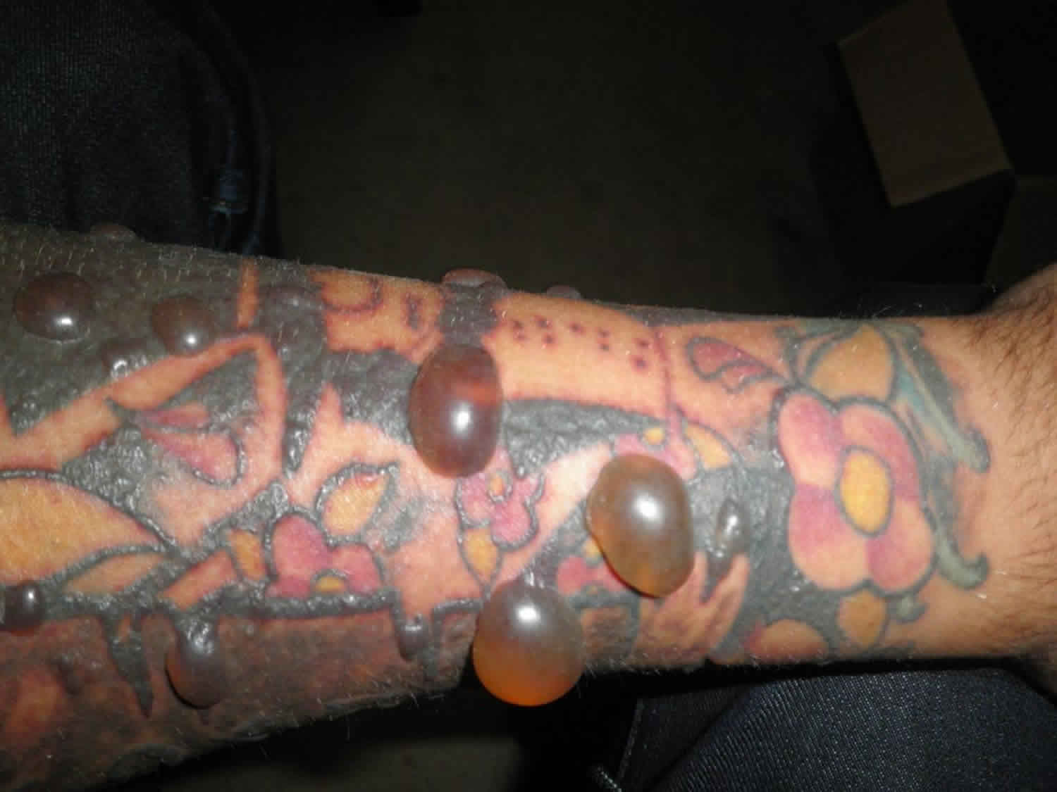
Signs of an infection: After getting a tattoo, it’s normal to see some redness and swelling. Your skin will feel sore, and you may see clear fluid oozing from your new tattoo. As your skin heals, it can itch and flake. Scabs may form. All of this can be part of your normal healing process.
If an infection develops, your skin reacts a bit differently. You may notice one or more of the following:
- Redness: It becomes darker or spreads instead of lightening and diminishing
- Pain: It continues or worsens instead of subsiding
- Rash of itchy, red, and painful bumps develop within the tattoo (shown here)
- Fever
- Chills and shivering
- Pus in the tattoo
- Open sore(s) in the tattoo
Take action: If you have any signs or symptoms of an infection, see your doctor or a board-certified dermatologist right away. The sooner treatment is started, the less damage it can do to your health and your tattoo.
Sun allergy rash
After getting a tattoo, some people develop a sun allergy on their inked skin. This reaction can happen every time the sun’s rays hit your tattoo.
Signs of a sun allergy: This allergy can appear within minutes of the sun hitting your tattoo or hours later. You may have a sun allergy on your inked skin if you notice any of the following:
- Swelling and redness around a tattoo
- Itchy rash of tiny bumps
- Blisters or hives
Take action: You can prevent a rash by protecting your skin from the sun. To protect your tattoo and your skin, dermatologists recommend that you:
- Apply sunscreen 15 minutes before going outside. To get the protection you need, use a broadspectrum sunscreen that offers SPF 30 or higher, broad-spectrum protection, and water resistance. You should apply sunscreen to all skin that will be exposed while you’re outdoors.
- Cover your tattoo with clothing before going outdoors. To test how well the clothing will protect your skin, hold the clothing up to a bright light. If you cannot see light through the fabric, the clothing offers good sun protection. Dermatologists still recommend applying sunscreen to all skin that will be bare while you’re outside.
- Seek shade. Staying in the shade is a simple way to reduce sun exposure.
Skin diseases
If you carry the genes for psoriasis, getting a tattoo can trigger a psoriasis flare or cause psoriasis to appear for the first time. Other skin diseases can also appear within or around a tattoo.
If a skin condition that appears, you’ll likely see signs of the disease within 10 to 20 days of getting the tattoo. The disease can also appear as early as 3 days after getting inked. Sometimes, it shows up years later.
Skin cancer can also form within a tattoo.
Signs of skin disease: Around the tattoo, you may see signs of one of the following skin conditions:
- Psoriasis
- Eczema
- Vitiligo
- Lichen planus
- Keloid
- Sarcoidosis
- Scars
- Skin cancer
Take action:
- If you have a tendency to scar or have ever had a scar that grew bigger than the wound causing it (a keloid), rethink getting a tattoo. Scarring can ruin the appearance of your tattoo.
- If you’ve already developed a scar or signs of a skin disease, make an appointment to see a board-certified dermatologist. A dermatologist can minimize the look of a scar, diagnose a skin disease, and develop a treatment plan for a skin disease.
MRI burn
While rare, a few people have developed a burn on tattooed skin during an MRI.
Signs of a reaction: If the ink used to create a tattoo or permanent makeup causes a burn, it’s likely to be mild. A few serious burns have been reported.
If you have a minor burn, it can cause:
- Pain
- Redness
- Swelling
Take action: If you have a tattoo or permanent makeup, you can still get an MRI. Doing the following can help prevent a burn:
- Tell the technician who is giving you the MRI that you have tattooed skin or permanent makeup.
- Ask the technician to stop the MRI if you feel burning or stinging during the MRI where you have a tattoo or permanent makeup.
Swollen lymph nodes
Ink usually spreads to the lymph nodes as your skin heals from getting the tattoo.
Signs of a reaction: Swelling in lymph nodes, usually near a tattoo. The largest groups of lymph nodes are found in your neck, armpits, and groin.
Take action: If you feel long-term swelling in any lymph nodes, dermatologists recommend that you:
- See your primary care doctor to rule out another possible cause. Swollen lymph nodes could be a sign of an infection or another health concern.
Dermatitis
Eczematous reactions to red tattoos—alongside lichenoid—are the most common type of reaction observed, being either allergic contact dermatitis or photo-allergic dermatitis 12. Photo-allergic reactions most commonly occur secondary to the cadmium subset of red pigment 13 and skin protection from ultraviolet light has been shown to exert protective effects 14.
One study in Denmark 15 found that approximately half of sunbathers with red or black tattoos in particular suffered from photo-allergic reactions. They hypothesised that reactive oxygen species may play a role in triggering a dermatitis-type reaction in such individuals.
One interesting study 16 carried out patch testing in 90 patients with tattoo reactions over a 4-year period. They found that red tattoo reactions did not predispose patients to have a positive patch test result with the common tested allergens. They hypothesised that the allergic result is due to haptenization (reaction of an antigenic compound (a hapten) with a carrier protein in order to stimulate an immune response), possibly due to ‘photochemical cleavage of red azo pigment’. To strengthen the argument that patch testing does not correlate with red tattoo reactions, Anthony and Harland 4 also found patch testing carried out prior to laser removal was negative to mercury at 48 and 96 hours.
The exact understanding of the mechanisms behind the eczematous reactions observed in red tattoos is still lacking with type 1–3 hypersensitivity reactions playing a role 17; however there is no conclusive evidence to date.
Lichenoid
In 1978 it was first hypothesized that tattoos may imitate a localized antigenic challenge, which in turn can cause a lichenoid pattern of reaction 18. Lichenoid reactions in red organic tattoos have been shown to elicit a cytotoxic inflammatory response of the tissue 19 with lichenoid basal damage 20 thought to be produced by a delayed cellular hypersensitivity to metal particles 21.
The current consensus for the most common tattoo reaction is conflicting, with some papers stating dermatitis is the most common type, and others lichenoid 22. Tattoo lichenoid reactions are most commonly associated with red pigment, particularly because of mercury 23.
Pseudolymphomatous
Pseudolymphoma is a term given to a histological entity and has been reported to occur as a complication of tattoos that is histologically indistinct from malignant T- or B-cell lymphoma 24; however the lymphoproliferative process is benign 25. The clinical presentation is a pruritic plaque within the tattoo, often initially mistaken for lymphoma. The distinguishing factor between pseudolymphoma and lymphoma is the polyclonal nature of the lymphocytes 24. Amann et al. 26 propose it can only be distinguished from lymphoma with the aid of electron microscopy, immunohistochemistry and molecular biology.
In one reported case a 35-year-old male reacted with infiltrated nodules within the red areas of his tattoo 2 months post tattooing 27, whilst another paper reported a pseudolymphomatous allergic immune reaction 6 months post initial tattooing 28.
Granulomatous
Similar to lichenoid reactions, granulomatous reactions are thought to occur as a result of delayed hypersensitivity reactions to the presence of the red pigment 10.
One case study found a granulomatous reaction in the red pigment of four multi-coloured tattoos; interestingly only one of these lesions contained the metal mercury—which is thought to commonly cause these reactions in red tattoos 29.
Allopurinol has proved to be useful in the treatment of red tattoo granulomas, with improvement of symptoms during a 6-month course and regression of results upon cessation of treatment 30.
Miscellaneous Reaction Patterns
The most common skin reactions to tattoo include a transient acute inflammatory reaction due to trauma of the skin with needles and medical complications such as superficial and deep local infections, systemic infections, allergic contact dermatitis, photodermatitis, granulomatous and lichenoid reactions, and skin diseases localized on tattooed area (eczema, psoriasis, lichen, and morphea) 31. These reactions may have different onset of symptoms immediately when the tattoo has been inked into it, days, months, or years later 32. Next to these inflammatory skin reactions you have to consider also the possibility of the development of cutaneous conditions such as pseudolymphomatous reactions 33 and pseudoepitheliomatous hyperplasia 34. The evolution in neoplastic lymphoma 35, squamous cell carcinoma, and Keratoacanthoma 36 is a rare outcome, since this neoplastic condition usually appears when they are fully evolved and not with “premalignant” condition.
There have also been several cases of systemic sarcoidosis presenting initially as a granulomatous tattoo reaction 37. One study looked at sarcoid development within cosmetic tattooing of the eyebrows and lips and observed that a granulomatous reaction containing a foreign body should not anticipate an exclusion of systemic sarcoidosis. A sarcoid within a tattoo is an example of the Koebner phenomenon 38. Whether a sarcoid is a reaction to the tattoo itself or coincidental with a systemic disease remains a conundrum. It is suggested that in case of a tattoo sarcoid a search for systemic sarcoidosis is warranted 39.
Finally there are reports of deep-seated infections manifesting within tattoos, such as leprosy, syphilis, tuberculosis and Mycobacterium chelonae 40.
Table 3. Dermatologic disorders and complications after tattooing
| Complications after tattooing | Clinical features | Onset of symptoms |
|---|---|---|
| Allergic disorders | Allergic dermatitis | Days to weeks |
| Photoallergic reaction | After sun exposure | |
| Skin infections | Erysipelas | First few days |
| Gangrene | = | |
| Sepsis | = | |
| Impetigo | = | |
| Ecthyma | = | |
| Cellulitis | = | |
| Tetanus | Weeks to years | |
| Lepra | = | |
| Syphilis | = | |
| Viral infections | Molluscum contagiosum | Weeks to months |
| Verruca vulgaris | = | |
| Hepatitis B, C | = | |
| AIDS | = | |
| Mycoses | Tinea cutis glabrae | After weeks |
| Zygomycoses | After months | |
| Tumors | Lymphoma | Years |
| Carcinoma basocellular | = | |
| Carcinoma spinocellular | = | |
| keratoacanthoma | = | |
| Melanoma | = | |
| Skin disease localized in tattooed area | Psoriasis | Weeks to years |
| Lichen planus | = | |
| Morphea | = | |
| Pseudolymphoma | = | |
| Pseudoepitheliomatous hyperplasia | = | |
Acute inflammatory reactions
This reaction is in direct response to the piercing of the skin with needles impregnated with pigment dyes prepared from metal salts. There may be transient redness and swelling of the area that disappears within 2–3 weeks. It is an expected side effect of the tattooing process.
Koebnerized skin reactions
The koebner phenomenon describes the appearance of new lesions of an existing skin disease within a cutaneous injury. A tattoo can result in psoriasis, lichen planus or vitiligo by this mechanism.
Skin infections
Infection is not common after tattooing. The following skin infections have been reported however, emphasizing the need to undergo the procedure in a clean environment using sterile equipment. Microorganisms have also been found in contaminated tattoo ink.
- Impetigo
- Cellulitis
- Herpes simplex
- Viral warts
- Atypical mycobacterial infection
Transmission of serious blood-borne infections may occur.
- Syphilis
- Leprosy
- Viral hepatitis
- Human immunodeficiency virus (HIV)
Eczematous hypersensitivity reactions
The two most common hypersensitivity reactions to tattoo pigments are allergic contact dermatitis and photoallergic dermatitis. The reaction usually appears as an inflamed red rash or may sometimes be scaly and flaky (exfoliative dermatitis). Red tattoo pigments cause the most reactions, particularly those made from mercury sulfide (cinnabar). Hypersensitivity reactions to pigments used to make black, blue, purple and green tattoos are much less common.
The components of tattoo ink are difficult to determine and undergo changes with time. There are no regulations for tattoo inks or colour additives, which contain inorganic pigments and carbon black, and/or organic pigments from various chemical classes. The table below lists some agents that have been used. There are many more.
Photo-aggravated reactions
Yellow tattoos created from cadmium sulfide are at most risk of causing hypersensitivity reactions when they are exposed to sunlight. Swelling and redness develop around the tattoo site. This phototoxic reaction caused by cadmium sulfide can also occur in red tattoos, as trace amounts of cadmium are added to brighten red tattoo pigment.
Granulomatous reactions
The term granuloma refers to the particular kind of cells that cause the reaction. A foreign body reaction to pigment may cause raised red bumps at the site of the tattoo that are made up of epithelioid cells, lymphocytes and a few giant cells. Most commonly red, but also, green, blue and purple pigment tattoos, and UV-visible tattoos have been associated with granulomatous reactions.
Lichenoid reactions
These types of reactions are much less common than eczematous hypersensitivity reactions. Their signs and symptoms are the same as those in lichen planus, although the reaction is usually confined to the red parts of the tattoo. Hence, red pigment is responsible for most lichenoid tattoo reactions.
Pseudolymphomatous reactions
These are usually the result of a delayed hypersensitivity reaction to tattoo pigment. Again, red pigment is the main cause but it has also been reported with green and blue pigments. Pseudolymphomas caused by tattoo pigment are characteristically plum to red coloured nodules and plaques. They need to be clinically distinguished from cutaneous lymphomas that may be the cause of serious malignant conditions.
Skin cancer
Squamous cell carcinoma has been described developing in tattoos, particularly in areas of red ink. Keratoacanthoma-like reactions to tattoos have also been described. Although the cause is unknown, it is thought that the ink may include carcinogens. In some cases, the tumors may be due to “pseudoepitheliomatous hyperplasia”, a benign proliferative reaction that mimics skin cancer.
Burns during magnetic resonance imaging
Magnetic resonance imaging (MRI scan) produces a magnetic field that has been reported to induce an electric current within tattoo ink containing iron oxide. Iron oxide is sometimes found in permanent cosmetic ink used to enhance lip lines and eyebrows. A MRI scan results in a minor burn at the tattooed site, ie, painful redness and swelling.
Tattoo ink allergy treatment
Multiple treatment options for red tattoo pigment reactions have been employed with little background evidence for their use. Medical treatment options have included allopurinol use as well as topical and intralesional corticosteroids 4 and secondary measures such as sun protection and antibiotics 30. Effective laser treatment has been demonstrated with both Q-switched Nd:YAG and erbium:YAG lasers 4. Anthony and Harland 4 demonstrated successful laser treatment in seven patients within an open non-randomised clinical trial and De Argila 42 presented a successful outcome of one case of a lichenoid tattoo reaction treated with five treatment sessions of erbium:YAG.
A carbon dioxide (CO2) laser has also been used. Kyanko et al. 43 treated two cases of red tattoo dermatitis with a CO2 laser in cases previously resistant to topical and intralesional corticosteroids. Madan found that the CO2 laser was particularly useful for red ink tattoo granulomas that were recalcitrant to conventional steroid treatment 44. Of note, the CO2 laser has also been reported to trigger the generalisation of localised tattoo dermatitis 45.
Finally, there is the option of surgical excision. The appropriateness of this will of course depend on the extent of the reaction and the size of the tattoo. Eczematous reactions have been successfully treated with excision and concomitant low-dose intralesional corticosteroids 46.
Tattoo Removal
Tattoos are most often treated with Q-switched lasers. In most cases, 5 to 12 treatments are required, at 6 to 8-week intervals. Complete clearance is not always possible.
The laser may be selected according to the colour of the tattoo pigment.
- Nd:YAG laser (1064 nm) for black and blue pigment
- Alexandrite laser (755 nm) for black, blue and green pigment
- Ruby laser (694 nm) for black, blue and green pigment
- Frequency doubled Nd:YAG laser (532 nm) for red, orange and purple pigment
- Flashlamp-pumped pulsed dye (510 nm) for red, purple, orange and yellow pigment
White and yellow pigment appears to be the most difficult to eradicate. Complications may include:
- Scarring
- Unwanted colour changes including darkening of tattoo e.g. brown to black (ferric oxide becomes ferrous oxide)
- Spreading of allergic reaction as tattoo granules disperse
Alternatively, ablative fractional resurfacing can be used.
References- Forbat E, Al-Niaimi F. Patterns of Reactions to Red Pigment Tattoo and Treatment Methods. Dermatol Ther (Heidelb). 2016;6(1):13–23. doi:10.1007/s13555-016-0104-y https://www.ncbi.nlm.nih.gov/pmc/articles/PMC4799043
- Garcovich S, Carbone T, Avitabile S, et al. Lichenoid red tattoo reaction: histological and immunological perspectives. Eur J Dermatol EJD. 2012;22(1):93–96.
- Lehmann G, Pierchalla P. Tattooing dyes. Dermatosen Beruf Umw Occup Environ. 1988;36(5):152–156.
- Antony FC, Harland CC. Red ink tattoo reactions: successful treatment with the Q-switched 532 nm Nd:YAG laser. Br J Dermatol. 2003;149(1):94–98. doi: 10.1046/j.1365-2133.2003.05342.x
- Vasold R, Naarmann N, Ulrich H, et al. Tattoo pigments are cleaved by laser light—the chemical analysis in vitro provide evidence for hazardous compounds. Photochem Photobiol. 2004;80(2):185–190. doi: 10.1562/2004-05-17-RA-170.1
- Kaur RB et al: Cutaneous allergic reactions to tattoo ink. Journal of Cosmetic Dermatology Volume 8, Issue 4, pages 295–300, December 2009
- Smith JD, Odom RB, Maibach HI. Contact allergy from cobalt chloride. Archives Dermatol 1975;111:1610-1
- Bagnato GF, De Pasquale R, Glacobbe O, Chirico G et al.Urticaria in a tattooed person. Allergol et Immunopathol 1992;27:32-3
- Litvinov IV, Sasseville D. Pyoderma gangrenosum triggered by red tattoo dye. CMAJ Can Med Assoc J J Assoc Medicale Can. 2014;186(12):935. doi: 10.1503/cmaj.140122
- Sweeney SA, Hicks LD, Ranallo N, et al. Perforating granulomatous dermatitis reaction to exogenous tattoo pigment: a case report and review of the literature. Am J Dermatopathol. 2013;35(7):754–756. doi: 10.1097/DAD.0b013e318209f117
- Greve B, Chytry R, Raulin C. Contact dermatitis from red tattoo pigment (quinacridone) with secondary spread. Contact Dermatitis. 2003;49(5):265–266. doi: 10.1111/j.0105-1873.2003.0225h.x
- Kaur RR, Kirby W, Maibach H. Cutaneous allergic reactions to tattoo ink. J Cosmet Dermatol. 2009;8(4):295–300. doi: 10.1111/j.1473-2165.2009.00469.x
- Goldstein N. Mercury–cadmium sensitivity in tattoos. A photoallergic reaction in red pigment. Ann Intern Med. 1967;67(5):984–989. doi: 10.7326/0003-4819-67-5-984
- Kazandjieva J, Tsankov N. Tattoos: dermatological complications. Clin Dermatol. 2007;25(4):375–382. doi: 10.1016/j.clindermatol.2007.05.012
- Hutton Carlsen K, Serup J. Photosensitivity and photodynamic events in black, red and blue tattoos are common: a ‘Beach Study’ J Eur Acad Dermatol Venereol. 2014;28(2):231–237. doi: 10.1111/jdv.12093
- Serup J, Hutton Carlsen K. Patch test study of 90 patients with tattoo reactions: negative outcome of allergy patch test to baseline batteries and culprit inks suggests allergen(s) are generated in the skin through haptenization. Contact Dermatitis. 2014;71(5):255–263. doi: 10.1111/cod.12271
- Ashinoff R, Levine VJ, Soter NA. Allergic reactions to tattoo pigment after laser treatment. Dermatol Surg. 1995;21(4):291–294. doi: 10.1111/j.1524-4725.1995.tb00175.x
- Taaffe A, Knight AG, Marks R. Lichenoid tattoo hypersensitivity. Br Med J. 1978;1(6113):616–618. doi: 10.1136/bmj.1.6113.616
- Garcovich S, Carbone T, Avitabile S, et al. Lichenoid red tattoo reaction: histological and immunological perspectives. Eur J Dermatol EJD. 2012;22(1):93–96
- Mortimer NJ, Chave TA, Johnston GA. Red tattoo reactions. Clin Exp Dermatol. 2003;28(5):508–510. doi: 10.1046/j.1365-2230.2003.01358.x
- Winkelmann RK, Harris RB. Lichenoid delayed hypersensitivity reactions in tattoos. J Cutan Pathol. 1979;6(1):59–65. doi: 10.1111/j.1600-0560.1979.tb00306.x
- Litak J, Ke MS, Gutierrez MA, et al. Generalized lichenoid reaction from tattoo. Dermatol Surg. 2007;33(6):736–740
- Sanghavi S, Dongre A, Khopkar U. Tattoo reactions—an epidemic on the surge: a report of 3 cases. Indian J Dermatol Venereol Leprol. 2013;79(2):231. doi: 10.4103/0378-6323.107644
- Mataix J, Silvestre JF. Cutaneous adverse reactions to tattoos and piercings. Actas Dermo Sifiliográficas Engl Ed. 2009;100(8):643–656.
- Campolmi P, Bassi A, Bonan P, et al. Cutaneous pseudolymphoma localized to black tattoo. J Am Acad Dermatol. 2011;65(5):e155–e157. doi: 10.1016/j.jaad.2011.06.039
- Amann U, Luger TA, Metze D. Lichenoid pseudolymphomatous tattooing reaction. Hautarzt Z Für Dermatol Venerol Verwandte Geb. 1997;48(6):410–413. doi: 10.1007/s001050050603
- Gardair Bouchy C, Kerdraon R, Kluger N, et al. Cutaneous lymphoid hyperplasia (pseudolymphoma) on the red dye of a tattoo. Ann Pathol. 2013;33(4):273–277. doi: 10.1016/j.annpat.2013.04.005
- Jaehn T, Kaiser A, Grüneis C, et al. Pseudolymphatic allergic immune reaction after tattooing. Organ Deutschsprachigen Arbeitsgemeinschaft Für Mikrochir Peripher Nerven Gefässe Organ Ver Dtsch Plast Chir. 2014;46(6):379–382
- Cruz FAM, Lage D, Frigério RM, et al. Reactions to the different pigments in tattoos: a report of two cases. An Bras Dermatol. 2010;85(5):708–711. doi: 10.1590/S0365-05962010000500019
- Godinho MM, Aguinaga F, Grynszpan R, et al. Granulomatous reaction to red tattoo pigment treated with allopurinol. J Cosmet Dermatol. 2015;14(3):241–245. doi: 10.1111/jocd.12149
- Sanghavi SA, Dongre AM, Khopkar US. Tattoo reactions-an epidemic on the surge: a report of 3 cases. Indian Journal of Dermatology, Venereology and Leprology. 2013;79(2):231–234.
- Sweeney SA, Hicks LD, Ranallo N, IV NS, Soldano AC. Perforating granulomatous dermatitis reaction to exogenous tattoo pigment: a case report and review of the literature. The American Journal of Dermatopathology. 2013;35(7):754–756.
- Campolmi P, Bassi A, Bonan P, et al. Cutaneous pseudolymphoma localized to black tattoo. Journal of the American Academy of Dermatology. 2011;65(5):e155–e157
- Breza TS, Jr., O’Brien AK, Glavin FL. Pseudoepitheliomatous hyperplasia: an unusual tattoo reaction. JAMA Dermatology. 2013;149(5):630–631
- Anandasabapathy N, Pulitzer M, Epstein W, Rosenman K, Latkowski J. Pseudolymphoma evolving into diffuse large B-cell lymphoma. Dermatology Online Journal. 2008;14(5) article 22
- Kluger N, Minier-Thoumin C, Plantier F. Keratoacanthoma occurring within the red dye of a tattoo. Journal of Cutaneous Pathology. 2008;35(5):504–507.
- Psaltis NM, Gardner RG, Denton WJ. Systemic sarcoidosis and red dye granulomatous tattoo inflammation after influenza vaccination: a case report and review of literature. Ocul Immunol Inflamm. 2014;22(4):314–321. doi: 10.3109/09273948.2014.891041
- Sowden JM, Cartwright PH, Smith AG, Hiley C, Slater D. Sarcoidosis presenting with a granulomatous reaction confined to red tattoos. Clin Exp Dermatol. 1992;17(6):446–448. doi: 10.1111/j.1365-2230.1992.tb00257.x
- Collins P, Evans AT, Gray W, et al. Pulmonary sarcoidosis presenting as a granulomatous tattoo reaction. Br J Dermatol. 1994;130(5):658–662. doi: 10.1111/j.1365-2133.1994.tb13116.x
- Preda VA, Maley M, Sullivan JR. Mycobacterium chelonae infection in a tattoo site. Med J Aust. 2009;190(5):278–279.
- Bassi A, Campolmi P, Cannarozzo G, et al. Tattoo-associated skin reaction: the importance of an early diagnosis and proper treatment. Biomed Res Int. 2014;2014:354608. doi:10.1155/2014/354608 https://www.ncbi.nlm.nih.gov/pmc/articles/PMC4132403
- De Argila D, Chaves A, Moreno J. Erbium:Yag laser therapy of lichenoid red tattoo reaction. J Eur Acad Dermatol Venereol. 2004;18(3):332–333. doi: 10.1111/j.1468-3083.2004.00936.x
- Kyanko ME, Pontasch MJ, Brodell RT. Red tattoo reactions: treatment with the carbon dioxide laser. J Dermatol Surg Oncol. 1989;15(6):652–656. doi: 10.1111/j.1524-4725.1989.tb03604.x
- Madan V. Dermatological applications of carbon dioxide laser. J Cutan Aesthetic Surg. 2013;6(4):175–177. doi: 10.4103/0974-2077.123393
- Zemtsov A, Wilson L. CO2 laser treatment causes local tattoo allergic reaction to become generalized. Acta Derm Venereol. 1997;77(6):497
- Biro L, Klein W. Unusual complications of mercurial (cinnabar) tattoo: Generalized eczematous eruption following laceration of a tattoo. Arch Dermatol. 1967;96(2):165–167. doi: 10.1001/archderm.1967.01610020057017


