What is trichomycosis pubis
Trichomycosis pubis also called trichobacteriosis pubis, is a bacterial infection of the pubic hair shaft, caused by Corynebacterium species, in sweat gland–bearing areas 1. Trichomycosis has been described most commonly in the axillary region (Trichomycosis axillaris) and is a common but underdiagnosed skin condition 2. While initially believed to be uncommon in the inguinal region, a series of papers has described trichomycosis pubis, a particular variant occurring in the inguinal area, and has suggested that the incidence of this disease is underestimated in the general population. Trichomycosis pubis is often a marker of poor hygiene.
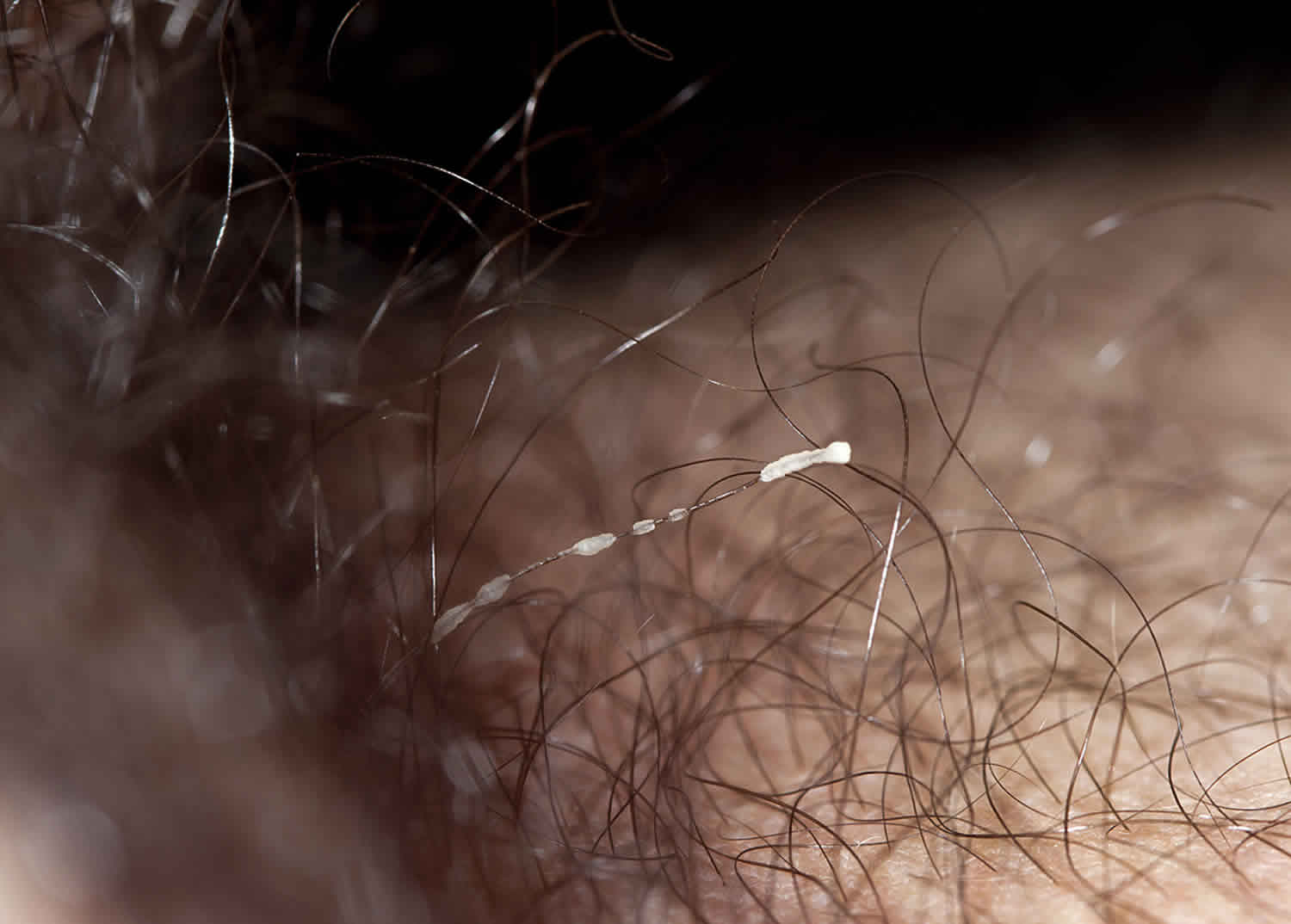
Trichomycosis pubis is often asymptomatic. Trichomycosis pubis is characterized by the appearance of concretions (hair-nodules), yellow, black or red granular nodules or concretions that stick to the hair shaft, which grow around the pubic hair shaft 3.
Due to the fact that the condition is asymptomatic and causes practically no discomfort 3, patients generally do not seek medical attention. However, when a careful, deliberate search is performed in the clinical setting, it tends to appear more frequently 4.
Is trichomycosis pubis STD?
No. Trichomycosis pubis is a benign condition that does not have any complications.
Trichomycosis pubis causes
Trichomycosis pubis is caused by the overgrowth of Corynebacterium (Corynebacterium tenuis, Corynebacterium propinguum, Corynebacterium flavescens) and Serratia marcescens bacteria. The concretions consist of tightly packed bacteria. The bacteria proliferate in moist areas of the body, thus mainly affect underarm hairs, and to a lesser extent, pubic hair. While as many as 33% of adults have colonization by these bacteria in the inguinal or axillary regions, factors such as hyperhidrosis initiate more extensive growth and clinical manifestations. Hence, disturbances in apo-eccrine sweat production and bacterial proliferation are crucial for development 5. The exact origin of the cement substance that creates the grossly visible nodules is debated. Electron microscopy studies favor origin from the causative agents, while others have favored elaboration from apocrine sweat 6. The actual nidus may be through the modification of apocrine sweat by elaborated cement substance to create the insoluble material that holds bacteria to the hair shaft. The white or yellowish, and less commonly, red or black, material on the hair contains an extremely high number of bacteria 7.
The bacteria cause malodour due to the metabolisation of testosterone in sweat into smelly compounds.
Trichomycosis infection begins when the causative agent comes in contact with the hair shaft, and the bacteria adhere to the surface, or the cuticle, of the hair, using a cement-like substance, the chemical composition of which is not yet known, that is insoluble in water as well as in the other principal solvents (i.e., acetone, ethanol) 8. Electron microscopy studies have clearly shown that the microorganism does not penetrate to the medulla’s cortex of the hair; instead, it only adheres strongly to the surface of the hair and develops slowly until it forms concretions around the hair shaft 9. Levit 10 has suggested that the adhesive substance is synthesized by both the apocrine glands of the human host and by the microorganism, which would explain why the disease develops in the areas of the body where it does (i.e., axillary, pubic and inter-gluteal hairs) 11.
Who gets trichomycosis pubis?
Trichomycosis pubis occurs in males and females of all races in temperate and tropical climates.
Contributing factors include:
- Humidity and warmth
- Crowded conditions
- Poor hygiene
- Hyperhidrosis
- Obesity
Studies in Panama and the United Arab Emirates revealed rates as high as 39% in patients attending a dermatology clinic 12. These results correlated with the notably higher incidence and prevalence in areas of high humidity, warmth, and poor hygiene. The only other study to mention incidence noted the presence of trichomycosis pubis upon examining institutionalized patients for trichomycosis axillaris in Edinburgh, Scotland, and noted that of the 609 men examined, 16 (2.6%) had pubic disease, of which three of the cases (0.5%) were not associated with axillary involvement. Ages of the males affected were 18 and 21 (3 patients) years and can be culled only from case reports. There is no universal consensus as to racial or sex preferences 13, although some reports note higher infection rates in men 14. Infection with Corynebacterium tenuis is also associated with poor hygiene, obesity, and hyperhidrosis 15. Human-to-human transmission has been noted and is particularly common in overcrowded groups such as soldiers, athletes, and among homosexuals 13.
Trichomycosis pubis symptoms
While many patients are asymptomatic, patients may present with reports of pubic rash, foul odor, or growths on the pubic hair. Colored sweat also has also been reported, resulting in a consideration of chromhidrosis.
Patients typically present with yellow, red, or black nodules or fine sheaths consisting of a bacterial biofilm encasing the hair shafts 5; yellow is most common, present in 95-98% of cases 16. Sweat in the region tends to be colored similarly. Lesions present in the inguinal region are often on the scrotum but occasionally on the base of the shaft of the penis. Lesions can be associated with erythema and itching, and superinfection with dermatophytes has been noted.
The primary complication appears in individuals who are immunocompromised and can develop septicemia secondary to colonization of catheters and surgical sites. Infection in hosts who are immunocompetent yields few long-term adverse effects.
Recurrence is common, but the simplicity of treatment and improved hygiene make follow-up care simple. In rare cases, shaving the pubic hair after treatment has been advocated to prevent recurrences.
Trichomycosis pubis diagnosis
- Trichomycosis pubis is mostly diagnosed by its clinical appearance.
- Wood lamp examination shows pale-yellow fluorescence.
- Potassium hydroxide preparation and Gram staining can identify the bacteria.
Trichomycosis pubis may resemble pubic lice (pediculosis) and Trichosporon aselie infections.
Trichomycosis pubis treatment
The fastest way to get rid of trichomycosis pubis is to clip the affected hairs or shave the area for a period of 2-3 weeks. Those patients who shave the affected area only once will generally experience a recurrence of the infection, since, the bacteria begin to develop the concretions once again as the hair grows back. Effective topical antibacterial preparations include clindamycin, erythromycin and fusidic acid. Topical treatments containing any of the following: 3% sulfur, 2% formalin, 1% mercury chloride (or mercuric chloride) or 2% sodium hypochlorite, as well as topical antibiotics with fusidic acid, erythromycin and clindamycin, may also be used 17. Benzoyl peroxide and sulfur soaps have also been reported to be effective in the treatment and prevention of trichomycosis 18. Anti fungal agents like naftifine and some azole derivatives (e.g., Clotrimazole powder) are effective as well and also curative 19.
Furthermore, rubbing while washing may disrupt the biofilm and increase the permeability of antiseptics, and shaving/clipping of the affected area and regular use of antiperspirants (including aluminum chloride) is often helpful and can reduce recurrences 5.
Recurrences of trichomycosis pubis are prevented by keeping the groin area dry and clean.
- Antiperspirants with aluminium chloride reduce sweating
- Antiseptics such as benzoyl peroxide gel or wash reduce bacterial colonization.
Trichomycosis pubis prognosis
Aside from the risk of recurrence, prognosis is excellent and treatment is effective. Morbidity is low, with most patients unaware of the colonization. When presenting, the most common reported symptom is a foul odor, and this may continue to cause problems, since trichomycosis often recurs.
References- Beigi RH, Meyn LA, Moore DM, Krohn MA, Hillier SL. Vaginal yeast colonization in nonpregnant women: a longitudinal study. Obstet Gynecol. 2004 Nov. 104 (5 Pt 1):926-30.
- Almazán-Fernández FM, Fernández-Crehuet Serrano P. Trichomycosis axillaris dermoscopy. Dermatol Online J. 2017 Jun 15. 23, 6.
- Bargman H. Trichomycosis of the scrotal hair. Arch Dermatol. 1984;120:299.
- McBride ME, Duncan WC. Trichomycosis axillaris. Arch Dermatol. 1972;105:459–60.
- Blaise G, Nikkels AF, Hermanns-Lê T, Nikkels-Tassoudji N, Piérard GE. Corynebacterium-associated skin infections. Int J Dermatol. 2008 Sep. 47 (9):884-90.
- White SW, Smith J. Trichomycosis pubis. Arch Dermatol. 1979 Apr. 115(4):444-5.
- Almazán-Fernández FM, Fernández-Crehuet Serrano P. Trichomycosis axillaris dermoscopy. Dermatol Online J. 2017 Jun 15. 23, 6
- Bonifaz A, Váquez-González D, Fierro L, Araiza J, Ponce RM. Trichomycosis (trichobacteriosis): clinical and microbiological experience with 56 cases. Int J Trichology. 2013;5(1):12–16. doi:10.4103/0974-7753.114704 https://www.ncbi.nlm.nih.gov/pmc/articles/PMC3746219
- Shelley WB, Miller MA. Electron microscopy, histochemistry, and microbiology of bacterial adhesion in trichomycosis axillaris. J Am Acad Dermatol. 1984;10:1005–14.
- Levit F. Trichomycosis axillaris: A different view. J Am Acad Dermatol. 1988;18:778–9
- Lestringant GG, Qayed KI, Fletcher S. Is the incidence of trichomycosis of genital hair underestimated? J Am Acad Dermatol. 1991;24:297–8
- Lestringant GG, Qayed KI, Fletcher S. Is the incidence of trichomycosis of genital hair underestimated?. J Am Acad Dermatol. 1991 Feb. 24(2 Pt 1):297-8.
- Bonifaz A, Ramírez-Ricarte I, Rodríguez-Leviz A, Hernández MA, Mena C, Valencia A. [Trichomycosis (trichobacteriosis) capitis in an infant: Microbiological, dermoscopic and ultrastructural features]. Rev Chil Pediatr. 2017 Apr. 88 (2):258-262.
- Rojas Mora E, Freites Martínez A, Hernández-Núñez A, Borbujo Martínez J. Trichomycosis axillaris: Clinical, Wood lamp, and dermoscopic diagnostic images. Actas Dermosifiliogr. 2017 Apr. 108 (3):264-266.
- Fernández-Crehuet P, Almazán-Fernández FM. [Trichomycosis axillaris]. An Pediatr (Barc). 2016 May. 84 (5):295.
- Bonifaz A, Ramírez-Ricarte I, Rodríguez-Leviz A, Hernández MA, Mena C, Valencia A. [Trichomycosis (trichobacteriosis) capitis in an infant: Microbiological, dermoscopic and ultrastructural features]. Rev Chil Pediatr. 2017 Apr. 88 (2):258-262
- Rho NK, Kim BJ. A corynebacterial triad: Prevalence of erythrasma and trichomycosis axillaris in soldiers with pitted keratolysis. J Am Acad Dermatol. 2008;58:S57–8.
- Navarrete-Dechent C, Fich F, Gonzalez S. Trichomycosis (trichobacteriosis) capitis misdiagnosed as poliosis: the utility of dermoscopy and why it should always be done. J Eur Acad Dermatol Venereol. 2017 Jun. 31 (6):e275-e276.
- Bonifaz A, Gómez-Daza F, Paredes V, Ponce RM. Tinea versicolor, tinea nigra, white piedra, and black piedra. Clin Dermatol. 2010;28:140–5.


