What is a vitrectomy
Vitrectomy is a microsurgical eye surgery used to treat problems of the eye’s retina and vitreous gel (also called vitreous humor). Vitreous gel (vitreous humor) is a thick, colorless, gel-like fluid that fills the large space in the middle of the eye, behind the lens. It helps the eyeball maintain its shape. In vitrectomy surgery, an ophthalmologist (eye specialist) may:
- remove blood or other substance keeping light from focusing properly on the retina
- remove scar tissue that is wrinkling or tearing the retina and causing poor vision
- help repair a retina that has detached (pulled away) from the eye wall
- remove a foreign object stuck inside the eye from an injury
During vitrectomy eye surgery, your ophthalmologist used small tools to remove some or all of the vitreous gel from the middle of your eye. This vitreous (vitreous humor) is replaced with either a salt water (saline) solution or a bubble made of gas or oil. It lightly presses the retina against the wall of the eye. You will need to keep your head in a certain position for most of the day and night while the eye heals. If an oil bubble is used, you will need another surgery to remove the oil after the eye has healed. After a while, the eye makes new fluid that fills in the space again.
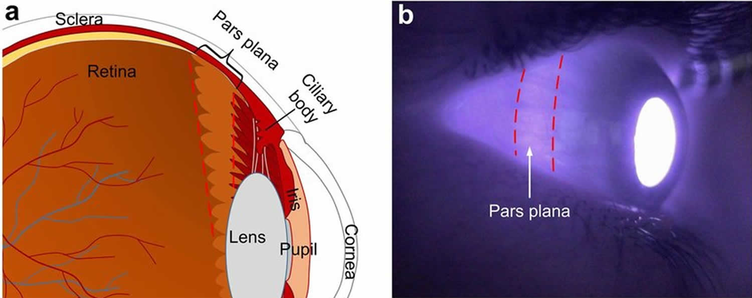
The initial step in vitrectomy is usually the placement of ports through very small incisions in the eye wall to the vitreous chamber (sclerotomy). Usually three ports are placed in this way: One for infusion, one for high in-tensity fiberoptic light sources to illuminate the vitreous chamber during surgery, and one for microsurgical instruments. The removal of the vitreous gel is achieved by miniature handheld cutting devices (vitrectomy probes), and by replacing the vitreous gel with special saline solu-tions. Although vitrectomy procedures are sometimes performed through incisions made near the front of the eye, most vitreoretinal surgeons enter the eye ball through an incision in the area of the “pars plana” (see Figure 3). This is an area of the ciliar body, which is divisible into two parts: the pars plana and the pars plicata anteriorly. The pars plana area is adjacent to the end of the retina and lacks of big vessels. Entering the eye through this location avoids damage to the retina and the lens.
Vitrectomy surgery lasts 2 to 3 hours. Your eye doctor will decide if only your eye will be numb or if you will also be asleep during surgery (local or general anesthesia).
Vitrectomy is usually done as outpatient surgery.
After the surgery, your eye may be swollen, red, or tender for several weeks. You might have some pain in your eye and your vision may be blurry for a few days after the surgery. You will need 2 to 4 weeks to recover before you can do your normal activities again. It may take longer for your vision to get back to normal.
Figure 1. Eye anatomy

Figure 2. Vitrectomy
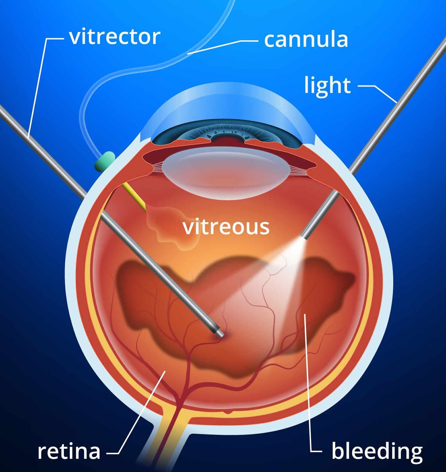
Figure 3. Pars Plana Vitrectomy
Vitrectomy indications
Your ophthalmologist may recommend a vitrectomy if you have one of these diseases or conditions:
- Diabetic retinopathy, with bleeding or scar tissue affecting the retina or vitreous gel
- Some forms of retinal detachment (when the retina lifts away from the back of the eye)
- Macular hole (a hole or tear in the macula)
- Macular pucker (wrinkles or creases in the macula)
- An infection in the eye called endophthalmitis
- Severe eye injury
- Certain problems during cataract surgery
- Vitreomacular traction
- Refractory macular edema
- Vitreous hemorrhage
- Tractional retinal detachment
- Rhegmatogenous retinal detachment
- Dislocated intraocular lens
- Refractory uveitis
- Retained lens material
- Intraocular foreign bodies
- Floaters
- Aqueous misdirection syndrome
What happens during a vitrectomy?
Vitrectomy is usually done in an outpatient surgery center. You will have a local or a general anesthesia to numb the eye. Surgery can take from one to several hours.
During surgery, the ophthalmologist will make a small cut (incision) in the white of the eye (sclera). He or she will use a microscope to see inside your eye. Your surgeon will use tiny tools to do one or more of these steps:
- remove all cloudy vitreous
- remove scar tissue from the retina
- remove any cataracts
- remove any object that should not be in the eye
- return the retina to its proper position against the back of the eye
- use a laser to repair a torn retina or other procedure
- place an air or gas bubble in your eye to help the retina remain in its proper position (bubble goes away on its own)
- place a silicone oil bubble in your eye (oil removed later during second surgery)
Following the surgery, you will be monitored as you rest and recover from anesthesia. Then you can go home.
Vitrectomy recovery
After vitrectomy surgery your ophthalmologist will prescribe medicine to help relieve pain. You will also be given eye drops to use for up to 4 weeks.
Your doctor will have you wear a patch on your eye for a few days to protect it. He or she will tell you when you can safely get back to doing your normal activities.
If a gas bubble was placed in your eye
At home, you may need to keep your head in a certain position for a while. You may need to keep your head in a face down (or side-facing) position for a specific period of time. Your ophthalmologist will tell you exactly how long to stay in that position. It is very important to follow these instructions to heal properly.
You cannot fly in an airplane until the gas bubble is gone. This is because a rapid altitude change can affect the size of the bubble.
Call your eye doctor right away if you notice any problems after surgery, such as:
- Decreasing vision.
- Signs of infection. These include increasing pain, redness, or swelling around the eye.
- Any discharge from the eye.
- Any new floaters, flashes of light, or other changes in your field of vision.
Activity
- Rest when you feel tired.
- Allow the eye to heal. Don’t do things that might cause you to move your head. This includes moving quickly, lifting anything heavy, or doing activities such as cleaning or gardening.
- If your doctor used an oil or gas bubble to hold the retina in place, keep your head in a certain position for most of the day and night for 1 to 3 weeks after the surgery. Make a plan for this part of your recovery, because it will be hard to do some daily activities. Your doctor will give you specific instructions.
- Do not lie on your back, or the bubble will move to the front of the eye and press against the lens instead of the retina.
- If your doctor used a gas bubble, avoid airplane travel until your doctor tells you it is safe. This is because the change in altitude may cause the gas bubble to expand and increase the pressure inside the eye.
- You will probably need to take 2 to 4 weeks off from work. It depends on the type of work you do and how you feel.
- You may drive when your vision allows it. If you are not sure, ask your doctor.
Ice and elevation
- Close your eye and put ice or a cold pack on it for 10 to 20 minutes at a time. Try to do this every 1 to 2 hours for the next 3 days (when you are awake) or until the swelling goes down. Put a thin cloth between the ice and your skin.
Medicines
- Your doctor will tell you if and when you can restart your medicines. He or she will also give you instructions about taking any new medicines.
- If you take aspirin or some other blood thinner, be sure to talk to your doctor. He or she will tell you if and when to start taking this medicine again. Make sure that you understand exactly what your doctor wants you to do.
- Be safe with medicines. Read and follow all instructions on the label.
- If the doctor gave you a prescription medicine for pain, take it as prescribed.
- If you are not taking a prescription pain medicine, ask your doctor if you can take an over-the-counter medicine.
- You will need to use eyedrops for up to 6 weeks.
Diet
- You can eat your normal diet. If your stomach is upset, try bland, low-fat foods like plain rice, broiled chicken, toast, and yogurt.
Other instructions
- You can shower and wash your hair and face. But don’t get any soap in your eye. You may want to use a wash cloth to wash your face. Some people wear swimming goggles.
- Wear sunglasses during the day. You may have to wear an eye patch or shield for a few days.
Call your local emergency number anytime you think you may need emergency care. For example, call if:
- You have a sudden loss of vision.
- You have severe eye pain.
Call your doctor now or seek immediate medical care if:
- Your vision gets worse.
- You have new or increasing eye pain.
- You have symptoms of an eye infection, such as:
- Pus or thick discharge coming from the eye.
- Redness or swelling around the eye.
- A fever.
- You have vision changes or see new flashes or floaters. (Flashes are “sparks” that you may see when you move your head. Floaters are shadows or dark objects that “float” across your field of vision.)
Watch closely for changes in your health, and be sure to contact your doctor or nurse call line if:
- You do not get better as expected.
What not to do after a vitrectomy
- Don’t do things that might cause you to move your head. This includes moving quickly, lifting anything heavy, or doing activities such as cleaning or gardening.
- If your doctor used an oil or gas bubble to hold the retina in place, keep your head in a certain position for most of the day and night for 1 to 3 weeks after the surgery. Make a plan for this part of your recovery, because it will be hard to do some daily activities. Your doctor will give you specific instructions.
- Do not lie on your back, or the bubble will move to the front of the eye and press against the lens instead of the retina.
- If your doctor used a gas bubble, avoid airplane travel until your doctor tells you it is safe. This is because the change in altitude may cause the gas bubble to expand and increase the pressure inside the eye.
Vitrectomy recovery time
You will probably need to take 2 to 4 weeks off from work. It depends on the type of work you do and how you feel.
Vitrectomy risks
Like any surgery, vitrectomy has risks. They include:
- Cataract, the most common complication
- Endophthalmitis
- Suprachoroidal hemorrhage
- Vitreous hemorrhage
- Retinal tear
- Retinal detachment
- Poor vision
- Glaucoma, when pressure builds up within your eye
- Optic neuropathy
- Phototoxicity
- Raised intraocular pressure, usually from gas or oil tamponade
- Hypotony (an intraocular pressure (IOP) of 5 mm Hg or less. )
Vitrectomy is generally related to the risk of post-operative endophthalmitis, hypotony, retinal detachment, mechanical trauma and, very rarely, detachment of trocar components.
Another possible risk after vitrectomy is getting a cataract in that eye. Cataract is especially likely to happen in people over age 50 who have vitrectomy. If you already had cataract surgery with a lens implant, vitrectomy will not harm your implanted lens.
Vitrectomy surgery often improves vision or keeps it from getting worse.


