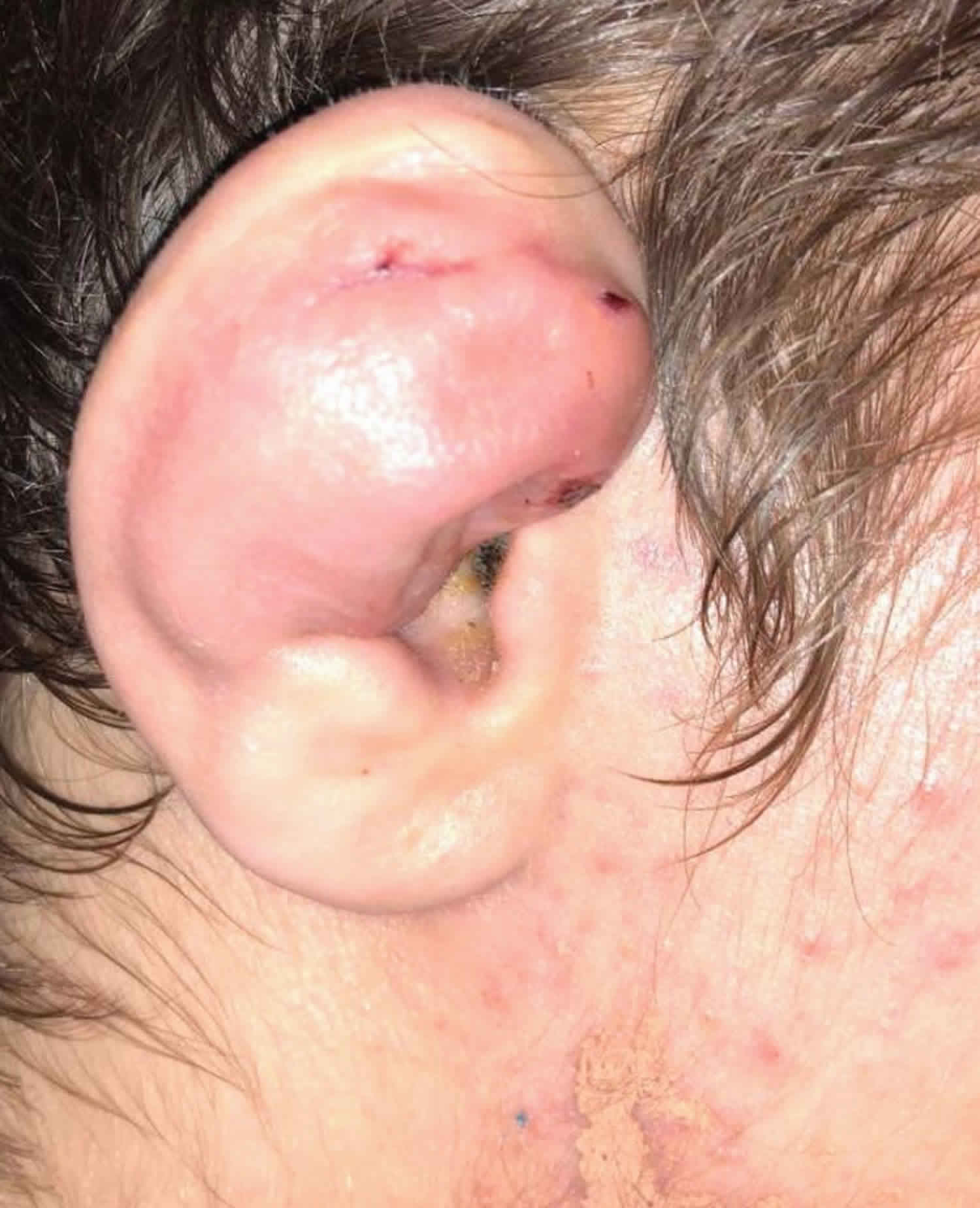
Wrestler’s ear
Wrestler’s ear also called cauliflower ear is a deformation of the ear typically caused by direct trauma to the auricula and surrounding tissue 1. Wrestler’s ear is classically experienced by wrestlers or boxers, the condition is caused by direct, blunt trauma where significant shearing forces lead to an auricular hematoma (a collection of blood underneath the perichondrium of the ear and typically occurs secondary to trauma) 2. The hematoma then disrupts the blood supply to auricular perichondrium and subsequently the ear’s cartilage. As a result, blood accumulates in the subperichondrial space and if left untreated can lead to necrosis, infection, and loss of cartilage. Cauliflower ear is the product of the fibrocartilage and fibrosis that occurs upon the destruction of the ears healthy cartilage once it loses its blood supply 3.
Contact sports such as wrestling, mixed martial arts, ultimate fighting, rugby, and boxing may more readily predispose to cauliflower ear. It could be deduced that males are at a higher risk than females; however, the exact ratio is not known. In a survey of college wrestlers, the incidence of auricular hematoma was found to be 52% for those refusing to wear headgear versus 26% who wore ear protection 4. This places them at a higher risk of developing cauliflower ear 5.
The exact prevalence of cauliflower ear is not noted in the literature. From studies conducted on wrestlers, researchers approximated that between 39-45% of athletes were affected with cauliflower ear 6.
It should be noted from the outset that cauliflower ear is potentially avoidable through the appropriate covering and protection of the ear during contact sports. This protection reduces or eliminates the types of blunt, shearing forces the ear experiences altogether and subsequently, the formation of an underlying hematoma. According to studies of wrestlers, the use of protective headgear can reduce the prevalence of auricular hematomas by up to 50% 7.
Management involves:
- Urgent aspiration of hematoma to prevent pinna deformity (i.e., wrestler’s ear or cauliflower ear)
- Pressure dressing applied after evacuation
- Close follow-up to monitor for reaccumulation
Wrestler’s ear causes
Auricular hematoma is typically caused by trauma. This can be from multiple forms of trauma, such as earring placement though is more common with a larger force or direct blow to the ear such as from a motor vehicle accident. It is most commonly secondary to contact sports such as wrestling, boxing, and martial arts 2
Historically, researchers have debated the exact mechanism behind the formation of cauliflower ear. From a series of experiments conducted in the mid-1970s when weights were dropped on the ears of rabbits, researchers concluded hematomas in the intercartilaginous space were responsible for the deformity. However, further testing (and more rabbits), showed blood accumulation in the subperichondrial space was responsible for disrupting circulation to the anterior ear from the posterior auricular and superficial temporal arteries. This buildup leads to the necrosis of the ear’s healthy cartilage. The hematoma is then replaced by chondroblasts which form neocartilage. This process sets into action a cascade of fibrosis and contracture and the subsequent development of cauliflower ear 6.
Wrestler’s ear symptoms
Patients present as having undergone trauma to the affected ear, usually during contact sports like wrestling, boxing, or increasingly, mixed martial arts. The ear appears red, swollen, and warm, while the patient may describe any combination of difficulty hearing, tinnitus, vision changes, pain, and/or a headache. If there is no concern for more underlying severe cranial pathology, then the practitioner should continue directly to treatment.
The strong relationship between trauma causing auricular hematomas should make practitioners suspicious of those individuals presenting with the signs described above, but who deny having undergone any reported trauma. Especially where children and the elderly are concerned, it is crucial that the possibility of abuse be entertained and screened for through a careful history and exam.
Wrestler’s ear diagnosis
Cauliflower ear is a clinical diagnosis that requires no formal testing or imaging.
Ultrasound can be utilized to evaluate ear swelling and to rule out an auricular abscess 3. If significant trauma has occurred, there is concern for a foreign body or an abscess or it is determined that it is important to evaluate middle or inner ear structures, CT or MRI can be ordered. CT and MRI should not be used routinely to evaluated auricular hematomas.
If there is evidence of erythema, warmth to the area, diffuse pain on palpation of cartilage, evidence of external auditory canal swelling, or drainage, then the diagnosis of auricular hematoma is less likely. Typically, hearing is not affected by isolated auricular trauma and if the patient has subjective hearing loss, then expanding the differential diagnosis is waranted. In summary, auricular hematomas are generally a clinical diagnosis.
Wrestler’s ear treatment
The management and treatment of cauliflower ear can take many forms depending on when the patient presents and the size and scope of the deformity. Any trauma to the head severe enough to injure the exterior ear mandates a thorough head and neck examination that includes the otoscopic inspection of the tympanic membranes. This exam ensures a more critical intracranial injury is not overlooked.
The primary treatment for cauliflower ear is prevention. The best possible outcomes require early identification and management of the hematoma before cartilage death has occurred. Typically, this requires a patient be seen within the first six hours of injury so that an emergency department doctor can aspirate the underlying auricular hematoma. This involves anesthetizing the distribution of the greater auricular nerve using lidocaine and epinephrine. This is followed by insertion of an 18-gauge needle into the area of greatest fluctuance. If the patient is unfortunate enough to present outside the 6-hour period where aspiration is likely to be successful, a more invasive incision is required to be sure to remove free blood, but also blood that has begun to coagulate.
By removing the pocket of blood, the perichondrium can reattach to the ear’s cartilage, preventing the loss of its valuable blood supply. Further aiding in the ear’s development of healthy cartilage, is the use of compressive dressings. A variety of compressive dressings are described in the literature including cotton bolsters, dental silicone, silicone rubber splints or even auricular stents, but no one technique has been studied and deemed superior.
Unfortunately, recurrence remains a frustrating complication of treating cauliflower ear. The most successful management of an auricular hematoma comes with the high likelihood that the blood will re-accumulate. For this reason, it’s important that outpatient providers refer patients to an otolaryngologist, an ear, nose, and throat (ENT) specialist for ongoing care and treatment.
For patients who present outside the window when auricular hematoma drainage is feasible, surgical intervention remains the mainstay of treatment. In general, a surgical referral is appropriate for any patient with an existing cauliflower ear, where extensive fibrosis has already occurred. Once the surgery is settled upon, the exact approach can vary based on the degree and location of the underlying deformity. Some texts go so far as to classify cauliflower ear into four distinct types, each requiring a unique reconstructive technique. The overall goal of surgery, however, remains the same regardless of surgical method, and that is to remove the damaging fibrocartilage without compromising the ear’s structural integrity or its natural contours. If damage to the ear is so severe that simply removing the malformed cartilage does not equate to the desired cosmetic outcome, costal cartilage can be used to provide greater structural integrity.
General procedure steps for auricular hematoma drainage
- Gather the necessary equipment and make sure there is appropriate lighting.
- Ensure adequate exposure and place patient in the supine position with the head of the bed elevated.
- The patients head should be turned so that the unaffected ear is facing towards the stretcher and the affected ear is towards the ceiling.
- Supplies should consist of an 11 blade or 15 blade scalpel and/or an 18-gauge needle with a 10 cc syringe, suction canister, tubing and suctioning instrument (Frasier), a hemostat, toothed forceps, suture supplies with scissors, bolster material, local anesthetic, and local skin cleansing material 2.
- Appropriate hand hygiene should be practiced and gloves should be worn during the procedure. Application of sterile gloves, gowning, headlamp use, and or Loupes to optimize vision are optional.
- After the patient is positioned properly, the ear is cleaned with a local cleansing agent such as povidone-iodine.
- Local anesthesia should then be injected or applied topically to the site where the incision or aspiration will be performed (e.g., lidocaine, bupivacaine, LET gel). For best results, the anesthetic can be injected in an auricular block pattern or directly into the site of the auricular hematoma. Several minutes after injection the level of local anesthesia should be assessed. This can be performed by grabbing the tissue of the planned incision with toothed forceps to determine if the area is numb. There are two methods that can potentially be used to drain the auricular hematoma. You should choose the method you will utilize prior to start of the procedure. One method is to incise and drain the hematoma using a scalpel the other is needle aspiration
Incision and drainage
- First complete steps 1 through 7 listed above under general procedure steps.
- Next make a linear incision can be made on the skin overlying the swelling or hematoma 2. The goal of the incision is to drain the fluid collection; however, making the incision in a cosmetically appealing site is ideal. Incision in areas of concavity will heal with a more aesthetically pleasing results compared to areas of convexity.
- After the incision is made, hemostats and suction can be used to evacuate the hematoma.
- Once all the hematoma is removed, the site can be irrigated with normal saline.
- A bolster dressing is applied. The bolster serves to close the dead space or potential space where the hematoma formed. When using dental rolls as a bolster, two rolls should be used. Each roll should be placed so it to runs parallel with the incision line on either side of the ear. Two vertical mattress sutures should be placed through the dental rolls to secure the bolster 2. A permanent suture material such as nylon is appropriate. The suture is ideally on a Keith Needle; however, this is not mandatory. Adequate bolster is applied when there is no potential space for accumulation of hematoma; however, it is important to make the sutures lose enough to preserve the vascular supply of the ear.
- Bacitracin can be applied to the incision site post procedure.
- It is important to remove all instruments and dispose of sharps appropriately once the procedure is deemed complete. Proper wound care instructions, follow-up, and disposition should be explained to the patient and/or family.
Needle Aspiration
- First complete steps 1 through 7 listed above under general procedure steps.
- The alternate procedure utilizes an 18-gauge needle to aspirate the hematoma. Some studies suggest that an 18-gauge needle may be acceptable for auricular hematoma evacuation when the hematoma is under 2 cm 2. If the needle aspiration technique is used a bolster should be applied to the affected area of the ear after complete removal of the hematoma.
- A bolster dressing is applied. The bolster serves to close the dead space or potential space where the hematoma formed. When using dental rolls as a bolster, two rolls should be used. Each roll should be placed so it to runs parallel with the incision line on either side of the ear. Two vertical mattress sutures should be placed through the dental rolls to secure the bolster 2. A permanent suture material such as nylon is appropriate. The suture is ideally on a Keith Needle; however, this is not mandatory. Adequate bolster is applied when there is no potential space for accumulation of hematoma; however, it is important to make the sutures loose enough to preserve the vascular supply of the ear.
- Bacitracin can be applied to the incision site post procedure.
- It is important to remove all instruments and dispose of sharps appropriately once the procedure is deemed complete. Proper wound care instructions, follow-up, and disposition should be explained to the patient and/or family.
Bolster options post hematoma evacuation
There are several variances in the type of bolster used, however, the goal is the same: eliminate the potential space for fluid to accumulate 2. Newer strategies include the use of splinting material that can be molded to the ear 8. A recent case reports using fibrin glue to secure the perichondrium to the cartilage to reduce the risk of separation 9. Bolster dressing can be removed after 5 to 7 days. Antibiotic use is left to the discretion of the physician. If cauliflower ear does form, excision with repair may be undertaken in the form of otoplasty; however, this will require referral to ENT or plastic surgery 2.
Postoperative and rehabilitation care
Patients with auricular hematomas can generally be managed as outpatients. Consultation and/or referral to otolaryngology or plastic surgery is recommended. Patients are instructed to take antibiotics if prescribed, pain medications as prescribed and follow-up as directed. The patient should limit physical activity for 10 to 14 days and avoid contact sports for 1 to 2 weeks. If a splint is used, this will need to be removed in 5 to 7 days. Antibiotics can be given while the splint is in place.
References- Kordi R, Maffulli N, Wroble RR, Wellby S, editors. Combat Sports Medicine. 1 ed. London: Springer; 2009.
- Greywoode JD, Pribitkin EA, Krein H. Management of auricular hematoma and the cauliflower ear. Facial Plast Surg. 2010 Dec;26(6):451-5.
- Krogmann RJ, King KC. Auricular Hematoma. [Updated 2019 Dec 13]. In: StatPearls [Internet]. Treasure Island (FL): StatPearls Publishing; 2020 Jan-. Available from: https://www.ncbi.nlm.nih.gov/books/NBK531499
- Ghanem T, Rasamny JK, Park SS. Rethinking auricular trauma. Laryngoscope. 2005 Jul;115(7):1251-5.
- Schuller DE, Dankle SD, Strauss RH. A technique to treat wrestlers’ auricular hematoma without interrupting training or competition. Arch. Otolaryngol. Head Neck Surg. 1989 Feb;115(2):202-6.
- Patel BC, Skidmore K, Hutchison J, et al. Cauliflower Ear. [Updated 2019 Jul 24]. In: StatPearls [Internet]. Treasure Island (FL): StatPearls Publishing; 2020 Jan-. Available from: https://www.ncbi.nlm.nih.gov/books/NBK470424
- Shilpa K, Leelavathy B, Lakshmi DV, Divya G. Unilateral Cauliflower Ear due to Leprosy or Trauma – A Diagnostic Challenge. Indian J Lepr. 2016 Jul-Sep;88(3):189-192.
- Choung YH, Park K, Choung PH, Oh JH. Simple compressive method for treatment of auricular haematoma using dental silicone material. J Laryngol Otol. 2005 Jan;119(1):27-31.
- Mohamad SH, Barnes M, Jones S, Mahendran S. A new technique using fibrin glue in the management of auricular hematoma. Clin J Sport Med. 2014 Nov;24(6):e65-7.


