What is acne keloidalis nuchae
Acne keloidalis nuchae also known as keloidal folliculitis (inflammation of hair follicle unit) or nuchal keloidal acne, is a chronic skin condition characterized by inflamed bumps and scars on the on the nape of the neck and occipital scalp 1, 2. These acne keloidalis nuchae is incorrect because acne keloidalis nuchae is not acne and the scars formed are not true keloids. The names are confusing, especially as acne can result in keloid scarring. Although it is not related to common acne (acne vulgaris), acne keloidalis nuchae initially appears as acne-like lesions of inflamed hair follicles (folliculitis) on the nape of the neck (nuchal area) and without treatment, can result in large scars (keloids).
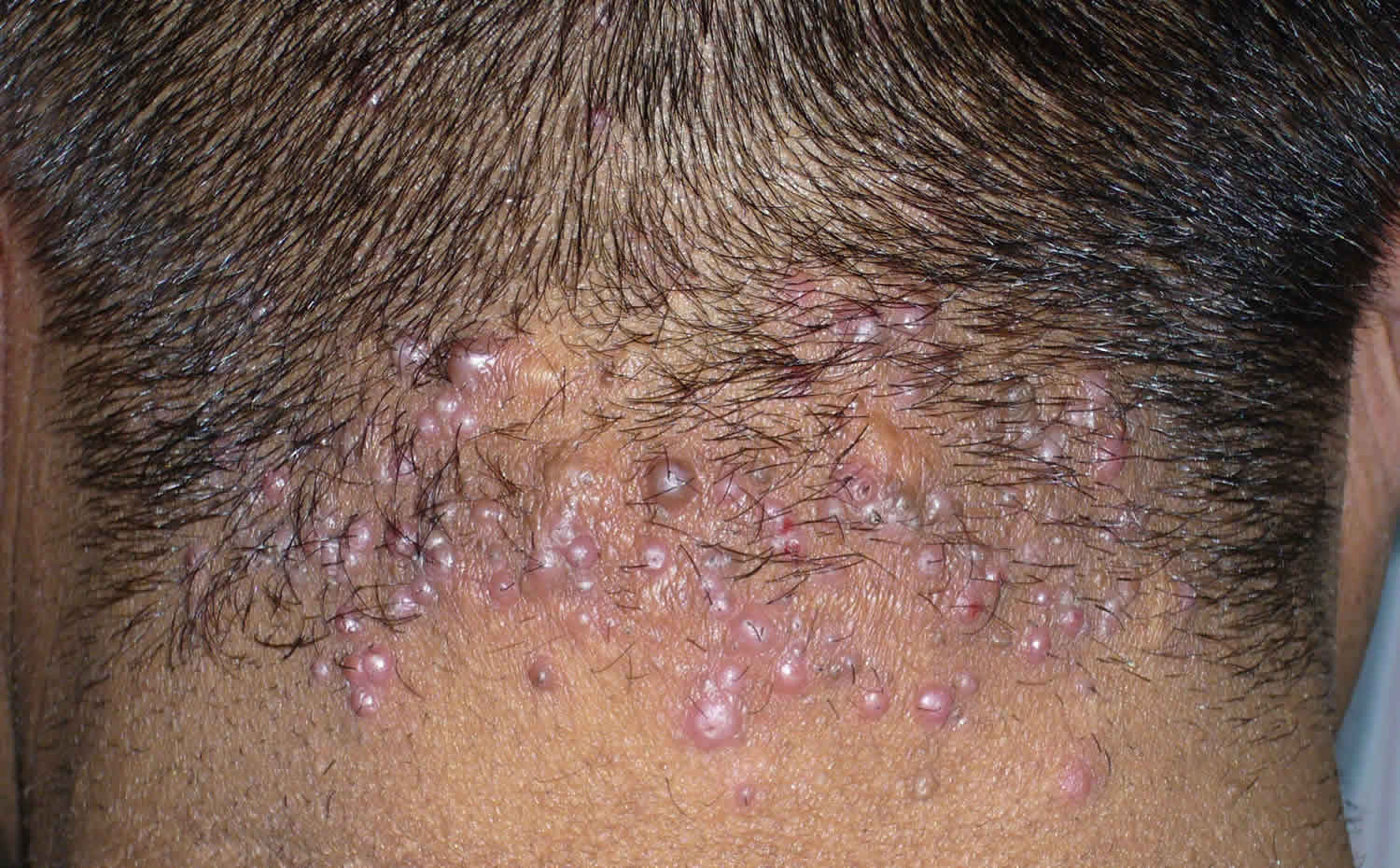
Characteristic findings on physical examination include follicular-based papular or pustular presentation with ingrown hairs and hypertrophic or keloidal scarring on the nape of the neck. Papules or pustules may form confluent plaques with draining sinus tracks. Involved follicles may show tufted hair folliculitis while intact follicles at the margins may demonstrate polytrichia. More advanced or progressive states may demonstrate keloidal formation.
In the United States, acne keloidalis nuchae occurs most commonly in young Afro-American men, followed in frequency by Hispanic, Asian descent and less often Caucasian men. Acne keloidalis may occur in women, but it is very uncommon in women with the male-female ratio is at least 20:1. In addition, acne keloidalis nuchae is very rarely seen in people prior to puberty or after middle age.
If you suspect you have acne keloidalis nuchae, you should seek help from your primary care provider or a dermatologist in order to prevent the possible formation of large scars and permanent hair loss to the involved areas.
Patients with acne keloidalis nuchae to avoid any causes of mechanical irritation to the posterior hairline: no plucking, pulling or close haircutting in the region, with recommended daily brushing of the area to lift hairs that may be regrowing into the surface of the skin.
Is acne keloidalis nuchae contagious?
The exact etiology of acne keloidalis nuchae is unclear, but it might be triggered by chronic irritation or occlusion of the follicles due to hair cutting practices (e.g., close shaves), trauma, friction (e.g., rubbing from shirt collars or helmets), heat, or humidity as a predisposing or exacerbating factors 3.
Acne keloidalis nuchae causes
The definitive cause of acne keloidalis nuchae is unknown. No specific genetic factor has been identified. Some researchers have concluded that acne keloidalis nuchae may begin with an injury during a close hair cut or use of a razor. In individuals with tightly curled hair, shaving or trimming the nape of the neck may initiate the presentation of acne keloidalis nuchae. It is thought to be a mechanical form of folliculitis, in which ingrown hair shafts irritate the wall of the hair follicle resulting in inflammation. This completely destroys the hair follicle and results in scarring.
The association of mild trauma to the hair follicle and subsequent development of a “pseudofolliculitis “ response in the posterior neck region supports an analogous mechanism of action and potential genetic predisposition. In one study, two thirds of the acne keloidalis nuchae-affected population had concomitant seborrheic dermatitis. The persistent irritation and potential trauma from excoriation may initiate the acne keloidalis nuchae process in those with seborrheic dermatitis.
The pathophysiologic mechanism responsible for acne keloidalis is analogous to that of pseudofolliculitis barbae. Basically, it represents a foreign-body reaction as a result of the hair penetrating the dermis. The actual penetration of the skin can occur through one of two pathways: through the stratum corneum after growing out of the follicle and curling back towards the skin, or by piercing the follicular wall directly.
Also, acne keloidalis nuchae might be triggered by infection (Demodex or bacteria) 3. Other potential contributory factors include autoimmunity, excess androgens or increased sensitivity to androgens, seborrhea, and medications (e.g., cyclosporine) 4.
Others argue that acne keloidalis nuchae is a primary skin disease unrelated to either ingrown hairs or bacterial infection.
An association with obesity and metabolic syndrome has been observed in some patients.
Acne keloidalis nuchae pathophysiology
The repeated friction, trauma, and infection of the skin overlying the nape of the neck leads to acne keloidalis nuchae. Herzberg et al. proposed a series of ultrastructural events that are responsible for the formation of acne keloidalis nuchae 5:
- Acute perifollicular inflammation followed by a weakening of the follicular wall at the level of the lower infundibulum, the isthmus, or both.
- Formation of acute and chronic granulomatous inflammation by the release of naked hair shaft which acts as a foreign body.
- The fibroblasts will produce new collagen and fibrosis.
- Distortion and occlusion of the follicular lumen by the fibrosis results in retention of the hair shaft in the follicle.
Acne keloidalis nuchae signs and symptoms
The most common locations of acne keloidalis nuchae include:
- Back of the neck (posterior neck)
- Lower back of the scalp (occipital scalp)
Initially, lesions of acne keloidalis nuchae appear as red or pus-filled bumps close to the hair-bearing area of the back of the neck (occipital scalp), which may be tender or very itchy and scratching can lead to secondary bacterial infection (Staphylococcus aureus). Sometimes there are pustules around the hair follicles (folliculitis). Over time, these inflamed bumps develop into small scars. Without treatment, the small scars may greatly enlarge into large, thick scars, or keloids. Areas of widespread scarring may be associated with hair loss and can form a band along the hairline. Tufted hairs may be present; these are multiple hair shafts emerging from a single follicular opening. Rarely, advanced acne keloidalis nuchae lesions can develop deep pockets of pus with connections to the surface of the skin, and a foul-smelling discharge may ooze from these sinus tracts.
Acne keloidalis nuchae diagnosis
The diagnosis of acne keloidalis nuchae is made clinically by finding follicular papules, pustules, and scars on the occipital scalp, but laboratory studies are done to look for bacteria. If pathogenic microorganisms are isolated, then the appropriate antibiotics should be started. The histology of folliculitis keloidalis nuchae is characteristic, should a biopsy be performed.
Acne keloidalis nuchae treatment
Overall, the treatment of acne keloidalis nuchae is difficult and unsatisfactory. Over the years, many treatments have been used with varying degrees of success. There is no first-line therapy, and it all depends on the personal preference of the treating physician. The prognosis of acne keloidalis nuchae is good if treatment is started early.
The following measures are sometimes helpful:
- Making sure clothing and equipment, such as high collars and helmets, do not rub the back of the neck
- Avoid a short or razor hair cut.
- Wash the affected area using an antimicrobial cleanser to reduce secondary infection.
- 2 to 4–week courses of topical steroids are useful if the papules are less than 3 mm in size
- Steroids injected into the lesions (intralesional injections) are more suitable for large papules and plaques.
- Oral tetracycline as an anti-inflammatory or other antibiotics for secondary infection
- Laser-assisted hair removal has been shown to improve folliculitis keloidalis. Best results occur if treatment is started early before significant scarring has developed.
- A three-month course of clindamycin and rifampicin antibiotics if infection persists
- Surgery to removing large thickened plaques or nodules
- Laser vaporisation or excision and electrosurgery are alternatives to surgery
- Oral isotretinoin
- Radiotherapy
Topical creams, lotions, or gels may include:
- A retinoid cream such as tretinoin, tazarotene, or adapalene.
- A prescription-strength steroid or cortisone preparation.
- An antibiotic such as clindamycin.
Oral medications may include:
- Antibiotic pills.
- A short course of steroids, such as prednisone (for severe or advanced cases only).
Procedures to reduce inflammation and reduce or remove scar tissue include:
- Steroid injections directly into the inflamed bumps or scars.
- Surgical excision of single bumps or larger scars.
- Laser destruction.
- Liquid nitrogen (freezing or cryotherapy).
The first part of treatment is to educate the patient on preventing progression of the disease. This means avoiding close shaving and frequent haircuts. Secondly, the individual should be told to avoid shirts with tight collars and restrictive athletic neck gear. Any ornaments like chains worn around the neck should be discontinued.
The treatment should be initiated at the time of diagnosis to avoid cosmetic disfigurement. One may use topical antimicrobial agents such as benzoyl peroxide or chlorhexidine to prevent secondary bacterial infections.
Mild keratolytic agents can help soften the coarse hairs.
The patient should be told to avoid the use of all hair product greases that can interfere with hair growth.
When the disease is in the early papular stage, it may respond to potent topical steroids with or without a retinoid.
If folliculitis is present, one may need to use minocycline or doxycycline. Moderate dose isotretinoin (20 mg daily) has an anti-inflammatory effect on folliculitis but does not affect indurated lesions.
Once the acute disease is under control, one can use maintenance therapy with a topical steroid, benzyl peroxide washes, and a topical retinoid.
If the papules are hard, intralesional injection of triamcinolone can help reduce the size and also soften the lesion. The injections can be very painful, and pretreatment with topical lidocaine may help. Steroid injections may result in hypopigmentation and skin atrophy. Some dermatologists use cryotherapy with liquid nitrogen to create edema before the injection of steroids.
Cryotherapy has also been used as solo therapy, but it is often not well-tolerated because of pain. The treatment also causes hypopigmentation and often requires months of treatment.
Surgical excision may be performed, but mild recurrences are not rare. Cryosurgery also seems to be effective in acne keloidalis nuchae, while vaporization with the CO2 laser is less effective than surgery.
Various other types of lasers have been used to treat acne keloidalis nuchae with varying success. The most effective lasers are the 1064 nm Nd: YAG laser and 810 nm diode laser. Most patients require several laser sessions in combination with topical steroids or retinoids to improve the cosmesis. Pulsed Dye laser (595 nm) has also been used in acne keloidalis nuchae but is less effective than the other lasers. Phototherapy using targeted ultraviolet B light three times a week for 8 to 10 weeks has also been effective.
Radiation therapy should only be reserved for recalcitrant cases.
Noninflamed papules
Topical retinoid such as tretinoin 0.1% cream, topical cortical steroid such as betamethasone diproprionate 0.05% gel or combination of both.
Inflamed papules with pustules
Bacterial cultures to rule out Staphylococcus aureus or methicillin-resistant Staphylococcus aureus (MRSA), topical clindamycin, oral tetracycline, doxycycline or minocycline incombination with Derma-Smoothe oil with monthly follow up to check progress. If no progress is noted, advance therapy. Papules could also be intralesionally injected with triamcinolone monthly with initial triamcinolone 3 mg/kg for small lesion up to triamcinolone 10 – 40 mg/kg for nodularity. Laser hair removal, such as the neodynium: YAG as in pseudofolliculitis barbae patients, is beneficial in acne keloidalis nuchae. In individuals with culture-positive S aureus, ensure that nasal carriage has been adequately assessed with culture and treated with topical mupirocin . Hibiclens soap could also be induced into the hygienic routine.
Nodules, plaque or keloid development
Once hypertrophic scarring has developed, treatment with oral or topical antibiotics is much less successful and measures to control formation of hypertrophic or keloidal scarring must be employed. Potent topical corticosteroid ointments may be helpful, but intralesional injection of triamcinolone can drastically reduce the bulk of scar tissue. Surgical excision with primary closure or for larger lesions, allowing them to heal by secondary intention. In severe cases with plaques larger than10 cm that were unresponsive to triamcinolone injections, monthly 5-fluorouracil injections have demonstrated excellent results.
Sinus tracts
Sinus tracts may respond to chloramphenicol 500mg in 30 g of fluocinonide ointment/cream, applied three times a day.
Acne keloidalis nuchae natural treatment
People who develop acne keloidalis nuchae should focus on avoiding irritation to the area in order to prevent the formation of additional lesions:
- Wash the area gently with non-irritating cleansers (no hard scrubbing!).
- Avoid head wear (such as sports helmets) and shirt collars that rub against the back of your neck.
- Avoid closely shaving of the back of your neck.
- For itchy lesions, try an over-the-counter cortisone cream.
Generally, persons with acne keloidalis nuchae should see their primary care doctor or a dermatologist for treatment in order to prevent progression of the condition.
References- Na K, Oh SH, Kim SK. Acne keloidalis nuchae in Asian: A single institutional experience. PLoS One. 2017 Dec 14;12(12):e0189790. doi: 10.1371/journal.pone.0189790
- Maranda EL, Simmons BJ, Nguyen AH, Lim VM, Keri JE. Treatment of Acne Keloidalis Nuchae: A Systematic Review of the Literature. Dermatol Ther (Heidelb). 2016 Sep;6(3):363-78. doi: 10.1007/s13555-016-0134-5
- Al Aboud DM, Badri T. Acne Keloidalis Nuchae. [Updated 2019 Mar 23]. In: StatPearls [Internet]. Treasure Island (FL): StatPearls Publishing; 2019 Jan-. Available from: https://www.ncbi.nlm.nih.gov/books/NBK459135
- Adotama P, Tinker D, Mitchell K, Glass DA, Allen P. Barber Knowledge and Recommendations Regarding Pseudofolliculitis Barbae and Acne Keloidalis Nuchae in an Urban Setting. JAMA Dermatol. 2017 Dec 01;153(12):1325-1326
- Herzberg AJ, Dinehart SM, Kerns BJ, Pollack SV. Acne keloidalis. Transverse microscopy, immunohistochemistry, and electron microscopy. Am J Dermatopathol. 1990 Apr;12(2):109-21.


