What is idiopathic guttate hypomelanosis
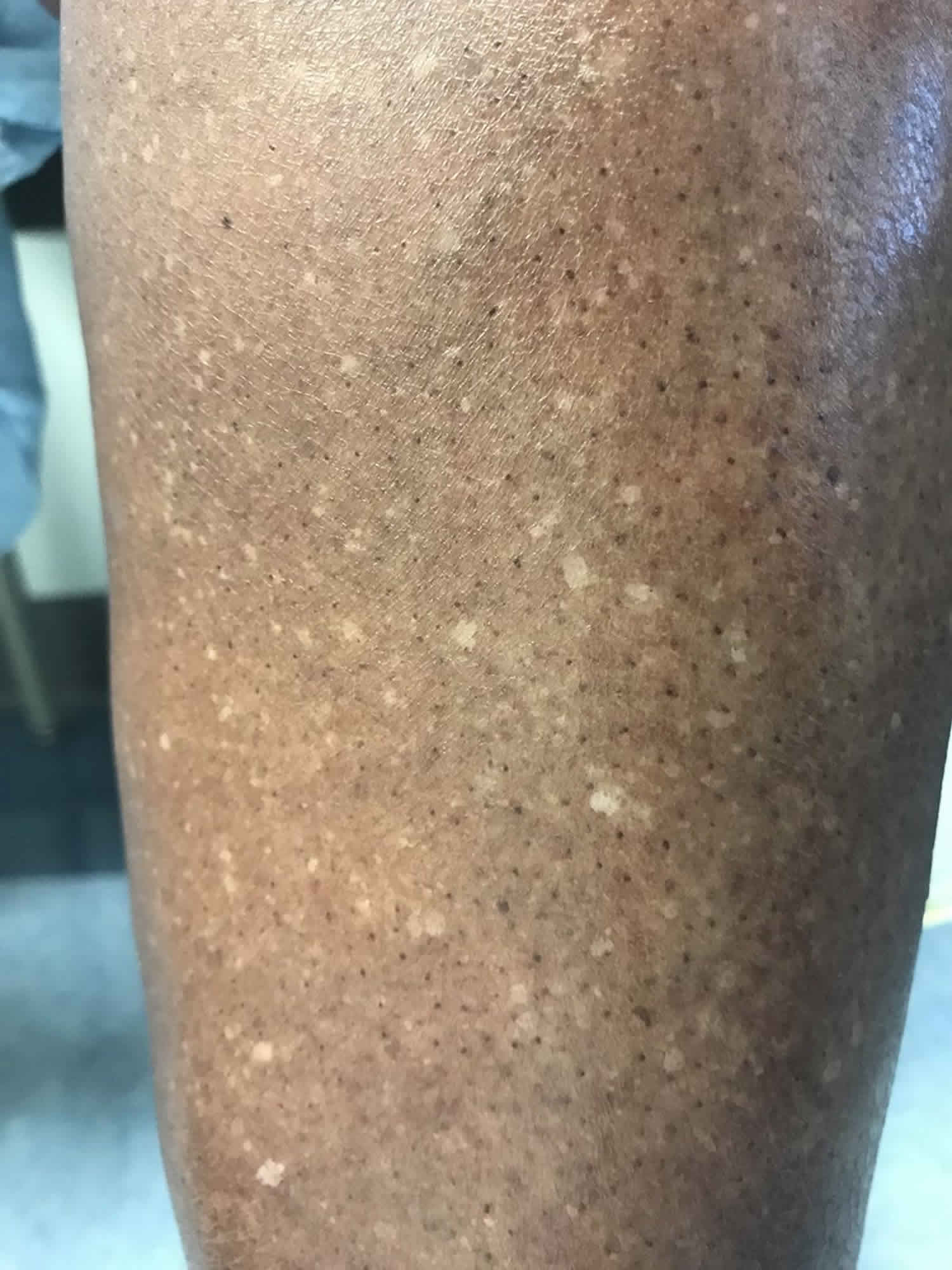
Idiopathic guttate hypomelanosis also called leukopathia symmetrica progressiva, is the name given to a common benign acquired skin condition that presents as small irregularly-shaped, well-defined hypopigmented porcelain white macules (2 to 5-mm flat white spots), predominantly on shins and forearms. ‘Idiopathic’ means the cause is unknown, ‘guttate’ means resembling tear-drops, and ‘hypomelanosis’ refers to the lighter color of the affected areas. The surface is smooth, without atrophy. Usually, the trunk and face are spared. Once present, lesions do not change in size.
Idiopathic guttate hypomelanosis progresses with increasing sun exposure and, to a lesser degree, with age.
Idiopathic guttate hypomelanosis is more common in those over 40 years of age and the prevalence increases with age; however, idiopathic guttate hypomelanosis may occur in young adults. They are more common in women than in men. Inherited factors may be relevant as the lesions appear to be more common in family members.
Idiopathic guttate hypomelanosis predominantly affects fair-skinned individuals, but it may occasionally arise in darker skin. Although most often found on the shins and sun-exposed parts of the forearms, guttate hypomelanosis may also arise on other sun-exposed areas including the face, neck and shoulders. The white marks are usually smooth with a reduction in the normal skin markings, but they may be slightly scaly.
While idiopathic guttate hypomelanosis is benign, this condition may be visually disturbing to some patients and treatment may be requested. Primary treatment should consist of educating patients on the importance of sun protection as the appearance of idiopathic guttate hypomelanosis points to signs of sun damage. Patients should be encouraged to wear sunscreen on a daily basis and avoid artificial tanning beds. Treatment modalities that may reduce the appearance of idiopathic guttate hypomelanosis include the topical steroids, tretinoin, pimecrolimus and dermabrasion.
Does idiopathic guttate hypomelanosis spread?
Idiopathic guttate hypomelanosis is most commonly a complaint of middle-aged, light-skinned women, but it is increasingly seen in both sexes and older dark-skinned people with a history of long-term sun exposure. Idiopathic guttate hypomelanosis is seen far more frequently in women, beginning around the age of 30 years. However, with increasing age and sun exposure, it is found almost equally in elderly men and women. Why idiopathic guttate hypomelanosis occurs earlier in young women than in young men is unknown. Fair-skinned women develop this condition first; later, with increasing age and exposure to sun, both sexes seem to be equally affected.
The cause is not known, but it appears to be related to the effect of the sun on melanocytes. Because pigmentation of the skin is due to an integration of melanocyte and keratinocyte function, an acquired defect of the epidermal melanin unit results in the observed hypopigmentation in idiopathic guttate hypomelanosis patients. In 1967, Hamada and Saito 1 found a 50% reduction in melanocytes.
Is idiopathic guttate Hypomelanosis dangerous?
Idiopathic guttate hypomelanosis is aesthetically displeasing. However, it is not a dangerous process 2. Once present, lesions do not remit.
Idiopathic guttate hypomelanosis causes
The exact cause of idiopathic guttate hypomelanosis is unknown. A skin biopsy demonstrates that there is no pigment (melanin) in the skin cells (keratinocytes). There is also a reduction in the number of pigment-producing cells (melanocytes). The skin is slightly thinner than normal and flattened out.
Histology reveals flattening of rete ridges at the dermal-epidermal junction, moderate to marked reduction of melanin granules in the basal and suprabasal layers, basket-weave hyperkeratosis, and a reduced number of DOPA-positive epidermal melanocytes.
The cause of idiopathic guttate hypomelanosis is speculative. It is thought to be an inevitable part of the ageing process, with a gradual reduction in melanocytes – a similar process to greying of hair. Other theories include:
- Sun damage – the lesions are a kind of white freckle
- Non-sun related seborrheic keratosis – degenerative scaly spots
Other possible causes include genetic factors like HLA-DQ3, phototherapy and autoimmunity.
Idiopathic guttate hypomelanosis does not appear to be due to trauma or viral infection. The white areas do not predispose to skin cancer.
Idiopathic guttate hypomelanosis symptoms
Idiopathic guttate hypomelanosis is a benign and asymptomatic skin manifestation characterized as diffuse hypopigmented macules, or white spots. The size of the lesions varies from 1-10mm, but are most commonly 1-3 mm in diameter. It is most commonly seen in fair-skinned individuals and appears to be related to cumulative sun exposure rather than just age. Interestingly, this condition manifests in women earlier than men, probably because their legs are more exposed. However, as age increases the incidence of idiopathic guttate hypomelanosis in women and in men is the same. idiopathic guttate hypomelanosis can also be seen in older dark-skinned individuals, but usually at more advanced age. The distribution of idiopathic guttate hypomelanosis can be seen along most exposed areas of the body. These include areas of the arms, legs, upper back, and face. Lesions are usually seen first along the anterior portion of the legs and then seen on the arms, back and face.
Idiopathic guttate hypomelanosis diagnosis
The diagnosis of idiopathic guttate hypomelanosis is made clinically by visual inspection alone. Biopsies can be done, but are usually unnecessary. Histology reveals flattening of rete ridges at the dermal-epidermal junction, moderate to marked reduction of melanin granules in the basal and suprabasal layers, basket-weave hyperkeratosis, and a reduced number of DOPA-positive epidermal melanocytes.
Idiopathic guttate hypomelanosis treatment
In most cases, treatment is not required as the marks are completely harmless. Attempts to destroy the lesions may leave brown marks or larger white marks, which may look worse than the original condition. Sun protection is very important.
The following measures may improve the appearance.
- Broad spectrum sunscreens (SPF 50+)
- Light cryotherapy
- Localized superficial dermabrasion or microdermabrasion
- Carbon dioxide laser
- Chemical peel (phenol and trichloroacetic acid)
- Pinch grafts of normally pigmented skin
- Topical tretinoin cream
- Topical steroids
- Topical pimecrolimus 1%
- Cosmetic cover-up.
Patients should be advised that treatment options are limited and results are mixed. Sun protection can help prevent development of new lesions. If treatment with cryotherapy is being considered, patients should be counseled regarding the risk of hypopigmentation post-treatment. Also, if a patient has a history of hypertrophic scars or keloids, a test site site should be done before treatment of all lesions with superficial dermabrasion.
Treatment for idiopathic guttate hypomelanosis is limited. Most treatments are anecdotal without any randomized controlled clinical trials. A conservative approach with observation and photoprotection may prevent development of new lesions.
Tretinoin cream used daily for 4 months showed some improvement in pigmentation in one study. In this study 0.025% tretinoin cream was used nightly for the first week, 0.05% nightly for the second week, and 0.1% nightly for the remainder of the study. Pimecrolimus 1% cream applied twice daily for 16 weeks demonstrated some improvement in pigmentation in another small study.
Cryotherapy has been reported to be useful in treating idiopathic guttate hypomelanosis. In one study, treated lesions showed repigmentation in 6-8 weeks and histologically demonstrated more DOPA-positive melanocytes than untreated lesions, but less than normal skin. Another small study, reported that treatment with local superficial dermabrasion caused repigmentation in 80% of treated patients. Carbon dioxide laser and superficial chemical peels have been tried as treatment options without much success.
In addition, cover-up make up and concealers made by brands such as Dermablend can be used to mask the hypopigmented macules.
References- Hamada T, Saito T. SENILE DEPIGMENTED SPOTS (IDIOPATHIC GUTTATE HYPOMELANOSIS). Arch Dermatol. 1967;95(6):665. doi:10.1001/archderm.1967.01600360111025
- Brown F, Crane JS. Idiopathic Guttate Hypomelanosis. [Updated 2018 Oct 27]. In: StatPearls [Internet]. Treasure Island (FL): StatPearls Publishing; 2019 Jan-. Available from: https://www.ncbi.nlm.nih.gov/books/NBK482182


