Buphthalmos
Buphthalmos is enlargement of the eyeball, which originates from the Greek word “ox-eyed” and is most commonly seen in infants at birth or soon after and young children, due to primary congenital glaucoma (onset at birth) or primary infantile glaucoma (onset after birth to 3 years) 1.
Other conditions causing buphthalmos:
- Aniridia: Characterized by complete or partial iris hypoplasia. Inheritance is autosomal dominant. Angle-closure is thought to be the cause of the glaucoma
- Neurofibromatosis type 1: Characterized by multiple neurofibromas, cafe au lait spots, iris Lisch nodules, and axillary/ inguinal freckling. Inheritance is autosomal dominant. Glaucoma is thought to be due to developmental anomalies of the anterior chamber angle.
- Sturge-Weber syndrome: Characterized by nevus flammeus of the face and meningeal angiomas. Glaucoma and associated angle anomalies are seen in 60% of cases. Angle developmental anomalies and raised episcleral venous pressure are the proposed causes of glaucoma 2.
Although buphthalmos is not a very common condition, it may be associated with significant visual loss. Many children require low vision aids and visual rehabilitation even after successful control of intraocular pressure (IOP) 3. Once considered a condition with a bleak prognosis, buphthalmos can now be managed reasonably well with the modern surgical procedures.
The main aim of treatment is to reduce intraocular pressure (IOP) to prevent progressive corneal opacification and glaucomatous optic atrophy and thereby preserve existing vision 4. The most definitive treatment of buphthalmos is surgical. Medical management is indicated to control the intraocular pressure to clear the cornea during surgery.
Many studies have put the average prevalence of buphthalmos at 1 in 30,000 births 5. Studies among the Slovak Romani population and studies conducted in South India and Saudi Arabia show a significantly higher prevalence of primary congenital glaucoma. In Saudi Arabia and the Romani population of Slovakia, primary congenital glaucoma is the most frequent cause of childhood blindness. The highest reported prevalence is in Slovakia (1:1250 live births), followed by Saudi Arabia (1:2500 live births) 6.
Buphthalmos causes
Buphthalmos occurs most commonly due to primary congenital glaucoma (onset at birth) or primary infantile glaucoma (onset after birth to 3 years). Other conditions which can cause buphthalmos includes raised intraocular pressure in early childhood, for example, Sturge-Weber syndrome, neurofibromatosis, aniridia, etc can also cause buphthalmos 1. Buphthalmos is usually not seen in glaucoma with onset after the age of 3 years. That is why juvenile onset (3 years to teenage years) glaucoma is not associated with buphthalmos 7.
Extensive growth of the human eye occurs in the first 5 years of life with the greatest increase in axial length seen in the first 4 years. High intraocular pressure causes an increase in axial length as well as an increase in the corneal diameter. This increase in the size of the eyeball in congenital glaucoma occurs due to the extreme softness and elasticity of the infantile eyeball. Increase in the size of the eyeball can cause axial myopia and increase in corneal diameter can cause thinning of cornea and breaks in the Descemet’s membrane. These breaks in the Descemet’s membrane are called Haab’s striae, which is a classical finding in patients with buphthalmos. A reduction in the number of endothelial cells in the cornea has also been reported. Severe congenital glaucoma has been associated with corneal haze, and studies report a correlation between corneal haze and other factors which determine the severity of the disease, for example, increased intraocular pressure, CD ratio, and corneal diameter.
Buphthalmos symptoms
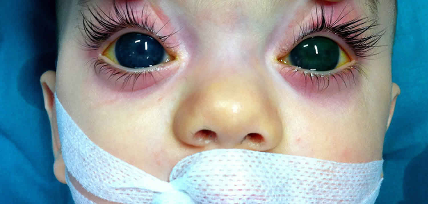
Buphthalmos occurs most commonly due to primary congenital glaucoma (onset at birth) or primary infantile glaucoma (onset after birth to 3 years). The classical symptoms of congenital and infantile glaucomas include tearing, photophobia, and irritability. The parents may notice a hazy cornea or an increase in the size of the cornea. On examination, clinicians notice that there is increased corneal diameter, deep anterior chamber, and an increase in the size of the globe. Corneal examination reveals the presence of corneal edema, with ruptures of Descemet’s membrane known as Haab’s striae. The intraocular pressure is elevated, and there may be optic disc cupping.
Buphthalmos diagnosis
The mainstay of evaluation of Buphthalmos patients is examination under anesthesia. The following examinations should be done:
- Refraction using streak retinoscope
- Corneal examination:
- Measurement of corneal diameter using calipers may show an increase. A corneal diameter greater than 12 mm is considered abnormal.
- Corneal edema may be seen due to raised intraocular pressure.
- Breaks in the Descemet’s membrane or Haab’s striae may be seen, which are typically horizontal or parallel to the limbus 8.
- Examination of the anterior segment may reveal other underlying causes of Buphthalmos like Aniridia and Lisch nodules of neurofibromatosis.
- Measurement of intraocular pressure in the first few minutes of anesthesia to avoid falsely low readings.
- Gonioscopy with a direct gonioscope may demonstrate anterior insertion of iris onto the trabecular meshwork, the “Loch Ness monster” phenomenon (vascular loops in the angle) and “Lister’s morning mist” (fine, fluffy tissue on the peripheral iris).
- Ophthalmoscopy may demonstrate the presence of optic disc cupping. It is interesting to note that optic disc cupping may be reversed with appropriate treatment in many cases of primary congenital glaucoma.
- Ultrasound biomicroscopy may be done in cases of an opaque cornea to evaluate the anterior segment structures.
- Clinical genetics consultation may be advised.
Buphthalmos treatment
The main aim of treatment is to reduce intraocular pressure (IOP) to prevent progressive corneal opacification and glaucomatous optic atrophy and thereby preserve existing vision 4. The most definitive treatment of buphthalmos is surgical. Medical management is indicated to control the intraocular pressure to clear the cornea during surgery.
Medical treatment
Medical therapy can be instituted with topical beta blockers, carbonic anhydrase inhibitors or prostaglandin analogs.
Surgical treatment
- Goniotomy: In this procedure, openings are created in the trabecular meshwork, thus reducing resistance to outflow.
- Trabeculectomy: The trabecular meshwork is incised by cannulating Schlemm’s canal with a probe.
- Trabeculectomy: A section of the trabecular meshwork and Schlemm’s canal is removed underneath a partial thickness scleral flap, thus creating a fistula draining aqueous to the subconjunctival space.
- Combined trabeculectomy and trabeculectomy: Involves removal of a block of sclera after performing trabeculectomy
- Glaucoma drainage implants
- Cyclodestructive procedures
In the postoperative period, the surgeon should correct any refractive errors and also manage amblyopia, if any. Lifelong monitoring of intraocular pressure (IOP) is indicated in these patients.
Buphthalmos prognosis
Over the years, the prognosis of children with buphthalmos has slightly improved. However, despite treatment, many children require low vision aids and visual rehabilitation 3. Once considered a condition with a bleak prognosis, buphthalmos can now be managed reasonably well with the modern surgical procedures.
References- Feroze KB, Patel BC. Buphthalmos. [Updated 2019 Nov 11]. In: StatPearls [Internet]. Treasure Island (FL): StatPearls Publishing; 2019 Jan-. Available from: https://www.ncbi.nlm.nih.gov/books/NBK430887
- Vasileiadis GT, Frangouli O. Unilateral congenital buphthalmos. BMJ Case Rep. 2015 Jun 03;2015
- Zagora SL, Funnell CL, Martin FJ, Smith JE, Hing S, Billson FA, Veillard AS, Jamieson RV, Grigg JR. Primary congenital glaucoma outcomes: lessons from 23 years of follow-up. Am. J. Ophthalmol. 2015 Apr;159(4):788-96.
- Aponte EP, Diehl N, Mohney BG. Medical and surgical outcomes in childhood glaucoma: a population-based study. J AAPOS. 2011 Jun;15(3):263-7.
- Aziz A, Fakhoury O, Matonti F, Pieri E, Denis D. [Epidemiology and clinical characteristics of primary congenital glaucoma]. J Fr Ophtalmol. 2015 Dec;38(10):960-6.
- Abu-Amero KK, Edward DP. Primary Congenital Glaucoma. In: Adam MP, Ardinger HH, Pagon RA, Wallace SE, Bean LJH, Stephens K, Amemiya A, editors. GeneReviews® [Internet]. University of Washington, Seattle; Seattle (WA): Sep 30, 2004.
- Alves M, Malki LT, Rocha EM. Buphthalmos development in adult: case report. Arq Bras Oftalmol. 2012 Oct;75(5):361-2.
- Mahelková G, Filous A, Odehnal M, Cendelín J. Corneal changes assessed using confocal microscopy in patients with unilateral buphthalmos. Invest. Ophthalmol. Vis. Sci. 2013 Jun 10;54(6):4048-53.


