Cecal volvulus
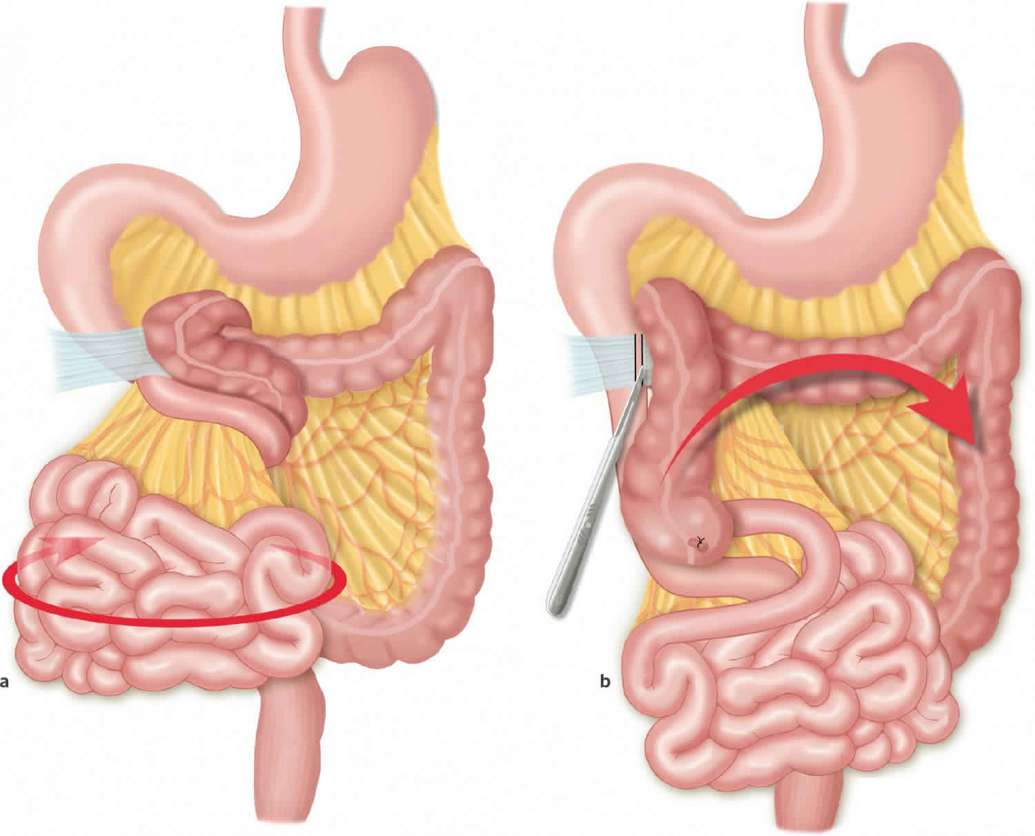
Cecal volvulus is a twisted loop of the bowel (axial twist of the cecum, ascending colon and terminal ileum) around their mesenteric pedicles 1. When a volvulus involves the cecum alone, it is also called a cecal bascule. This occurs when a highly mobile cecum traverses from a caudad to cephalad direction 2. However, the term cecal volvulus is a misnomer because, in most patients with cecal volvulus, the twisted loop of the bowel is located in the ascending colon above the ileocecal valve 3. In general, a partial malrotation is necessary for cecal volvulus to occur, because the cecum and parts of the ascending colon are involved. Cecal volvulus is essentially a closed-loop bowel obstruction that may lead to vascular compromise with consequent gangrene and perforation, requiring early diagnosis to reduce the high mortality rate 4. According to the several studies, cecal volvulus accounts for 10–60 % of all colonic volvulus 5 and can be divided into two major subgroups: the first is loop axial ileocolic volvulus, which accounts for 90 % of cases, and the second is cecal bascule, which accounts for the remaining cases 6. The classic ileocolic volvulus is a clockwise or counterclockwise rotation of the cecum with distal ileum in an oblique pattern. However, the counterclock-type is more commonly seen. In cecal bascule, there is an upward folding of the cecum, either anteriorly or posteriorly 7.
Cecal volvulus as an uncommon cause of acute intestinal obstruction. Cecal volvulus incidence is 2.8–7.1 cases per million annually 8. Most of the cecal volvulus reports are from Asia 8, and cecal volvulus occurs less frequently than sigmoid volvulus 9, which is also common in Asia, as well as in Turkey 10. Although there are many different cause and predisposing factors for cecal volvulus, exact cause is most likely multifactorial in presence of mobile cecum. Cecal volvulus clinical presentation is highly variable, ranging from intermittent episodes of abdominal pain to abdominal catastrophe depending on pattern, severity and duration of cecal volvulus causing intestinal obstruction. Due to its rarity and nonspecific presentation, preoperative diagnosis is rarely achieved in most cases. Abdominal radiographs as an initial diagnostic test are usually abnormal and can detect cecal volvulus in half of cases. Nowadays, computerized tomography (CT scan) is used for more accurate diagnosis and differentiation from other acute emergent conditions. Perioperative mortality of cecal volvulus is approximately 0–40% depending on the bowel viability or gangrene, as well as the type of the therapeutic procedure 11. Early diagnosis is essential in order to reduce the high mortality rate 9. Resection with right hemicolectomy and primary ileocolic anastomosis has been recommended for surgical treatment of cecal volvulus.
Cecal volvulus key points
- Cecal volvulus is much rare compared to sigmoid volvulus.
- Cecal volvulus is not so common in North America but since it carries a very high mortality, it is important that healthcare workers be aware of the disorder.
- With cecal volvulus, the torsion is usually in a clockwise direction.
- Vascular compromise is more common in cecal volvulus compared to sigmoid volvulus.
- The plain x-rays are usually adequate for diagnosis.
- The options for treatment include endoscopic decompression, cecopexy or a right hemicolectomy.
- With decompression alone, recurrence rates are very high.
- Because most patients have numerous comorbidities, the initial management is best done in a critical care unit. The patient must be hydrated and cleared for surgery by the internist. Depending on patient age and comorbidity, the type of procedure will vary. Hence a gastroenterologist and a general surgeon must be in communication to offer the patient the best treatment available.
- There are only small case series and isolated reports on outcomes of patients managed with cecal volvulus. For those who have delayed diagnosis, the outcomes are poor. Even those who undergo timely surgery have high morbidity as a consequence of their age.
Figure 1. Cecal volvulus abdominal X-ray
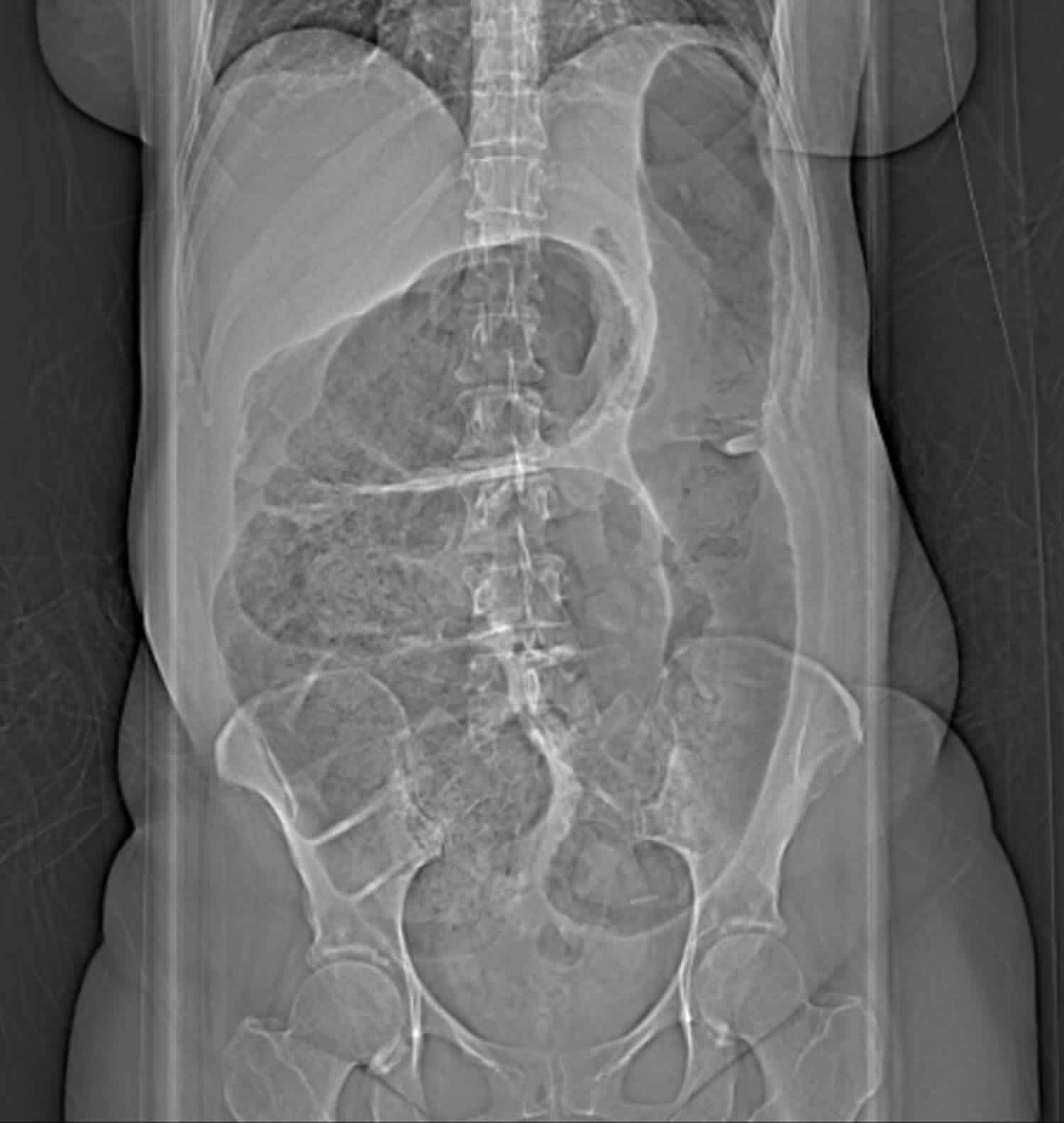
Footnote: Abdominal xray of a cecal volvulus revealing a dramatic dilation of bowel extending from the right lower quadrant moving upwards to the left upper quadrant of the abdomen.
[Source 12 ]Figure 2. Cecal volvulus
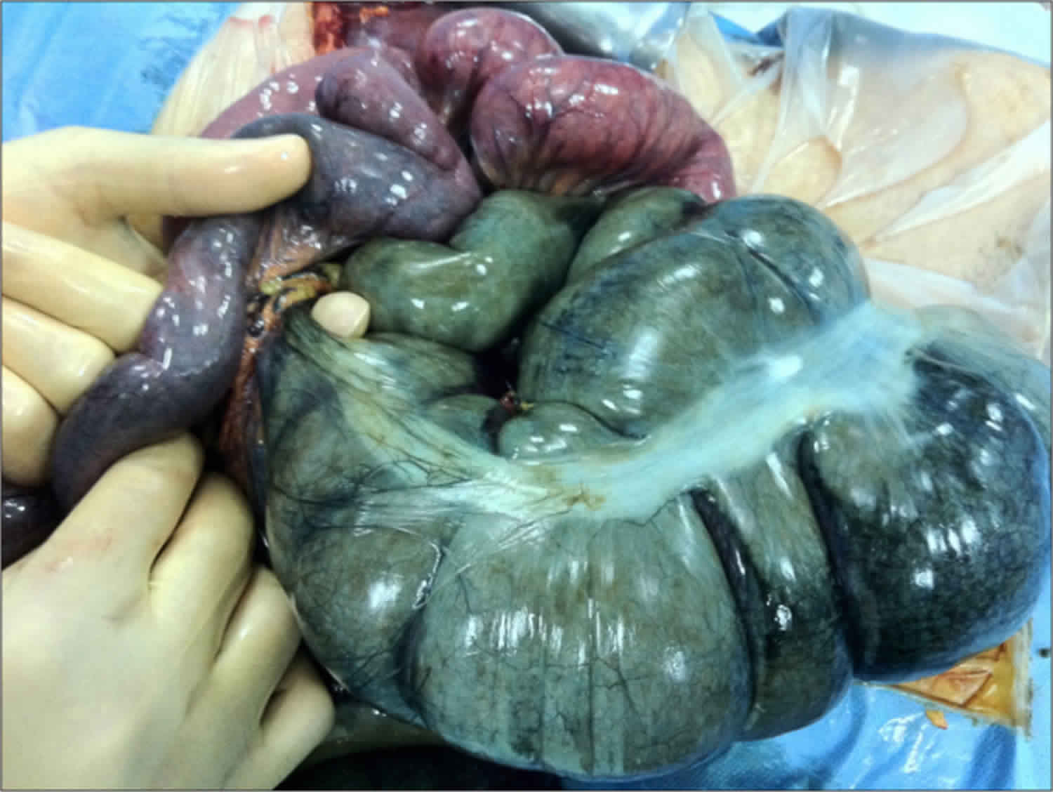
Footnote: Operative appearance shows twisted and gangrenous terminal ileum and cecum/ascending colon.
[Source 13 ]Cecal volvulus types
In general, there are three different types of cecal volvulus 12:
- Type 1: This cecal volvulus forms by a clockwise axial twisting or torsion of the cecum along the long axis. The location of the cecal volvulus is in the right lower quadrant.
- Type 2: This cecal volvulus develops from a twisting or torsion of a portion of the cecum and a portion of the terminal ileum. The location of the cecum gets displaced to an ectopic location (typically left upper quadrant) and is relocated in an inverted orientation. Traditionally, but not for all cases, a type 2 cecal volvulus will encounter a counterclockwise twist.
- Type 3: This cecal volvulus also known as cecal bascule, is the upward folding of the cecum. There is no axial twisting like with type 1 and type 2.
Type 1 and type 2, which involve axial torsion, account for approximately 80% of all cecal volvuli. Cecal bascules account for the remaining 20% of cecal volvuli.
Cecal volvulus causes
Many factors have been referred as correlated to cecal volvulus development, mainly anatomical predispositions such as incomplet intestinal rotation, and previous abdominal operations 14. Cecal volvulus predominantly affects female patients 40–60 years of age 10. Patients that have psychiatric conditions or are institutionalized and taking psychotropic drugs have a higher incidence of colonic volvulus. The use of psychotropic drugs can cause hindered intestinal mobility and predispose patients to volvuluses 15.
Cecal volvulus symptoms
Abdominal pain, distension, nausea, vomiting, and diarrhea or constipation are the main clinical features of cecal volvulus 11, but unfortunately clinical symptoms, signs, and routine laboratory tests are not specific enough to lead to a prompt diagnosis 11.
Cecal volvulus diagnosis
Although abdominal radiography may show the features of an intestinal obstruction, including widespread small intestinal air-fluid levels and/or distended cecum in the right abdomen, making the cecal volvulus diagnosis is difficult or impossible in most of the cases 9. Doppler ult may lead to make a definite diagnosis by showing twisted mesenteric vessels 10 and CT may be more diagnostic by demonstrating cecal distension, cecal apex in left upper quadrant, mesenteric whirl, ileocecal twist, and small bowel distension 16. Despite the identified diagnostic features, cecal volvulus is rarely diagnosed correctly at the time of presentation due to the low incidence of the disease 11.
Cecal volvulus treatment
Surgical intervention is the only treatment of cecal volvulus 8. Laparoscopic surgery is preferred to open surgery but sometimes the urgency of the situation may not allow it 17. Other inventions such as a barium enema or a colonoscopy, can offer a non-operative reduction of a cecal volvulus 12. However, these modalities are rarely successful. For these non-operative treatments, there is a high risk of perforations and should not be attempted. Colonic necrosis can be miss up to approximately 20% to 25% of the time when non-operative modalities are used. If there is intestinal gangrene, resection is inevitable. In nongangrenous cases, it is sufficient to simply untwist the cecum or additionally to perform a cecopexy by fixing it to the abdominal wall 11, and laparoscopic technique is preferred 18.
Cecal volvulus surgery
Surgical treatment and will vary based on patient stability and findings seen intraoperatively. Intraoperatively, the surgeon will ascertain if there is bowel compromise or if the bowel is viable. These findings will help dictate appropriate surgical intervention 19.
For patients who are stable with no bowel compromise, an ileocolic resection or a right hemicolectomy should be performed. In patients that receive an ileocolic resection, an additional colopexy to tack the right remnant colon to the posterior peritoneum to minimize the recurrence of another volvulus.
For patients who are hemodynamically unstable without bowel compromise, a cecopexy should be performed in conjunction with a cecostomy tube placement or cecopexy can be done alone.
For patients who are stable with bowel, the surgeon should proceed with a right hemicolectomy or ileocolic resection followed by an ileocolic anastomosis.
For patients who are unstable with bowel, the surgeon should proceed with a right hemicolectomy or ileocolic resection with an ileostomy creation. Later, once the patient is stabilized, the ileostomy may be reversed.
Cecal volvulus prognosis
Cecal volvulus is a potentially life threatening disorder. If the treatment is delayed, it carries a mortality in excess of 30%. Most studies indicate that the time to treat should be within 24-72 hours after diagnosis. This much time is required for hydration and any investigations. Even after cecal volvulus is treated, patients have high morbidity due to a prolonged ileus, wound infection, respiratory failure, and bowel obstruction 20.
Common complications after cecal volvulus treatment include:
- Wound infection
- Sepsis
- Anastomotic leak
- Colocutaneous fistula
- Pelvic or abdominal abscess
Postoperative and rehabilitation care
Patients often require a prolonged stay in the hospital. Most patients are elderly and frail. If the ileus is prolonged, they often require IV fluids for a few days. DVT (deep vein thrombosis) prophylaxis and physical therapy are recommended.
References- Hasbahceci M, Basak F, Alimoglu O. Cecal volvulus. Indian J Surg. 2012;74(6):476–479. doi:10.1007/s12262-012-0432-9 https://www.ncbi.nlm.nih.gov/pmc/articles/PMC3537995
- Lung BE, Yelika SB, Murthy AS, Gachabayov M, Denoya P. Cecal bascule: a systematic review of the literature. Tech Coloproctol. 2018 Feb;22(2):75-80.
- Cecal Volvulus Imaging. https://emedicine.medscape.com/article/364967-overview
- Perret RS, Kunberger LE. Case 4: Cecal volvulus. AJR Am J Roentgenol. 1998 Sep. 171(3):855, 859, 860.
- Mulas C, Bruna M, García-Armengol J, Roig JV. Management of colonic volvulus. Experience in 75 patients. Rev Esp Enferm Dig. 2010;102:239–248. doi: 10.4321/S1130-01082010000400004
- Madiba TE, Thomson SR. The management of cecal volvulus. Dis Colon Rectum. 2002;45:264–267. doi: 10.1007/s10350-004-6158-4
- Tirol FT. Cecocolic torsion: classification, pathogenesis, and treatment. JSLS. 2005;9:328–334.
- Bandurski R, Zareba K, Kedra B. Cecal volvulus as a rare cause of intestinal obstruction. Pol Przegl Chir. 2011;83:515–7.
- Khaniya S, Shakya VC, Koirala R, et al. Cecal volvulus: a twisted tale. Trop Doct. 2010;40:244–6.
- Takada K, Hamada Y, Sato M, et al. Cecal volvulus in children with mental disability. Pediatr Surg Int. 2007;23:1011–4.
- Pulvirenti E, Palmieri L, Toro A, Di Carlo I. Is laparotomy the unavoidable step to diagnose caecal volvulus? Ann R Coll Surg Engl. 2010;92:27–9.
- Le CK, Qaja E. Cecal Volvulus. [Updated 2019 Feb 28]. In: StatPearls [Internet]. Treasure Island (FL): StatPearls Publishing; 2019 Jan-. Available from: https://www.ncbi.nlm.nih.gov/books/NBK470305
- Atamanalp, S. S., Ozogul, B., & Kisaoglu, A. (2012). Cecal volvulus: a rare cause of intestinal obstruction. The Eurasian journal of medicine, 44(2), 115–116. doi:10.5152/eajm.2012.25 https://www.ncbi.nlm.nih.gov/pmc/articles/PMC4261296
- Arulmolichelvan A, Sivaraman A, Muthukrishnan A. Cecal volvulus associated with intestinal malrotation presenting as postoperative intestinal obstruction. Med Princ Pract. 2012;21:389–91.
- Le CK, Cooper W. Volvulus. [Updated 2019 Mar 13]. In: StatPearls [Internet]. Treasure Island (FL): StatPearls Publishing; 2019 Jan-. Available from: https://www.ncbi.nlm.nih.gov/books/NBK441836
- Rosenblat JM, Rozenblit AM, Wolf EL, DuBrow RA, Den EI, Levsky JM. Findings of cecal volvulus at CT. Radiology. 2010;256:169–75.
- Ramírez-Ramírez MM, Villanueva-Sáenz E, Ramírez-Wiella-Schwuchow G. [Elective laparoscopic right colectomy for caecal volvulus: case report and literature review]. Cir Cir. 2017 Jan – Feb;85(1):87-92.
- Jones RG, Wayne EJ, Kehdy FJ. Laparoscopic detorsion and cecopexy for treatment of cecal volvulus. Am Surg. 2012;78:251–2.
- Miura da Costa K, Saxena AK. A systematic review of the management and outcomes of cecal and appendiceal volvulus in children. Acta Paediatr. 2018 Dec;107(12):2054-2058.
- Gomes CA, Soares C, Catena F, Di Saverio S, Sartelli M, Gomes CC, Gomes FC. Laparoscopic Management of Mobile Cecum. JSLS. 2016 Oct-Dec;20, 4


