Cholesterol embolism
Cholesterol embolism also known as cholesterol embolism syndrome is caused by embolization of cholesterol crystals from atherosclerotic plaques lining the walls of major arteries often called hardening of the arteries 1. Cholesterol embolism syndrome should be suspected in a patient who develops worsening renal function 2, hypertension, distal ischemia, or acute multisystem dysfunction after an invasive arterial procedure 3. Cholesterol emboli may also occur spontaneously. The diverse manifestations of cholesterol embolism syndrome make the diagnosis challenging. Cholesterol embolism is a disease of persons ranging from middle-aged to elderly, with a minimum age of 50 years. The risk is greater for men than it is for women. As the population ages, the incidence of cholesterol embolism syndrome will increase.
Atherosclerosis is a disease in which plaque builds up inside your arteries. Plaque is a sticky substance made up of fat, cholesterol, calcium, and other substances found in the blood. Over time, plaque hardens and narrows your arteries. That limits the flow of oxygen-rich blood to your body.
The risk factors for cholesterol embolization syndrome include advanced age, smoking, hypertension, diabetes mellitus, hyperlipidemia, and peripheral vascular disease. Cholesterol embolization syndrome is seen following invasive vascular procedures as in our patient, thrombolytic therapy, or anticoagulants but may occur spontaneously as well.
Atherosclerosis can lead to serious problems, including:
- Coronary artery disease. These arteries supply blood to your heart. When they are blocked, you can suffer angina or a heart attack.
- Carotid artery disease. These arteries supply blood to your brain. When they are blocked you can suffer a stroke.
- Peripheral arterial disease. These arteries are in your arms, legs and pelvis. When they are blocked, you can suffer from numbness, pain and sometimes infections.
Since the abdominal aorta is the most common location of atherosclerosis, the kidneys, visceral organs, and lower extremities are commonly involved 4. Atheromas of other arteries, such as the ascending aorta or the carotid, may result in emboli, producing neurologic, retinal, or cardiac symptoms 5.
Estimates of the incidence of cholesterol embolic disease are usually based on autopsy data. Tissue sections from patients with the following diseases or procedures indicate the incidence of atheroembolic events:
- Aortic aneurysms (31%)
- Abdominal aortic aneurysm repair (up to 77%)
- Severe aortic disease (13-16%)
- Mild aortic disease (1-2%)
Of patients undergoing angiography, 25-30% may have atheroembolic events, whereas 2.5-3% of patients undergoing percutaneous transluminal coronary angioplasty (PTCA) vein grafts and 1.4-3% of patients undergoing renal artery angioplasty or cardiac catheterization have been reported to have clinical signs of atheroemboli. Cholesterol embolism syndrome has been reported as occurring months after thrombolytic therapy for stroke, but the true incidence is unknown 6.
Cholesterol embolism key points:
- Cholesterol crystal embolization is a poorly described multisystem disorder with a high mortality.
- It can occur up to eight weeks after invasive arterial procedures.
- Common features include abdominal pain and bleeding, renal failure, and purpura.
- It should be considered in the differential diagnosis of vasculitic disorders.
- Pulmonary features, usually haemoptysis and dyspnoea, are rare, but when present are usually fatal.
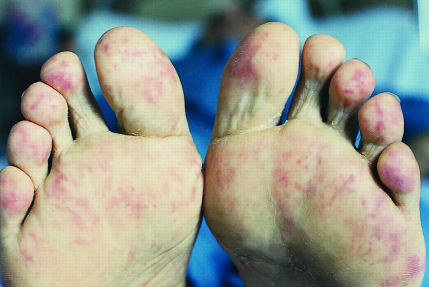
Figure 1. Cholesterol emboli foot
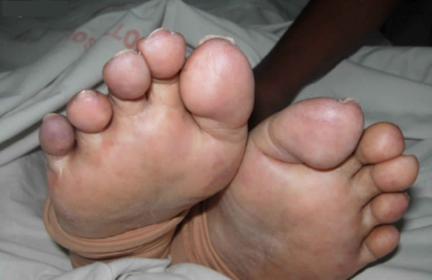
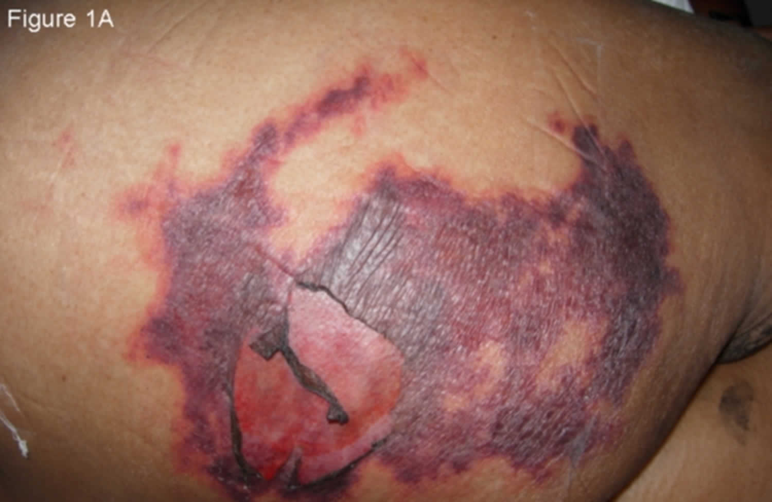
Footnote: An 81-year-old woman with chronic kidney disease and hypertension underwent an endovascular repair for an abdominal aortic aneurysm. Twenty-four hours later she complained of bilateral calf muscle pain and developed altered sensorium with deterioration of renal function. On the third post-operative day she developed symmetrical purpuric macules with erythematous margins of sizes ranging from 4 x 5 cm to 10 x 7 cm on the gluteal region (Figure 1A) and bluish reticulated patches on the soles and tips of toes (Figure 1). This was followed by melena. Abnormal laboratory parameters included a decrease in hemoglobin from 10.2 gm/dL (at admission) to 6.5 gm/dL, leukocytosis of 13,700/mm³, serum creatinine of 2 mg/dL (0.5-1.4 mg/dL) increasing to 3.1 mg/dL, serum urea, 85 mg/dL (15-40 mg/dL) increasing to 116 mg/dL, amylase, 212 U/L (28-100 U/L), creatine kinase, 1274 U/L (45-195 U/L), serum aspartate aminotransferase, 126 U/L (8-40 U/L), serum alanine aminotransferase, 43 U/L (5-35 U/L), and erythrocyte sedimentation rate, 87 mm/hour. Doppler sonography of both lower limbs for deep vein thrombosis and a CT scan of the brain were normal. A skin biopsy from the periphery of the lesion on the gluteal area showed an arteriole in the subcutis with typical needle-shaped (cholesterol) clefts and an unremarkable epidermis and dermis confirming the diagnosis of cutaneous cholesterol embolization syndrome. She was treated with hemodialysis and supportive management and she recovered.
[Source 1 ]Cholesterol embolism causes
Any risk factor for atherosclerotic disease is a risk factor for cholesterol embolism. Atherosclerosis often called hardening of the arteries, is the buildup of fatty substances in the arteries, which can lead to heart disease and stroke. It’s a slow, complex disease that typically starts in childhood and progresses with age.
Deposits of fatty substances, cholesterol, cellular waste products, calcium and other substances build up in the inner lining of an artery. They cause the formation of blood clots that can block blood flow or break off and travel to another part of the body. View a detailed animation of atherosclerosis.
If a clot blocks a blood vessel that feeds the heart, it causes a heart attack. If it blocks a blood vessel that feeds the brain, it causes a stroke. If blood supply to the arms or legs is reduced or blocked, it can cause difficulty walking and eventually gangrene.
Males and people with a family history of premature cardiovascular disease have an increased risk of atherosclerosis. Other risk factors include:
- High blood cholesterol
- Cigarette smoking and exposure to tobacco smoke (the chemicals in cigarettes can cause damage to blood vessels accelerating the development of atherosclerosis)
- High blood pressure (it damages the lining of blood vessels making them susceptible to atherosclerosis)
- Diabetes mellitus
- Obesity
- Physical inactivity
The inner lining of the artery, called the endothelium, can be damaged due to high cholesterol and triglyceride levels, cigarette smoke, high sugar levels and other factors. High blood pressure can also cause damage. Once the damage is done, atherosclerosis begins and a plaque forms.
Fats, cholesterol, platelets, cellular debris and calcium begin to deposit in the artery walls. These substances may stimulate the cells of the artery wall to produce still other materials. More cells accumulate, and many divide. At the same time, fat builds up within and around these cells, and connective tissue forms. This buildup is called plaque. It usually affects large and medium-size arteries. These cells and surrounding material thicken the endothelium significantly. The artery’s diameter shrinks and blood flow decreases, reducing oxygen supply.
Key components of cholesterol embolism syndrome include the following:
- Proximal large-caliber arterial plaque
- Plaque rupture with embolization of debris
- Mechanical occlusion of small arteries
- Intense foreign-body inflammation
- End-organ damage from mechanical obstruction
- Inflammatory vascular changes
Any organ system, with the exception of the lungs, may be directly affected. Cholesterol embolism syndrome has two mechanisms of action.
With the first mechanism, cholesterol crystals spontaneously break off from severely atherosclerotic plaques and shower into downstream organs, occluding arterioles 100-200 μm in diameter. The crystals induce an inflammatory foreign-body reaction and adventitial fibrosis, which eventually obliterate the vessel lumen. Local vasospastic mediators compound tissue ischemia and produce progressive, irreversible organ damage.
With the second mechanism, larger cholesterol plaques break off and occlude larger arteries, causing tissue infarction with acute organ dysfunction. This can occur after local trauma to the atherosclerotic plaque, such as that caused by angiography or aortic trauma, or it can occur after destabilization of the protective clot overlying the plaque, which can occur as a result of anticoagulation.
Cholesterol crystal embolization (see the image below) occurs from the arterial system, and crystals are trapped in the arterioles, where they either immediately occlude the vessels or induce an intense inflammatory response that leads to tissue ischemia. Crystals do not travel to the lungs; however, inflammatory mediators released by ischemic tissue may result in acute lung injury.
Preoperative risk factors for cholesterol embolism syndrome after coronary artery bypass surgery (CABG) include the following:
- Age greater than 60 years
- Hypertension
- Cerebrovascular disease
- Aortoiliac disease
- Mitral annular calcification
Although the other factors have been well known for some time, it was only comparatively recently that the association between mitral annular calcification and aortic atherosclerosis was identified.
Identifying patients at risk and making efforts to minimize aortic-wall trauma help reduce the chance of cholesterol embolism. The risk that cholesterol embolism will develop may be reduced by taking a brachial or axillary approach in patients known to have severely ulcerated aortic plaque, using soft flexible catheters, and avoiding high-pressure jets of contrast material.
Cholesterol embolism prevention
If an invasive radiologic procedure is necessary, the risk of inducing cholesterol embolism must be considered. If the patient is at high risk, with known or suspected severe aortic atherosclerosis or aortic aneurysm, the Judkins (ie, brachial) approach or a radial artery approach may be used for introducing the catheter into the aorta. However, some investigators found that the approach made no difference, which led them to suspect the ascending aorta as a major source of atheroemboli.
Gentle handling of the severely diseased aorta during cardiac or aortic surgery can reduce the risk of cholesterol embolism. Careful clamping techniques and careful selection of aortotomy sites may minimize disruption of the atherosclerotic plaque.
Cholesterol embolism symptoms
The diagnosis of cholesterol embolism must be considered in patients older than 50 years who have atherosclerotic disease and who present with multisystem dysfunction after undergoing an invasive vascular procedure or receiving an anticoagulant or thrombolytic agent within the past several months. Cholesterol embolization syndrome should be considered in any patient presenting with cutaneous features and renal failure in the period of up to eight weeks after an invasive vascular procedure. Patients may have unexplained fever, weight loss, myalgias, or anorexia for weeks or months after a procedure before presenting with acute renal failure, hyperkalemia, gastrointestinal (GI) bleeding, or stroke. Lung involvement is uncommon but when present appears to confer an extremely poor prognosis. All patients with the classic triad of livedo reticularis, acute renal failure, and eosinophilia should undergo evaluation for cholesterol embolism, including a funduscopic examination.
Cholesterol embolism symptoms include the following:
- Fever
- Weight loss
- Hypermetabolic state
Cardiovascular manifestations include the following:
- Tachycardia (fast heart rate)
- Uncontrolled or accelerating hypertension
- Congestive heart failure
- Myocardial infarction (heart attack)
- Intact peripheral pulses with livedo reticularis and tissue ischemia – These findings suggest small-vessel occlusion, such as cholesterol embolization, in a patient at risk (see Figure 1A above)
Neurologic manifestations include the following:
- Hollenhorst plaques in retinal arteries
- Hemispheric ischemic stroke
- Paraplegia
- Confusion
- Delirium
Kidney manifestations include the following:
- Oliguria
- Acute renal failure
Skin manifestations include the following:
- Gangrene, nodules, purpura, cyanosis, ulcerations (in 35-90% of patients)
- Livedo reticularis
- Infarction of perineal area
- Ischemic patches involving lower extremities more often than upper
- Blue toe syndrome and splinter hemorrhages 7.
In one study, cutaneous manifestations were reported in 88.5% of patients 5. These included livedo reticularis (49%), gangrene (35%), cyanosis (28%), ulceration (17%), nodules (10%), and purpura (9%) 8. Skin biopsy is diagnostic in 92% of the cases 9 and it must include an area between middle to deep dermis. The needle-shaped spaces left by the dissolved crystals within the lumen of arterioles from affected skin are characteristic.
Gastrointestinal manifestations include the following:
- Minor or major bleeding
- Abdominal pain
- Bowel infarction
- Pancreatitis
- Acalculous cholecystitis
Other manifestations are as follows:
- Musculoskeletal – Myalgias (muscle pain)
- Endocrine – Adrenal insufficiency
- Pulmonary – Acute respiratory distress syndrome (ARDS)
Cholesterol embolism complications
Cholesterol embolism can directly affect all organs except the lungs, resulting in complications that range from mild dysfunction to complete organ failure. Supportive care of organ dysfunction may be necessary and may include hemodialysis, bowel resection, cholecystectomy, and pancreatitis management.
Cholesterol embolism diagnosis
Laboratory studies to be considered in the workup for cholesterol embolism include the following:
- Complete blood count (CBC) – Leukocytosis with left shift is nonspecific; eosinophilia strongly suggests atheroembolization and is present in as many as 80% of patients with cholesterol embolism syndrome
- Chemistry – Elevated blood urea nitrogen (BUN) and creatinine levels are present in virtually all cases of cholesterol embolism syndrome
- Urinalysis – Microscopic hematuria, proteinuria, and hyaline casts are common; pyuria actually may be eosinophiluria, a major clue for the diagnosis of cholesterol embolism syndrome
- Tissue-specific laboratory tests – Muscle injury causes an elevated creatine kinase (CK) level; myocardial, pancreatic, and hepatobiliary involvement produces increases in cardiac enzymes, amylase, and hepatobiliary enzymes
- Inflammatory mediators – Nonspecific findings include hypocomplementemia, positive rheumatoid factor (RF), antinuclear antibodies (ANAs), and elevated C-reactive protein(CRP) and sedimentation rates; one study found a CRP level higher than 1.0 mg/dL to be an independent predictor of cholesterol emboli in patients with coronary artery disease (coronary heart disease) 10
Imaging studies
Angiography
Contrast angiography of involved organs may be performed to rule out more treatable causes of tissue ischemia, such as polyarteritis nodosa. Angiography may induce atheroembolism.
Echocardiography
Transesophageal echocardiography (TEE) is an increasingly well accepted imaging tool for detecting atheromatous lesions in the ascending and thoracic aorta 11. Protruding mobile atheromatous masses have been associated with a higher incidence of stroke or cholesterol embolism in patients who undergo cardiac bypass or patients who receive anticoagulants. Transesophageal echocardiography (TEE) may eventually be performed in all patients undergoing bypass before aortic cannulation. It also may be performed in all patients with ischemic stroke with an unclear cause.
Computed tomography
Thin sections viewed on nonenhanced dual helical (spiral) computed tomography (CT) may be useful for rapid and noninvasive detection of protruding aortic atheroma 12. This test can help visualize areas that are poorly imaged on transesophageal echocardiography (TEE), such as the distal ascending aorta and arch. One study suggests 87% sensitivity, 82% specificity, and 84% overall accuracy.
Magnetic resonance imaging
Data on magnetic resonance imaging (MRI) and atheromatous plaque are relatively sparse, but a reasonable expectation is that MRI should exhibit good sensitivity in this setting.
Biopsy
Demonstration of cholesterol crystals in occluded arterioles is the only definitive test for cholesterol embolism. Skin 13, renal, muscle, or gastrointestinal tract biopsy may reveal crystal ghosts inside vessels. Often, multiple samples may be necessary to demonstrate the crystals.
Cholesterol embolism treatment
Cholesterol embolization syndrome treatment is supportive 14. Hemodynamic monitoring, including pulmonary artery catheterization, may be helpful for fluid and vasopressor adjustments. If acute respiratory distress syndrome (ARDS) occurs, mechanical ventilation may be required for a prolonged period. Dialysis should be started when indicated because patients can recover limited renal function. Aggressive nutritional and metabolic support is essential because these patients often lose considerable lean body mass to ongoing catabolism.
Pharmacologic therapy has not been particularly successful in patients with cholesterol embolism syndrome. Vasodilator therapy with calcium-channel blockers may help relieve the local ischemia resulting from vasospasm, but angiotensin-converting enzyme (ACE) inhibitors should not be used, because of their negative effects on renal afferent arterioles and the glomerular filtration rate (GFR).
Patients presumed to have vasculitis have been treated with high-dose steroids and anti-inflammatory agents, with anecdotal reports of recovery. However, steroids may predispose patients to infectious, metabolic, and nutritional complications and difficulties with wound healing. In a report of four cases of cholesterol embolism after cardiac catherization that were associated with deteriorating renal function, low-dose (0.3 mg/kg/day) corticosteroid therapy yielded improved renal function in three of the four patients 15.
The use of anticoagulants is controversial because anticoagulants and thrombolytics have been shown to induce atheroemboli. Anecdotal reports of treatment with apheresis, as well as with iloprost, statins, colchicine, or combinations of these drugs with steroids, reported improvement in some cases 16.
A study by Ishiyama et al found that low-density-lipoprotein apheresis (LDL-A) reduced the incidence of maintenance dialysis and mortality at 24 weeks in 49 patients with cholesterol crystal embolism 17. In a subsequent study, the same group found that LDL-A plus corticosteroids restored deteriorated renal function better than corticosteroids alone did in patients with cholesterol crystal embolism 18.
Further invasive vascular procedures and anticoagulant or thrombolytic therapies should be avoided. If such treatments are unavoidable, downstream protection devices to trap atheromatous debris after stenting or angioplasty are suggested 19.
Surgical care
Surgical therapy (eg, aortic aneurysm resection) may be necessary to remove the source of atheroembolic material. Stent-grafting may be a less invasive method to reduce risk of embolization 20.
Damaged tissue should be protected and allowed to demarcate for several months. Surprisingly, a majority of the damaged area may recover. Necrotic tissue should be debrided, and establishing vascular access for dialysis also may be necessary.
In severe cases, lumbar sympathetic block (rarely, surgical sympathectomy) has been used to avoid impending lower-extremity tissue loss resulting from intense vasoconstriction.
Cholesterol embolism prognosis
Patients with multisystem cholesterol embolism syndrome have a poor prognosis. The mortality of acute multisystem organ failure resulting from cholesterol embolism syndrome is 58-90%. Jucgla et al 21 found an overall incidence of 58% at 15 months, increasing to 65% if visceral organs were involved. Preexisting chronic renal insufficiency had a relative risk of death of 4.54.
The mortality of severe cholesterol embolism is 90% at 3 months. Mild cases with renal dysfunction, with or without skin findings, have a mortality of 16%.
Cholesterol crystal showers can become stabilized, leaving patients with varying degrees of organ dysfunction. Renal function can recover if no further insults occur, even to the point where dialysis can be discontinued. However, patients remain at risk for recurrence of emboli.
References- Patro, N., George, R., Singh, P., & Joseph, G. (2012). Cutaneous cholesterol embolization syndrome: A case report. Dermatology Online Journal, 18(7). Retrieved from https://escholarship.org/uc/item/3d11w0pb
- Li X, Bayliss G, Zhuang S. Cholesterol Crystal Embolism and Chronic Kidney Disease. Int J Mol Sci. 2017 May 24. 18, 6.
- Cholesterol embolism. https://emedicine.medscape.com/article/460428-overview
- Fine MJ, Kapoor W, Falanga V. Cholesterol crystal embolization: a review of 221 cases in the English literature. Angiology. 1987; 38: 769-784.
- Jucgla A, Moreso F, Muniesa C, Moreno A, Vidaller A. Cholesterol embolism: still an unrecognized entity with a high mortality rate. J Am Acad Dermatol. Nov 2006; 55(5): 786-93.
- Higo S, Hirama A, Ueda K, Mii A, Kaneko T, Utsumi K, et al. A patient with idiopathic cholesterol crystal embolization: effectiveness of early detection and treatment. J Nippon Med Sch. 2011. 78 (4):252-6.
- Willens HJ, Kramer HJ, Kessler KM. Transesophageal echocardiographic findings in blue toe syndrome exacerbated by anticoagulation. J Am Soc Echocardiogr. 1996 Nov-Dec. 9(6):882-4.
- Falanga V, Fine MJ, Kapoor WN. The cutaneous manifestations of cholesterol crystal embolization. Arch Dermatol. 1998; 122: 1194-8.
- Pennington M, Yeager J, Skelton H, Smith KJ. Cholesterol embolization syndrome: cutaneous histopathological features and the variable onset of symptoms in patients with different risk factors. Br J Dermatol. 2002; 146: 511-517.
- Fukumoto Y, Tsutsui H, Tsuchihashi M, Masumoto A, Takeshita A,. The incidence and risk factors of cholesterol embolization syndrome, a complication of cardiac catheterization: a prospective study. J Am Coll Cardiol. 2003 Jul 16. 42(2):211-6.
- Adler Y, Shohat-Zabarski R, Vaturi M, Shapira Y, Ehrlich S, Jortner R. Association between mitral annular calcium and aortic atheroma as detected by transesophageal echocardiographic study. Am J Cardiol. 1998 Mar 15. 81(6):784-6.
- Tenenbaum A, Garniek A, Shemesh J, Fisman EZ, Stroh CI, Itzchak Y. Dual-helical CT for detecting aortic atheromas as a source of stroke: comparison with transesophageal echocardiography. Radiology. 1998 Jul. 208(1):153-8.
- Manganoni AM, Venturini M, Scolari F, Tucci G, Facchetti F, Graifemberghi S. The importance of skin biopsy in the diverse clinical manifestations of cholesterol embolism. Br J Dermatol. 2004 Jun. 150(6):1230-1.
- Belenfant X, Meyrier A, Jacquot C. Supportive treatment improves survival in multivisceral cholesterol crystal embolism. Am J Kidney Dis. 1999 May. 33(5):840-50.
- Masuda J, Tanigawa T, Nakamori S, Sawai T, Murata T, Ishikawa E, et al. Use of corticosteroids in the treatment of cholesterol crystal embolism after cardiac catheterization: a report of four Japanese cases. Intern Med. 2013. 52 (9):993-8.
- Sevillano-Prieto ÁM, Hernández-Martínez E, Caro-Espada J, Molina-Gómez M, Gutiérrez-Martínez E, Morales-Ruiz E, et al. Cholesterol atheroembolism and combined treatment with steroids and iloprost. Nefrologia. 2012. 32 (6):824-8.
- Ishiyama K, Sato T, Taguma Y. Low-Density Lipoprotein Apheresis Ameliorates Renal Prognosis of Cholesterol Crystal Embolism. Ther Apher Dial. 2015 Aug. 19 (4):355-60.
- Ishiyama K, Sato T, Yamaguchi T, Taguma Y. Efficacy of low-density lipoprotein apheresis combined with corticosteroids for cholesterol crystal embolism. Clin Exp Nephrol. 2017 Apr. 21 (2):228-235.
- Lowe HC, Houser SL, Aretz T, MacNeill BD, Oesterle SN, Palacios IF. Significant atheromatous debris following uncomplicated vein graft direct stenting: evidence supporting routine use of distal protection devices. J Invasive Cardiol. 2002 Oct. 14(10):636-9.
- Carroccio A, Olin JW, Ellozy SH, Lookstein RA, Valenzuela R, Minor ME. The role of aortic stent grafting in the treatment of atheromatous embolization syndrome: results after a mean of 15 months follow-up. J Vasc Surg. 2004 Sep. 40(3):424-9.
- Jucgla A, Moreso F, Muniesa C, Moreno A, Vidaller A. Cholesterol embolism: still an unrecognized entity with a high mortality rate. J Am Acad Dermatol. 2006 Nov. 55 (5):786-93.


