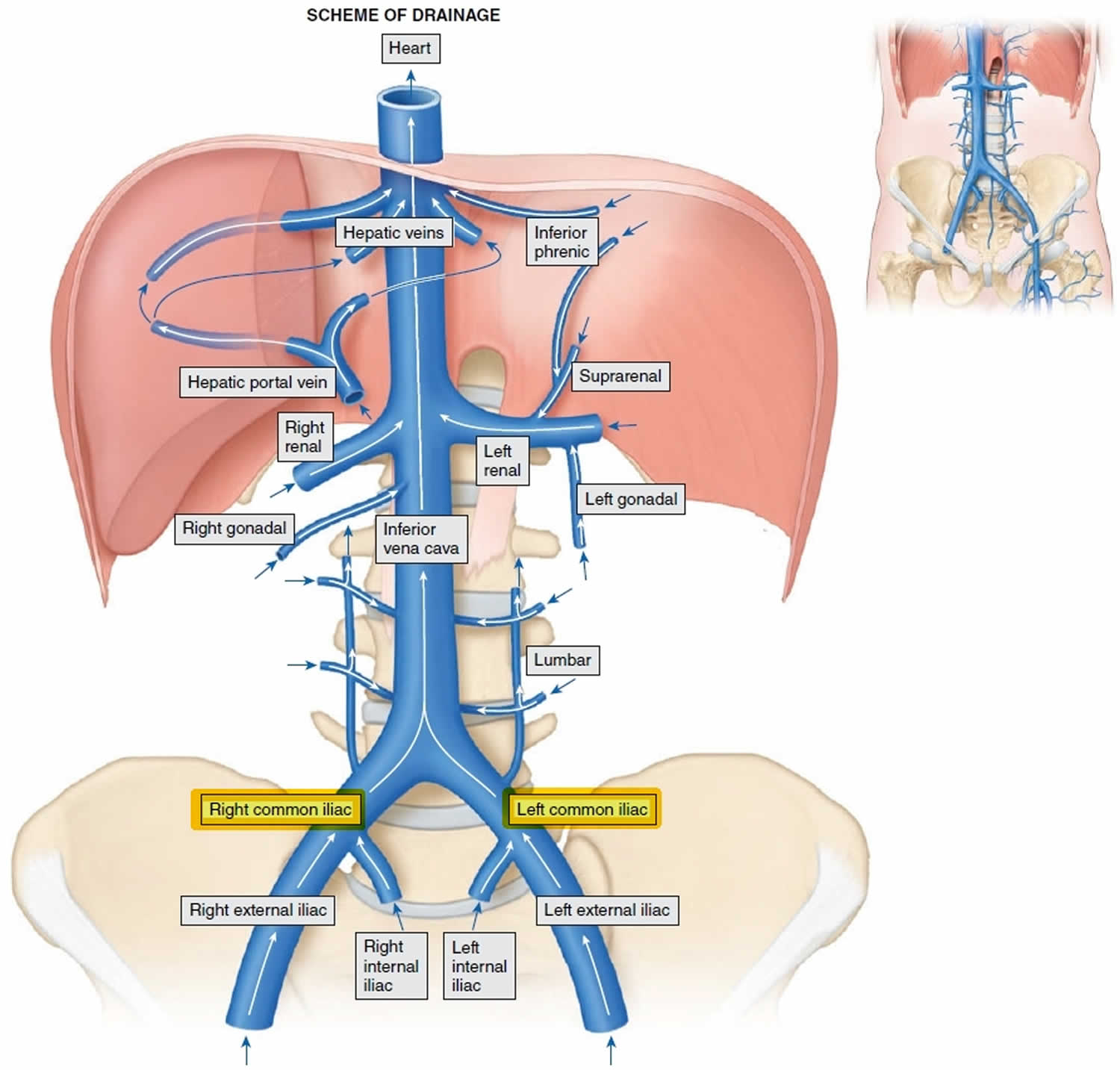
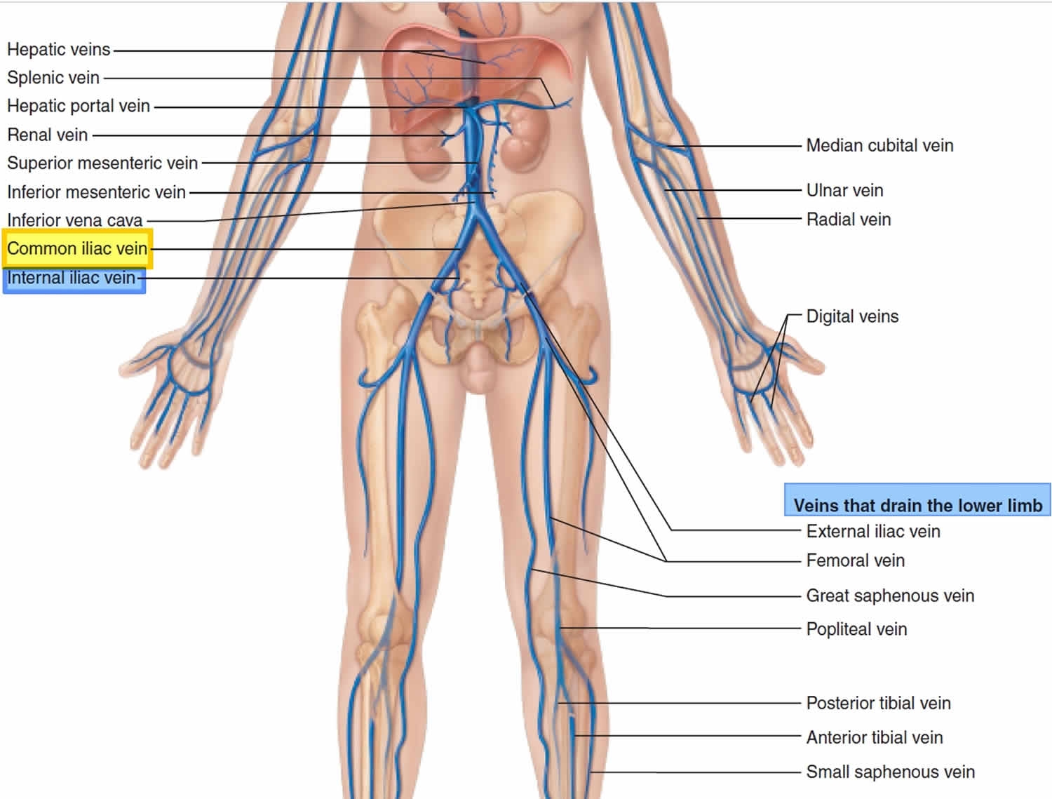
Common iliac vein
Common iliac vein is formed by the union of the internal and external iliac veins anterior to sacroiliac joint and anastomose anterior to fifth lumbar vertebra to form inferior vena cava (IVC). Right
common iliac is much shorter than left and is also more vertical, as inferior vena cava sits to right of midline. The primary function of these veins is to drain deoxygenated blood and return this blood to the heart.
- Common iliac veins drain the pelvis, external genitals, and lower limbs.
- Internal iliac veins drain the muscles of pelvic wall and gluteal region, pelvic viscera, and external genitals.
- External iliac veins drain the lower abdominal wall anteriorly, cremaster muscle in males, and external genitals and lower limb.
Each set of external and internal iliac veins on the right and left side of the body form an associated left or right common iliac vein. The left common iliac vein and right common iliac vein, together, form the inferior vena cava (IVC) 1. For pelvic veins, the left and right common iliac vein have differing pathways. The right common iliac vein moves in a straight course to form the inferior vena cava, while the left common iliac vein must initially join the right common iliac vein at the confluence with the inferior vena cava. Therefore, the left common iliac vein’s pathway is not as linear 1. The left common iliac vein is also longer than the right and receives drainage from the median sacral vein. The iliolumbar and lateral sacral veins drain into both common iliac veins 2. Frequently containing 1-2 valves, the external iliac vein receives from the following: the inferior epigastric vein, the deep circumflex iliac vein, and other pelvic veins. The inferior epigastric vein joins the external iliac vein superior to the inguinal ligament. The common femoral vein transitions to form the external iliac vein, which occurs at the level of the inguinal ligament. The external iliac vein receives the deep circumflex iliac vein and inferior epigastric vein via the same venous tributaries as the external iliac artery 1. The internal iliac veins drain parietal and visceral plexuses (two independent networks that form the intrapelvic venous system) through the use of valveless interconnected pathways. It also functions as the main venous drainage system of small pelvic veins. The internal iliac vein also receives venous drainage from the middle rectal veins, pudendal veins, obturator, and the inferior and superior vesical veins 3.
Anatomists have found variants in iliac veins. Right and left common iliac veins usually form by the joining of an external and internal iliac vein. The union of these veins forms a single inferior vena cava. There have been variations relative to the forming of the inferior vena cava by other tributaries 4.
The internal iliac vein can lie either medially or laterally. Normal iliac vein anatomy is defined as follows: a bilateral common iliac vein joined to form a right-sided inferior vena cava and a bilateral common iliac vein formed by internal and ipsilateral external iliac vein at a low position. About 79.1% have normal internal iliac vein anatomy, assuming they meet the criterion above 5.
Similar to the internal iliac vein, the left gastric vein yields many anatomical variations as well. Further research is needed to determine detailed anatomical findings of the left gastric vein branches and their relationship to the lower esophagus 6.
- Black CM. Anatomy and physiology of the lower-extremity deep and superficial veins. Tech Vasc Interv Radiol. 2014 Jun;17(2):68-73.
- Mehta K, Iwanaga J, Tubbs RS. Absence of the Right Common Iliac Vein with the Right Internal Iliac Vein Arising from the Left Common Iliac Vein: Case Report. Cureus. 2019 Apr 30;11(4):e4575.
- Kamina P, Chansigaud JP. [Functional anatomy of the pelvic veins in women]. Phlebologie. 1989 Jul-Oct;42(3):363-79; discussion 380-4.
- Ito T, Ikeda Y. A case of double inferior vena cava with renal, ovarian and iliac vein variation. Anat Sci Int. 2018 Jan;93(1):139-143.
- Beran BD, Shockley M, Arnolds K, Sprague ML, Zimberg SE, Tzakis A, Falcone T. Anatomy of the Internal Iliac Vein: Implications for Uterine Transplant. J Minim Invasive Gynecol. 2018 Feb;25(2):329.
- Maruyama H, Kobayashi K, Kiyono S, Ogasawara S, Ooka Y, Suzuki E, Chiba T, Kato N. Left gastric vein-based noninvasive test for esophageal varices: a same-day comparison of portal hemodynamic assessment with endoscopic appearance. Clin Transl Gastroenterol. 2018 May 25;9(5):154.


