Cranial electrotherapy stimulation
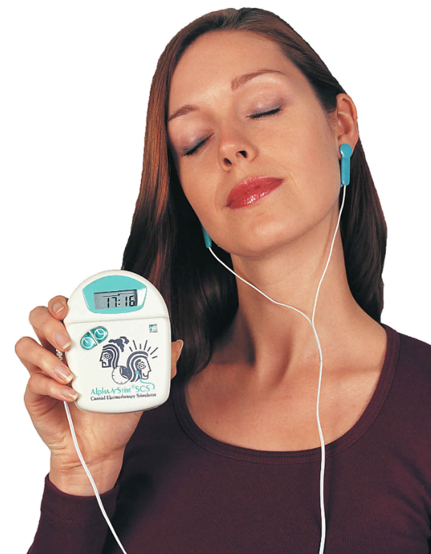
Cranial electrotherapy stimulation (CES) uses a battery powered pulse generator device that applies low-intensity electrical current (0-4mA) via electrodes to a patient’s earlobes, mastoid processes, zygomatic arches, or the maxillo-occipital junction to stimulate the cranium and brain with a current that cannot usually be sensed by the consumer (below four milliamps) to treat insomnia, depression, or anxiety 1. Cranial electrotherapy stimulation are prescription devices that are sold by or on the order of a licensed healthcare professional. The U.S. Food and Drug Administration (FDA) has recognized cranial electrotherapy stimulation as a class III device (premarket approval) for treatment of depression, anxiety, insonmia and chronic pain 2. In humans, the mechanism of action of cranial electrotherapy stimulation is not fully understood. Cranial electrotherapy stimulation devices is purported to stimulate the production of neurotransmitters, such as serotonin and GABA, associated with reducing the symptoms of depression, anxiety and insomnia 3. However, the evidence from 26 randomized controlled trials is insufficient to support conclusions that cranial electrotherapy stimulation has clinically important effects on headache, fibromyalgia, neuromuscular pain, depression, PTSD, or insomnia 4. Furthermore, there is low-strength evidence for a possible beneficial effect of modest size in patients who have anxiety with depression. Cranial electrotherapy stimulation is probably safe, in that no serious side effects have been reported in randomized controlled trials, although reporting bias is present 4.
Cranial electrotherapy stimulation (CES) is related to but distinct from other forms of transcranial electrical stimulation including electroconvulsive therapy, transcranial direct current stimulation (tDCS), and high-definition transcranial direct current stimulation 1. The different versions of transcranial electrical stimulation vary in the placement of electrodes, the intensity of the current, and the waveform of the current.
It remains unclear how the electrical current from cranial electrotherapy stimulation may alter brain activity. Forty-two to 46% of the applied cranial electrotherapy stimulation current enters the brain, with the highest levels of current recorded in the thalamus 5. One theory suggests that the cranial alternating current stimulation interferes with ongoing brain wave oscillations by introducing cortical noise 6. In vitro studies of rat brain slices show that high-frequency (50–200 Hz) sinusoidal alternating current stimulation suppresses activity in cell bodies and axons 7. Perhaps the most investigated effects to date of cranial electrotherapy stimulation have come from electroencephalographic (EEG) studies, which have found recordings to be altered during and after treatment with cranial electrotherapy stimulation. Alpha EEG waves were slowed following cranial electrotherapy stimulation in monkeys, and this change was associated with a reduction in adverse reactions to stressful stimuli 8. Applying cranial electrotherapy stimulation at 0.5- and 100-Hz with simultaneous EEG resulted in a downward shift in mean alpha frequency, with greater effect for 100-Hz stimulation 9. Cranial electrotherapy stimulation also results in a decrease in alpha band median frequency and beta band power fraction 10. These changes are similar to EEG changes in trained meditators, and may be associated with a relaxed state 11. Although it remains unclear if these alterations in brain wave oscillation patterns are a cause or effect of improved clinical states, pulsed current may interrupt nervous system function.
Results from this study 12 suggest that 0.5- and 100-Hz cranial electrotherapy stimulation causes cortical brain deactivation in midline prefrontal and parietal regions. In addition, 100-Hz stimulation significantly altered connectivity within the default mode network. Cranial electrotherapy stimulation appears to result in similar cortical deactivation patterns for 0.5- and 100-Hz, but is associated with stronger alterations in functional connectivity for 100-Hz stimulation. Moreover, cortical deactivation patterns differed from those associated with current intensity, suggesting that cortical deactivation may depend more on frequency than intensity of stimulation.
Cranial electrotherapy stimulation devices have been legally marketed in the United States for well over 30 years with new devices receiving FDA market authorization beginning in 1977, to treat the symptoms of depression, anxiety and insomnia 13. Cranial electrotherapy stimulation has never been indicated for the treatment of a specific diagnosis, but rather to treat symptoms associated with many underlying conditions. Cranial electrotherapy stimulation devices have been safely used, and are often prescribed, in conjunction with drug therapy. Cranial electrotherapy stimulation has not caused any significant harm to patients in the more than 30 years it has been on the market. In fact, less than 10 adverse events have been reported to FDA over that time.
The Neurological Devices Panel that discussed original classification for the cranial electrotherapy stimulator device in 1977 and 1978 ultimately recommended that the device be classified into class III because satisfactory device effectiveness had not been demonstrated 14. The Neurological Devices Panel considered information from the National Research Council, which reviewed 88 published studies on cranial electrotherapy stimulation and concluded that the device has not been shown to be effective in treating any of the conditions for which it was prescribed. In addition, the panel indicated that it was not possible to establish an adequate performance standard for cranial electrotherapy stimulation because the characteristics of the electrical current necessary for potential effectiveness were not known. The Neurological Devices Panel believed that general controls would not provide sufficient control over these characteristics, and that the device presented a potential unreasonable risk of illness or injury to the patient if the practitioner relied on the device, and it was ineffective in treating the patient’s illness. Therefore, the Neurological Devices Panel recommended that premarket approval (Class III) was necessary to assure the safety and effectiveness of cranial electrotherapy stimulation devices.
In support of a subsequent proposed rule in 1993 for classification of cranial electrotherapy stimulation into class III, FDA performed a literature review and identified additional studies that had been performed for cranial electrotherapy stimulation. After a review of the scientific literature, FDA concluded that the effectiveness of cranial electrotherapy stimulation had still not been established by adequate scientific evidence 14.
FDA has performed a literature search for studies of cranial electrotherapy stimulation published after the 1993 proposed rule (January 1, 1993, to present). Many studies were excluded from further review because they were conducted on very specific populations (e.g., alcoholics or other types of substance abuse), and therefore were not representative of the general population suffering from insomnia, anxiety, or depression. Six studies were identified for further review 15, 16, 17, 18, 19, 20. FDA also identified two relevant meta-analyses 21, 22.
The Bystritsky et al.15 was conducted open-label, and on only 12 subjects. The study involved observational baseline versus post-treatment without a control and therefore provided insufficient evidence of safety and effectiveness. The Heffernan study 16 concludes that a single cranial electrotherapy stimulation treatment may have physiologic effects; however, no outcomes of anxiety, depression or insomnia were measured and the study was conducted on only 20 subjects. The Overcash study 17 was a retrospective study design and used an anxiety rating scale that was not validated. The Voris study 18 analyzed only a subgroup of “psychiatric subjects” which included many types of anxiety disorders as well as non-anxiety psychiatric disorders. The subgroup represents a diagnostically heterogeneous group. The subgroup analysis was not pre-specified and the number of subjects per subgroup was not specified. The Hyun study 19 was a randomized controlled trial of 60 subjects. However, the indication under investigation was preoperative anxiety, which may not be indicative of an Axis I anxiety disorder. Moreover, the outcome measure, a 5-point Likert scale rating of anxiety, was not a standardized validated rating instrument. The Winick study 20, which was a randomized controlled trial of 33 subjects with anxiety prior to dental procedures and utilized a 7-point Likert scale, suffers from the same limitations as the Hyun study 19.
The O’Conner meta-analysis 21 examined the effect of cranial electrotherapy stimulation on reduction of primary and secondary withdrawal symptoms among various chemically dependent populations. The results of this analysis do not relate to the question of safety and effectiveness since the labeled indications for cranial electrotherapy stimulation currently include insomnia, depression, or anxiety, and not withdrawal symptoms of chemical dependence. The Klawansky meta-analysis 22 was based on an examination of literature on cranial electrotherapy stimulation versus sham treatment. Although the analysis showed cranial electrotherapy stimulation to be more effective than sham therapy for anxiety, the study populations showed great heterogeneity of diagnostic categories (e.g., in many cases anxiety was not the primary diagnosis, but rather one of a number of symptomatic outcome measures collected during a trial). Therefore, it is unclear whether the finding can be generalized to support the effectiveness of cranial electrotherapy stimulation in homogeneous populations of individuals suffering from anxiety, depression, or insomnia. Also, many of the studies evaluated in the Klawansky meta-analysis involved insufficient blinding 22.
FDA has concluded from a review of the scientific literature and the information provided that the effectiveness of cranial electrotherapy stimulation has not been established by adequate scientific evidence and the Agency continues to agree with the panel’s recommendation.
Cranial electrotherapy stimulation side effects
Cranial electrotherapy stimulation side effects may include:
- Worsening of the condition being treated—If the device is not effective and the patient is not treated in a conventional manner, the patient’s psychological condition may worsen.
- Skin irritation—The electrodes or the conductive cream used with the electrodes may cause skin irritation.
- Headaches—Reported cases of adverse effects of cranial electrotherapy stimulation devices include headaches following treatment with electrical stimulation.
- Potential risk of seizure—electrical stimulation of the brain may result in seizures, particularly in patients with a history of seizure.
- Blurred vision—placement of electrodes over the eyes may cause blurred vision.
- Potential adverse effects from electrical stimulation of the brain— The physiological effects associated with electrical stimulation of the brain by these devices have not been studied systematically; therefore, adverse effects which may be caused by these electrical stimuli remain unknown.
- Shekelle P, Cook I, Miake-Lye IM, et al. The Effectiveness and Risks of Cranial Electrical Stimulation for the Treatment of Pain, Depression, Anxiety, PTSD, and Insomnia: A Systematic Review [Internet]. Washington (DC): Department of Veterans Affairs (US); 2018 Feb. Available from: https://www.ncbi.nlm.nih.gov/books/NBK493132
- https://www.regulations.gov/document?D=FDA-2011-N-0504-0001
- Tan G, Rintala DH, Jensen MP, et al. Efficacy of cranial electrotherapy stimulation for neuropathic pain following spinal cord injury: a multi-site randomized controlled trial with a secondary 6-month open-label phase. J Spinal Cord Med. 2011;34(3):285-296. doi:10.1179/2045772311Y.0000000008 https://www.ncbi.nlm.nih.gov/pmc/articles/PMC3127367
- Shekelle P, Cook I, Miake-Lye IM, et al. The Effectiveness and Risks of Cranial Electrical Stimulation for the Treatment of Pain, Depression, Anxiety, PTSD, and Insomnia: A Systematic Review [Internet]. Washington (DC): Department of Veterans Affairs (US); 2018 Feb. EXECUTIVE SUMMARY. Available from: https://www.ncbi.nlm.nih.gov/books/NBK493136
- Jarzembski W, Sances AJ. Evaluation of specific cerebral impedance and cerebral current density. Ann. NY Acad. Sci. 1970;170:476–490.
- Zaghi S, Acar M, Hultgren B, Boggio PS, Fregni F. Noninvasive brain stimulation with low-intensity electrical currents: putative mechanisms of action for direct and alternating current stimulation. Neuroscientist. 2009;16:285–307.
- Jensen AL, Durand DM. Suppression of axonal conduction by sinusoidal stimulation in rat hippocampus in vitro. J. Neural Eng. 2007;4:1–16.
- Jarzembski WB. Electrical stimulation and substance abuse treatment. Neurobehav. Toxicol. Teratol. 1985;7:119–123.
- Schroeder MJ, Barr RE. Quantitative analysis of the electroencephalogram during cranial electrotherapy stimulation. Clin. Neurophysiol. 2001;112:2075–2083.
- Itil T, Gannon P, Akpinar S, Hsu W. Quantitative EEG analysis of electrosleep using analog frequency analyzer and digital computer methods. Dis. Nerv. Syst. 1972;33:376–381.
- Banquet JP. Spectral analysis of the EEG in meditation. Electroencephalogr. Clin. Neurophysiol. 1973;35:143–151.
- Feusner JD, Madsen S, Moody TD, et al. Effects of cranial electrotherapy stimulation on resting state brain activity. Brain Behav. 2012;2(3):211-220. doi:10.1002/brb3.45 https://www.ncbi.nlm.nih.gov/pmc/articles/PMC3381625
- https://www.regulations.gov/document?D=FDA-2012-P-0260-0001
- Requirement for Premarket Approval for Cranial Electrotherapy Stimulator. https://www.regulations.gov/document?D=FDA-2011-N-0504-0001
- Bystritsky A, L. Kerwin, J. Feusner, “A Pilot Study of Cranial Electrotherapy Stimulation for Generalized Anxiety Disorder,” Journal of Clinical Psychiatry, 69(3): 412-417, 2008.
- Heffernan, Michael, “The Effect of a Single Cranial Electrotherapy Stimulation on Multiple Stress Measures,” The Townsend Letter for Doctors and Patients, 147: 60-64, 1995.
- Overcash, Stephen J., “Cranial Electrotherapy Stimulation in Patients Suffering From Acute Anxiety Disorders,” American Journal of Electromedicine, 16(1): 49-51, 1999.
- Voris, Marshall D, “An Investigation of the Effectiveness of Cranial Electrotherapy Stimulation in the Treatment of Anxiety Disorders Among Outpatient Psychiatric Patients, Impulse Control Parolees and Pedophiles,” Manuscript submitted for publication. Delos Mind/Body Institute, Dallas and Corpus Christi, TX: 1-19, 1995.
- Hyun J.K., Y.K. Woon, S.L. Yoon, et al., “The Effect of Cranial Electrotherapy Stimulation on Preoperative Anxiety and Hemodynamic Responses.” Korean Journal of Anesthesiology, 55: 657-61, 2008.
- Winick, R.L., “Cranial Electrotherapy Stimulation (CES): A Safe and Effective Low Cost Means of Anxiety Control in a Dental Practice,” General Dentistry, 47(1): 50-55, 1999.
- O’Connor M.E., F. Bianco, R. Nicholson, “Meta-analysis of Cranial Electrostimulation (CES) in Relation to the Primary and Secondary Symptoms of Substance Withdrawal,” Presented at the 12th annual meeting of the Bioelectromagnetics Society, June 14, 1991.
- Klawansky S., A. Yeung, C. Berkey, et al., “Meta-analysis of Randomized Controlled Trials of Cranial Electrostimulation,” The Journal of Nervous and Mental Disease, 183(7): 478-485, 1995.


