Disseminated herpes zoster
Disseminated herpes zoster also called disseminated zoster or disseminated shingles, is usually defined as a generalized eruption of more than 10-12 extra dermatomal herpes zoster vesicles occurring 7-14 days after the onset of classic dermatomal herpes zoster 1. Herpes zoster also called shingles, is a localized, blistering and painful rash caused by reactivation of latent varicella-zoster virus (VZV). Herpes zoster is characterized by dermatomal distribution, that is the blisters are confined to the cutaneous distribution of one or two adjacent sensory nerves. This is usually unilateral, with a sharp cut-off at the anterior and posterior midlines. Herpes zoster often affects people with weak immunity. Disseminated herpes zoster typically occurs 4-11 days after the onset of localized shingles. Disseminated shingles may be confined to the skin or it may affect the viscera as well. Typically, disseminated shingles is clinically indistinguishable from varicella (chickenpox). Disseminated zoster most commonly affects immunocompromised patients and only rarely the immunocompetent population. It is estimated that cutaneous disseminated herpes zoster occurs in approximately 2% of herpes zoster cases in the general population but has been observed in as many as 35% of patients who are hospitalized or immunocompromised.
Anyone that has previously had varicella (chickenpox) may subsequently develop zoster. Zoster can occur in childhood but is much more common in adults, especially older people. People with various kinds of cancer have a 40% increased risk of developing zoster. People who have had zoster rarely get it again; the chance of getting a second episode is about 1%.
Disseminated shingles often is an indication of depressed cell-mediated immunity caused by various underlying clinical situations, including malignancies, lymphoproliferative malignancies, radiation therapy, cancer chemotherapy, organ transplant recipients, patients with AIDS and long-term use of systemic corticosteroids (short-term use of low-to-moderate doses of corticosteroids does not increase the incidence of dissemination). Disseminated zoster may be an early clinical sign of underlying human immunodeficiency virus (HIV) infection in high-risk populations. Patients in whom herpes zoster has disseminated must be observed carefully for the development of pneumonitis and encephalitis, which can be life-threatening 1.
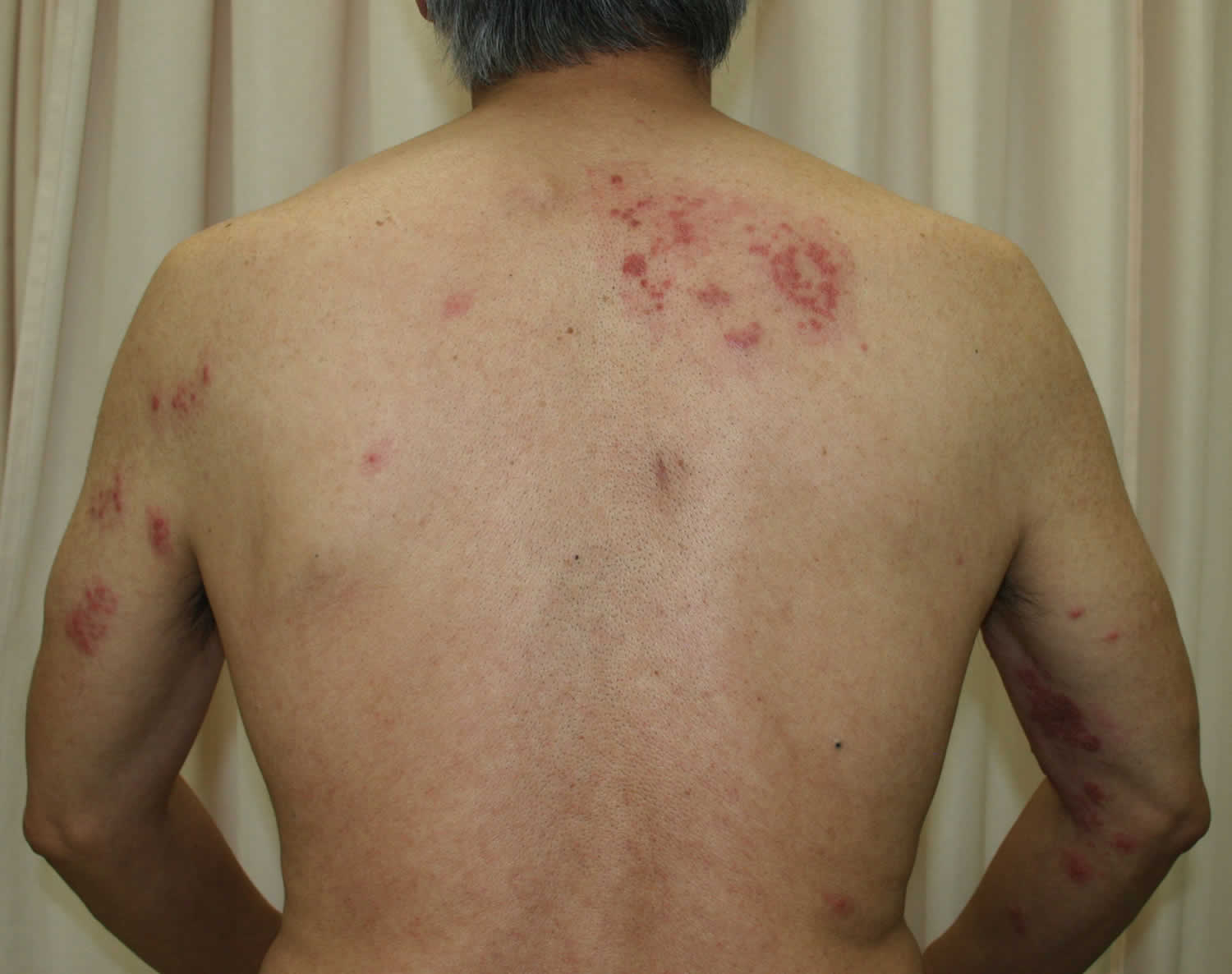
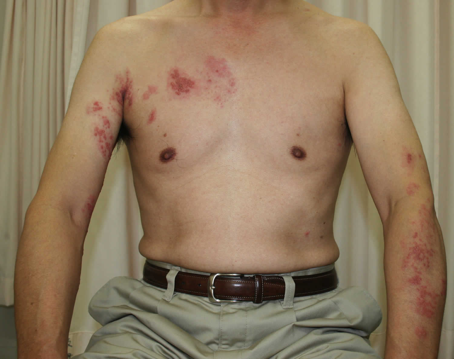
Figure 1. Disseminated shingles
Disseminated zoster symptoms
Disseminated herpes zoster also called disseminated zoster or disseminated shingles, is usually defined as a generalized eruption of more than 10-12 extra dermatomal herpes zoster vesicles occurring 7-14 days after the onset of classic dermatomal herpes zoster 1.
Herpes zoster (shingles) may begin with a systemic response (eg, fever, anorexia, and lassitude), though symptoms frequently are mild. Symptoms typically include prodromal sensory phenomena along 1 or more skin dermatomes lasting 1-10 days (average, 48 hours), which usually are noted as pain or, less commonly, itching or paresthesias 2. Prodromal pain typically is described as muscle or toothachelike in origin but may simulate headache, iritis, pleurisy, brachial neuritis, cardiac pain, appendicitis or other intra-abdominal disease, or sciatica. This simulation can result in incorrect tentative diagnoses; however, the dermatomal distribution usually helps clarify the diagnosis.
The prodromal interval of pain prior to onset of cutaneous findings has been believed to represent spread of varicella-zoster virus (varicella-zoster virus) particles along sensory nerves; however, approximately 10% of patients report the simultaneous onset of pain and rash.
After the onset of prodromal symptoms, the following signs and symptoms occur:
- Patchy erythema, occasionally accompanied by induration, in the dermatomal area of involvement
- Regional lymphadenopathy, either at this stage or subsequently
- Grouped herpetiform vesicles developing on the erythematous base (the classic finding)
- Pain in the dermatomal area of involvement may remain the same as in prodrome or may change in character and intensity with the onset of other symptoms; many patients describe the pain as burning, throbbing, or stabbing in nature; it may be severe, mild, constant, rare, or felt as another sensation such as pruritus; the involved area may be tender to palpation 3. The pain may be just in one spot, or it may spread out. The patient usually feels quite unwell with fever and headache. The lymph nodes draining the affected area are often enlarged and tender. Within one to three days of the onset of pain, a blistering rash appears in the painful area of skin. It starts as a crop of red papules. New lesions continue to erupt for several days within the distribution of the affected nerve, each blistering or becoming pustular then crusting over.
- Vesicular involution – Vesicles initially are clear but eventually cloud, rupture, crust, and involute, a process that may be greatly accelerated by treatment
The first sign of herpes zoster is usually pain, which may be severe, relating to one or more sensory nerves. The chest (thoracic), neck (cervical), forehead (ophthalmic) and lumbar/sacral sensory nerve supply regions are most commonly affected at all ages. The frequency of ophthalmic herpes zoster increases with age. Herpes zoster occasionally causes blisters inside the mouth or ears, and can also affect the genital area. Sometimes there is pain without rash—herpes zoster “sine eruptione”—or rash without pain, most often in children.
Pain and general symptoms subside gradually as the eruption disappears. In uncomplicated cases, recovery is complete within 2–3 weeks in children and young adults, and within 3–4 weeks in older patients.
After vesicular involution, slow resolution of the remaining erythematous plaques, typically without visible sequelae – Note, however, that scarring can occur if deeper epidermal and dermal layers have been compromised by excoriation, secondary infection, or other complications
Unfortunately, resolution of the associated pain does not always accompany resolution of erythema and vesiculation. Postherpetic neuralgia (postherpetic neuralgia), which usually is confined to the area of original dermatomal involvement, can persist for weeks, months, or years and is often severe. Severe prodromal pain and the density of the herpetic eruption have been characterized as risk factors and predictors for postherpetic neuralgia 4.
The reason why some patients with zoster experience postherpetic neuralgia and others do not is not fully understood, but it is clear that patients who are older (> 60 years), particularly those who are debilitated or arteriosclerotic, are affected far more frequently than younger patients are. In addition, postherpetic neuralgia is observed more frequently after cases of herpes zoster ophthalmicus (herpes zoster ophthalmicus) and in instances of upper-body dermatomal involvement.
Other less common postherpetic complications include hyperesthesia or, more rarely, hypoesthesia or anesthesia in the area of involvement.
Herpes zoster oticus (Ramsay Hunt syndrome) may produce an acute jugular foramen syndrome 5. It is characterized by acute-onset dysphagia and dysphonia, often accompanied or preceded by cranial, cervical, or pharyngeal pain. Herpetic vesicles on the skin or mucosa may or may not occur, may be noted late after onset, or may go undetected.
Disseminated zoster complications
Disseminated zoster may be seen in immunocompromised patients. In such cases, hematogenous spread may result in the involvement of multiple dermatomes. Visceral involvement can also occur. Hong and Elgart 6 have reported gastrointestinal complications.
Post-herpetic neuralgia is defined as persistence or recurrence of pain in the same area, more than a month after the onset of herpes zoster. Postherpetic neuralgia is the most common complication, affecting as many as 50% of patients older than 60 years. It may develop as a continuation of the pain that accompanies acute zoster, or it may develop after apparent resolution of the initial zoster reactivation. The pain may last for months to years 7. The underlying pathophysiology of postherpetic neuralgia may involve peripheral nerve damage or continued viral activity. It is particularly likely if there is facial infection. Post-herpetic neuralgia may be a continuous burning sensation with increased sensitivity in the affected areas or spasmodic shooting pain. The overlying skin is often numb or exquisitely sensitive to touch. Sometimes, instead of pain, the neuralgia results in a persistent itch (neuropathic pruritus).
Herpes zoster involving the first branch of the Trigeminal nerve (the fifth cranial nerve) or herpes zoster ophthalmicus, may be associated with conjunctivitis, keratitis, corneal ulceration, iridocyclitis, glaucoma, and decreased visual acuity or blindness. With ocular involvement, long-term antiviral treatment may be required.
Guillain-Barré syndrome is a rare complication from reactivation of latent varicella-zoster virus. Cresswell et al 8 reported that Guillain-Barré syndrome also resulted from primary varicella-zoster virus infection in an adult.
Varicella-zoster virus is being investigated for its possible role in least some cases of chronic fatigue syndrome 9.
In children, varicella-zoster virus infection may produce a facial palsy 10; it may also result in zoster sine herpete (a condition in which dermatomal distribution pain occurs in the absence of an antecedent rash), more frequently in children than adults 11. Ramsay Hunt syndrome (otic herpes zoster or herpes zoster oticus) tends to be found more often in school-aged children, whereas zoster sine herpete is more likely to be found in preschool children.
Childhood varicella zoster virus infections have been linked with an increased risk of arterial ischemic stroke and may precipitate a childhood arterial ischemic stroke 12.
Westenend and Hoppenbrouwers 13 reported fatal hemorrhagic encephalitis in an otherwise healthy female.
Ramsay Hunt syndrome is also known as otic herpes zoster, which is when varicella-zoster virus is reactivated in the geniculate ganglia and causes skin lesions in the face and ears, accompanied with peripheral facial weakness. Varicella-zoster virus also invades the 8th cranial nerve causing tinnitus, vertigo, and hearing impairment. Generally, the infection first appears as neurosensory abnormalities 2-15 days after the first occurrence of skin eruptions, but in some cases the symptoms appear before the skin lesions or when there are no skin lesions, making it difficult to diagnose. Facial palsy is most severe 1 week after the occurrence and then gradually recovers, with poor prognosis being observed in cases with advanced age, elevated arterial blood pressure, vertigo, or diabetes. Ramsay Hunt syndrome may also produce an acute jugular foramen syndrome 5. Treatment is generally the administration of high dose corticosteroids and antiviral agents, but for this case, steroids were not administered due to concern for immune suppression. Fortunately, there was early recovery and no severe complications. There are numerous studies which report that there were good nervous functions and improved recovery rates in groups where antiviral agents were administered together than in groups where corticosteroids were administered exclusively. When there is paralysis, a high dose of corticosteroids plays the role of accelerating the recovery from facial palsy by decreasing the degeneration of the facial nerve through the reduction of neural edema and by the alleviation of vascular spasms 14.
Disseminated zoster may be associated with a secondary bacterial infection at the site of the rash (typically streptococcal or staphylococcal). Necrotizing fasciitis is a possible and devastating complication.
Meningoencephalitis secondary to herpes zoster is more likely to be seen in immunocompromised patients than in immunocompetent patients. Other central nervous system complications may include myelitis, cranial nerve palsies, and granulomatous angiitis. Granulomatous angiitis may result in the development of a cerebrovascular accident.
Acute liver failure has also been reported as a very complication of disseminated zoster 15.
Disseminated zoster diagnosis
A disseminated herpes zoster infection can be diagnosed when 20 or more blisters develop systemically within a week of showing skin symptoms found in typical herpes zoster infections 16.
In most patients, confirming the diagnosis via laboratory testing usually has no utility, because most tests are time consuming, lack specificity, or are unavailable outside of research facilities. In select patient populations, however, the presentation can be atypical and may require additional testing. This is particularly true in immunocompromised patients.
Zoster has been considered a harbinger of other occult disease (eg, malignancies in older patients). Both zoster and malignancy are common in elderly individuals; therefore, most experts currently consider the association to be purely coincidental. There also appears not to be an increased incidence of malignancy in children with herpes zoster. Approximately 3% of pediatric cases occur in children with malignancies. Accordingly, a malignancy workup is not indicated in an otherwise healthy child who has herpes zoster.
Laboratory studies
Although varicella-zoster virus can be cultured, its growth rate is usually too slow for culturing to make a timely contribution to diagnosis.
One of the least expensive and simplest laboratory diagnostic methods for varicella-zoster virus and other herpesviruses is the Tzanck smear. The Tzanck smear is performed by obtaining a scraping from the base of a fresh vesicular lesion after it has been unroofed, spreading and drying the collected material on a glass slide, staining the result with Giemsa, and examining the material with a microscope for the characteristic presence of multinucleated giant cells.
The Tzanck smear confirms that the lesion is herpetic but cannot differentiate between varicella-zoster virus and other herpesviruses. Further, this test has a limited sensitivity compared with other diagnostic methods, such as polymerase chain reaction (PCR) assay. Therefore, a negative result does not rule out a herpes virus infection and should not preclude empiric treatment in patients 17.
When acute diagnostic confirmation is desired, modern tests, such as direct fluorescent antibody (DFA) testing or PCR (if available), are preferred to the Tzanck smear. Direct fluorescent antibody testing of vesicular fluid or a corneal lesion can yield the varicella-zoster virus antigen. Both direct fluorescent antibody and PCR have far greater sensitivity and specificity than the Tzanck smear and allow differentiation between herpes simplex virus (HSV) and varicella-zoster virus infections.
PCR assay of vesicular fluid or a corneal scraping can yield the varicella-zoster virus nucleic acid. Detection of varicella-zoster virus DNA in plasma can facilitate the early recognition of varicella-zoster virus infection in immunocompromised hosts 18. In cases of zoster sine herpete, DNA analysis by means of PCR appears more useful than the standard assays, especially for early diagnosis.
Disseminated herpes zoster is seen approximately 7 times more frequently in patients with HIV infection; therefore, when clinically indicated, an HIV test should be ordered.
Other studies
No imaging tests are indicated in typical cases of cutaneous herpes zoster infection. Magnetic resonance imaging (MRI) may be used in cases of myelopathy or encephalopathy as a means of excluding other etiologies.
A small percentage of patients, particularly those with cranial nerve involvement, may develop headache and neck stiffness, necessitating a lumbar puncture to exclude meningitis. Because the inflammatory response involves the leptomeninges, cerebrospinal fluid (CSF) may show increased protein and a pleocytosis.
Because the diagnosis can almost always be made on clinical grounds, skin biopsy is seldom necessary; as a rule, it is reserved for cases that are difficult to diagnose (eg, atypical lesions).
Disseminated shingles treatment
Patients with disseminated shingles or severe immunosuppression or who are unresponsive to therapy should be transferred to a higher level of care. If consultation is required but not available at the initial facility, patients should be transferred to a tertiary care medical center.
Medications used include steroids, analgesics, anticonvulsants, and antiviral agents. Surgical care is not generally indicated, though it may be required to treat certain complications (eg, necrotizing fasciitis). Rhizotomy (surgical separation of pain fibers) may be considered in cases of extreme, intractable pain.
Management of acute herpes zoster may include:
- Rest and pain relief
- Protective ointment applied to the rash, such as petroleum jelly.
- Oral antibiotics for secondary infection
Post-herpetic neuralgia may be difficult to treat successfully. It may respond to any of the following.
- Early use of antiviral medication
- Local anaesthetic applications
- Topical capsaicin
- Tricyclic antidepressant medications such as amitriptyline
- Anti-epileptic medications gabapentin and pregabalin
- Transcutaneous electrical nerve stimulation or acupuncture
- Botulinum toxin into the affected area
Nonsteroidal anti-inflammatories and opioids are generally unhelpful.
Herpes zoster in the setting of malignancy or organ transplantation
Intravenous acyclovir remains the therapy of choice for disseminated zoster in severely immunocompromised patients, including (1) allogeneic hematopoietic stem cell transplant recipients within 4 months of transplantation, (2) hematopoietic stem cell transplant recipients with moderate to severe acute or chronic graft-versus-host disease, or (3) any transplant recipient receiving aggressive antirejection therapy 19. In addition, any transplant recipient with suspected visceral dissemination (e.g., encephalitis or pneumonitis) should receive intravenous acyclovir. The recommended dose is 10 mg/kg (or 500 mg/m²) every 8 hours. When the infection is controlled, intravenous administration can be stopped, and oral antiviral medication can be initiated for the remainder of the course of therapy.
Initial clinical trials with intravenous acyclovir for localized or disseminated herpes zoster in immunocompromised patients demonstrated that treatment halts disease progression and reduces the duration of viral replication 20. Subsequent studies of bone marrow transplant recipients proved that acyclovir, in addition to promoting faster disease resolution, is highly effective at preventing varicella-zoster virus dissemination 21. Because most varicella-zoster virus-related fatalities result from disseminated infection, the ability to prevent dissemination has markedly reduced the rate of death due to herpes zoster in transplant recipients.
Herpes zoster in HIV-seropositive patients
Prospectively acquired data to guide clinicians when selecting antiviral therapy for herpes zoster in HIV-seropositive patients are currently limited. Nearly 300 HIV-infected patients with herpes zoster were enrolled in controlled studies comparing orally administered acyclovir (800 mg 5 times daily) with sorivudine (40 mg once daily) 22. Times to cessation of new vesicle formation, total crusting, and resolution of herpes zoster-associated pain were 3–4 days, 7–8 days, and ∼60 days, respectively 22. Although sorivudine was never marketed, these studies clearly confirmed the efficacy and safety of oral antiviral therapy for herpes zoster in patients with HIV infection. Famciclovir was judged to be effective and safe for treatment of herpes zoster in patients with AIDS when evaluated in a small, open-label clinical trial 23. Valacyclovir has not been systematically evaluated as a treatment for herpes zoster in HIV-infected patients, although preliminary data 24 and anecdotal clinical experience suggest therapeutic benefit. Patients who have herpes zoster ophthalmicus should always be treated to reduce the risk of serious ocular complications, even when presenting >72 hours after rash onset 25. Because of the documented risk of relapsing infection, herpes zoster in HIV-seropositive patients should be treated until all lesions have healed, which is often longer than the standard 7- to 10-day course. What impact herpes zoster therapy may have on the risk of subsequent complications, such as CNS infection or retinitis, is unknown. Adjunctive therapy for herpes zoster with corticosteroids has not been evaluated in HIV-infected patients and is not currently recommended. Long-term administration of anti-VZV medications to prevent recurrences of herpes zoster is not routinely recommended for HIV-infected patients.
Although rare, acyclovir-resistant VZV has been reported in immunocompromised patients, especially those with HIV infection, and results from mutations in the thymidine kinase gene. The presence of atypical lesions or a failed clinical response should prompt evaluation for drug susceptibility to determine whether resistance has developed. When acyclovir resistance occurs, treatment with alternative medications (e.g., intravenous foscarnet or cidofovir) is required.
Management of acute pain in herpes zoster and post herpetic neuralgia is similar in immunocompromised and immunocompetent patients, although NSAIDs that are not cyclooxygenase 2–specific inhibitors are contraindicated in thrombocytopenic patients.
Herpes zoster ophthalmicus and herpes zoster retinitis
Therapy for herpes zoster ophthalmicus is similar to that for herpes zoster elsewhere in the body but should include the care of an ophthalmologist familiar with the disease. Treatment includes the following 26: (1) approved dosages of famciclovir or valacyclovir for 7–10 days, preferably started within 72 hours of rash onset (with intravenous acyclovir given as needed for retinitis), to resolve acute disease and inhibit late inflammatory recurrences 27; (2) pain medications, as discussed above; (3) cool to tepid wet compresses (if tolerated); (4) antibiotic ophthalmic ointment administered twice daily (e.g., bacitracin-polymyxin), to protect the ocular surface; (5) topical steroids (e.g., 0.125%–1% prednisolone 2–6 times daily) prescribed and managed only by an ophthalmologist for corneal immune disease, episcleritis, scleritis, or iritis; (6) no topical antivirals, because they are ineffective; (7) mydriatic/cycloplegia as needed for iritis (e.g., 5% homatropine twice daily); and (8) ocular pressure–lowering drugs given as needed for glaucoma (e.g., latanaprost once daily and/or timolol maleate ophthalmic gel forming solution every morning). Systemic steroids are indicated in the presence of moderate to severe pain or rash, particularly if there is significant edema, which may cause orbital apex syndrome through pressure on the nerves entering the orbit 26. The dosage is commonly 20 mg of prednisone administered (together with an oral antiviral agent) orally 3 times daily for 4 days, twice daily for 6 days, and then once daily every morning for 4 days.
Therapy for chronic problems includes the following:
- Lubricating, preservative-free artificial tear gels or tears administered 4 times daily, antibiotic ointment administered once daily, and, possibly, lateral tarsorrhaphy to protect the corneas (which are often hypesthetic/anesthetic as a result of neuronal damage) from breakdown;
- Continuous-wear, therapeutic soft contact lenses and antibiotic drops (e.g., polymyxin-trimethoprim given 4 times daily as needed for corneal ulceration);
- Topical steroids and antibiotics for inflammatory disease (iritis, episcleritis, scleritis, and immune keratitis);
- Dilation for iritis;
- Glaucoma therapy as needed; and
- Surgical management as needed—for example, for amniotic membrane transplantation, tissue-adhesive seal ulcers, keratoprosthesis, and glaucoma trabeculectomy.
Chronic pain management is generally similar to that for postherpetic neuralgia in other dermatomes and includes gabapentin or pregabalin, tricyclic antidepressants (TCAs), opioid analgesics, and lidocaine gel (which is preferable to the lidocaine patch for periophthalmic use). Local nerve blocks or sympathetic blocks can be used for pain that is refractory to first-line therapy, although there are no controlled studies of these treatments 28.
The optimal antiviral therapy for VZV-induced, rapidly progressive herpetic retinal necrosis in immunocompromised patients remains undefined. Responses to intravenous acyclovir or ganciclovir have been inconsistent and disappointing, with 49%–67% of involved eyes progressing to no light perception 29. Several case reports have reported improved preservation of vision in patients treated with a combination of intravenous ganciclovir plus foscarnet, with or without intravitreal antivirals 30. Cidofovir has also been used successfully in a small number of patients 31. The optimal duration of induction therapy and options for long-term maintenance therapy have not been established.
Acute retinal necrosis in immunocompetent patients is a less virulent disease and responds better to antiviral therapy. For such patients, acyclovir is clearly beneficial for preserving useful vision 32. A suggested antiviral regimen for acute retinal necrosis in the otherwise healthy host is intravenous acyclovir (10–15 mg/kg every 8 hours for 10–14 days) followed by oral valacyclovir (1 g 3 times daily for 4–6 weeks), although this treatment approach has not been studied in a controlled fashion.
Neurologic complications of herpes zoster
The role of antiviral agents in the management of neurologic complications of herpes zoster has not been evaluated in a controlled fashion. For those diseases in which viral replication likely plays an important role in pathogenesis (e.g., meningitis, encephalitis, and myelitis), therapy with intravenous acyclovir is recommended; this approach is supported by benefits noted in anecdotal experience. For such conditions as delayed contralateral hemiparesis, in which the role of active viral replication is less clear, the value of antiviral therapy is uncertain, but the potential benefits of antiviral therapy outweigh any potential risks 33.
Renal failure
Dosages should be adjusted if renal insufficiency is present when acyclovir (creatinine clearance, <25 mL/min), famciclovir (creatinine clearance, <60 mL/min), or valacyclovir (creatinine clearance, <50 mL/min) is used. Because brivudin undergoes hepatic as well as renal excretion, dosage reduction for renal insufficiency is less critical, but hepatic function must be considered when this antiviral agent is used. Dosages of gabapentin and pregabalin should be reduced when renal function is impaired.
References- What is disseminated herpes zoster (shingles)? https://www.medscape.com/answers/1132465-40996/what-is-disseminated-herpes-zoster-shingles
- Goh CL, Khoo L. A retrospective study of the clinical presentation and outcome of herpes zoster in a tertiary dermatology outpatient referral clinic. Int J Dermatol. 1997 Sep. 36(9):667-72.
- [Guideline] Dworkin RH, Johnson RW, Breuer J, Gnann JW, et al. Recommendations for the management of herpes zoster. Clin Infect Dis. 2007 Jan 1. 44 Suppl 1:S1-26.
- Makharita MY. Prevention of Post-herpetic Neuralgia from Dream to Reality: A Ten-step Model. Pain Physician. 2017 Feb. 20 (2):E209-E220.
- Ono N, Sakabe A, Nakajima M. [Herpes zoster oticus-associated jugular foramen syndrome]. Brain Nerve. 2010 Jan. 62(1):81-4.
- Hong JJ, Elgart ML. Gastrointestinal complications of dermatomal herpes zoster successfully treated with famciclovir and lactulose. J Am Acad Dermatol. 1998 Feb. 38(2 Pt 1):279-80.
- Kost RG, Straus SE. Postherpetic neuralgia–pathogenesis, treatment, and prevention. N Engl J Med. 1996 Jul 4. 335(1):32-42.
- Cresswell F, Eadie J, Longley N, Macallan D. Severe Guillain-Barré syndrome following primary infection with varicella zoster virus in an adult. Int J Infect Dis. 2010 Feb. 14(2):e161-3.
- Shapiro JS. Does varicella-zoster virus infection of the peripheral ganglia cause Chronic Fatigue Syndrome?. Med Hypotheses. 2009 Nov. 73(5):728-34.
- Ogita S, Terada K, Niizuma T, Kosaka Y, Kataoka N. Characteristics of facial nerve palsy during childhood in Japan: frequency of varicella-zoster virus association. Pediatr Int. 2006 Jun. 48(3):245-9.
- Zoster sine herpete. It would be rash to ignore it. Peter G.E. Kennedy. Neurology Feb 2011, 76 (5) 416-417; DOI: 10.1212/WNL.0b013e31820a0d5d
- Grose C. Biological Plausibility of a Link Between Arterial Ischemic Stroke and Infection with Varicella-Zoster Virus or Herpes Simplex Virus. Circulation. 2016 Jan 26.
- Westenend PJ, Hoppenbrouwers WJ. [Fatal varicella-zoster encephalitis; a rare complication of herpes zoster]. Ned Tijdschr Geneeskd. 1998 Mar 21. 142(12):654-7.
- Zainine R, Sellami M, Charfeddine A, Beltaief N, Sahtout S, Besbes G. Ramsay Hunt syndrome. Eur Ann Otorhinolaryngol Head Neck Dis. 2012;129:22–25.
- Acute Liver Failure due to Disseminated Varicella Zoster Infection. Case Reports in Hepatology Volume 2018, Article ID 1269340 https://doi.org/10.1155/2018/1269340
- Yoon KJ, Kim SH, Lee EH, Choi JH. Disseminated herpes zoster in an immunocompetent elderly patient. Korean J Pain. 2013;26(2):195–198. doi:10.3344/kjp.2013.26.2.195 https://www.ncbi.nlm.nih.gov/pmc/articles/PMC3629351
- Ozcan A, Senol M, Saglam H, Seyhan M, Durmaz R, Aktas E, et al. Comparison of the Tzanck test and polymerase chain reaction in the diagnosis of cutaneous herpes simplex and varicella zoster virus infections. Int J Dermatol. 2007 Nov. 46(11):1177-9.
- Kalpoe JS, Kroes AC, Verkerk S, Claas EC, Barge RM, Beersma MF. Clinical relevance of quantitative varicella-zoster virus (VZV) DNA detection in plasma after stem cell transplantation. Bone Marrow Transplant. 2006 Jul. 38(1):41-6.
- Robert H. Dworkin, Robert W. Johnson, Judith Breuer, John W. Gnann, Myron J. Levin, Miroslav Backonja, Robert F. Betts, Anne A. Gershon, Maija L. Haanpää, Michael W. McKendrick, Turo J. Nurmikko, Anne Louise Oaklander, Michael N. Oxman, Deborah Pavan Langston, Karin L. Petersen, Michael C. Rowbotham, Kenneth E. Schmader, Brett R. Stacey, Stephen K. Tyring, Albert J. M. van Wijck, Mark S. Wallace, Sawko W. Wassilew, Richard J. Whitley, Recommendations for the Management of Herpes Zoster, Clinical Infectious Diseases, Volume 44, Issue Supplement_1, January 2007, Pages S1–S26, https://doi.org/10.1086/510206
- Balfour HH, Bean B, Laskin OL, et al. Acyclovir halts progression of herpes zoster in immunocompromised patients, N Engl J Med, 1983, vol. 308, pg. 1448-53
- Shepp DH, Dandliker PS, Meyers JD. Treatment of varicella-zoster infection in severely immunocompromised patients: a randomized comparison of acyclovir and vidarabine, N Engl J Med, 1986, vol. 314, pg. 208-12
- Gnann JWJr, Crumpacker CS, Lalezari JP, et al. Sorivudine versus acyclovir for treatment of dermatomal herpes zoster in human immunodeficiency virus-infected patients: results from a randomized, controlled clinical trial, Antimicrob Agents Chemother, 1998, vol. 42, pg. 1139-45
- Sullivan M, Skiest D, Signs D, Young C. , Famciclovir in the management of acute herpes zoster in HIV+ patientsPresented atFourth Conference on Retroviruses and Opportunistic Infections, Washington, DC, 22-26 January 1997
- Brentjens MH, Torres G, He J, Lee PC, Tyring SK. , A double-blind randomized study of the use of 2 grams vs. 1 gram valacyclovir TID for 7 days in the treatment of acute herpes zoster in immunocompromised individualsPresented atAnnual Meeting of the American Academy of Dermatology, San Francisco, March 2003
- Holland GN. Acquired immune deficiency syndrome and ophthalmology: the first decade, Am J Ophthalmol, 1992, vol. 114, pg. 86-95
- Pavan-Langston D. Foster S, Azar D, Dohlman C. Viral disease of the ocular anterior segment: basic science and clinical disease, The Cornea, 2005, 4th ed.PhiladelphiaLippincott Williams & Wilkins, pg. 297-397
- Harding SP, Porter SM. Oral acyclovir in herpes zoster ophthalmicus, Curr Eye Res, 1991, vol. 10 Suppl, pg. 177-82
- Severson EA, Baratz KH, Hodge DO, Burke JP. Herpes zoster ophthalmicus in Olmsted County, Minnesota: have systemic antivirals made a difference?, Arch Ophthalmol, 2003, vol. 121, pg. 386-90
- Johnston WH, Holland GN, Engstrom RE, Rimmer S. Recurrence of presumed varicella-zoster virus retinopathy in patients with acquired immune deficiency syndrome, Am J Ophthalmol, 1993, vol. 116, pg. 42-50
- Scott IU, Luu KM, Davis JL. Intravitreal antivirals in the management of patients with acquired immunodeficiency syndrome with progressive outer retinal necrosis, Arch Ophthalmol, 2002, vol. 120, pg. 1219-22
- Zambarakji HJ, Obi AA, Mitchell SM. Successful treatment of varicella zoster virus retinitis with aggressive intravitreal and systemic antiviral therapy, Ocul Immunol Inflamm, 2002, vol. 10, pg. 41-6
- Palay DA, Sternberg PJr, Davis J, et al. Decrease in the risk of bilateral acute retinal necrosis by acyclovir therapy, Am J Ophthalmol, 1991, vol. 112, pg. 250-5
- Gnann JWJr. Varicella-zoster virus: atypical presentations and unusual complications, J Infect Dis, 2002, vol. 186 Suppl 1, pg. 91-8


