Epidermal cyst
Epidermal cyst is also called epidermoid cyst, epidermal inclusion cyst, follicular infundibular cyst or keratin cyst, is a closed sac under the skin, or a skin lump, filled with dead skin cells 1. Epidermal inclusion cysts are extremely common, benign, not contagious, and can appear to resolve on their own. Epidermoid cysts often occur in areas where hair follicles have been inflamed and are usually common in conjunction with acne 2. Without definitive treatment, epidermal cysts can reoccur. Epidermal cysts may return if they are not completely removed by surgery.
Sometimes, epidermal cysts are called sebaceous cysts. This is not correct because the contents of the two types of cysts are different. Epidermal cysts are filled with dead skin cells, while true sebaceous cysts are filled with yellowish oily material 3. A true sebaceous cyst is called a steatocystoma. Epidermal inclusion cysts also do not originate from sebaceous glands; therefore, epidermal inclusion cysts are not sebaceous cysts. The term “sebaceous” cyst should not be used when describing an “epidermoid” cyst. Unfortunately, in practice, the terms are often used interchangeably 3.
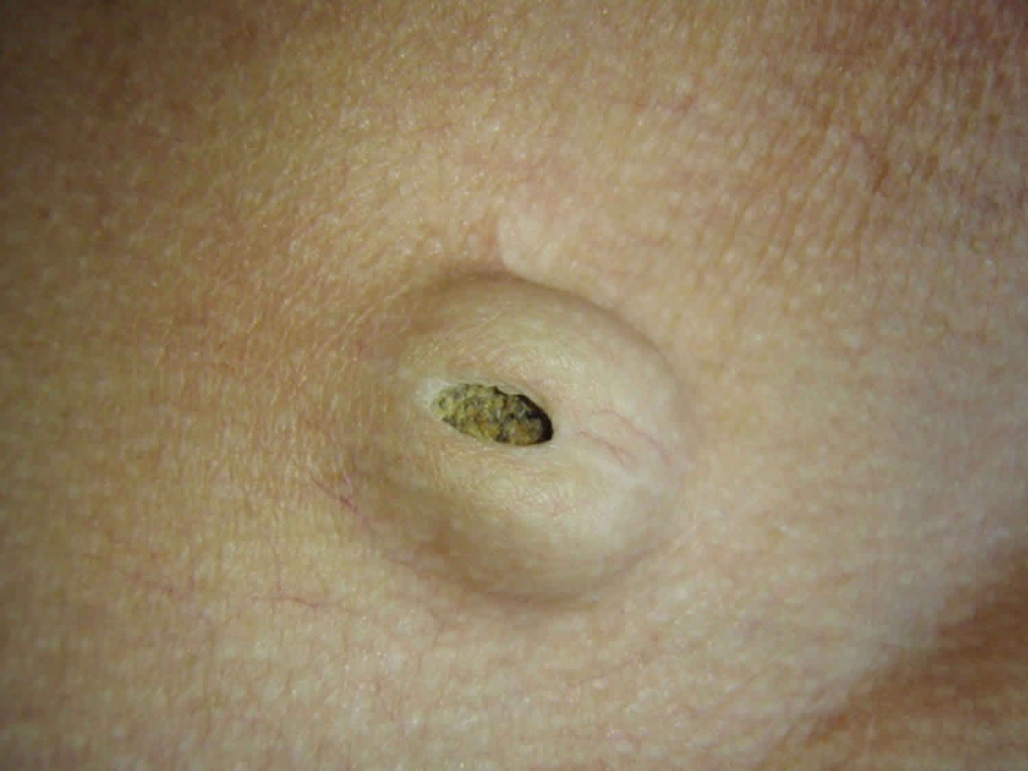
Epidermal inclusion cysts can occur anywhere on the body, typically present as nodules directly underneath the patient’s skin, and often have a visible central punctum. Epidermoid cysts are usually freely moveable. The size of these cysts can range from a few millimeters to several centimeters in diameter. Lesions may remain stable or progressively enlarge over time. There are no reliable predictive factors to tell if an epidermal inclusion cyst will enlarge, become inflamed, or remain quiescent 1. Epidermoid cysts may become infected and form painful abscesses. Infected and/or fluctuant epidermal cysts tend to be larger, erythematous, and more noticeable to the patient. Due to the inflammatory response, epidermoid cyst will often become painful to the patient and may present as a fluctuant filled nodule below the patient’s skin. The center of epidermoid cysts almost always contains keratin and not sebum. This keratin often has a “cheesy” appearance.
Epidermoid cysts are more common in men than women, with a ration of 2:1. Epidermal inclusion cysts occur more frequently in patients in their 20s to 40s. Epidermal inclusion cysts by themselves are usually not genetically linked. They can be hereditary in rare syndromes such as Gardner syndrome, nodular elastosis with cysts and comedones (Favre-Racouchot syndrome), and basal cell nevus syndrome (Gorlin syndrome) 1. Elderly patients with chronic sun-damaged skin areas have a higher likelihood of developing epidermoid cysts. Patients on BRAF inhibitors such as imiquimod and cyclosporine have a higher incidence of epidermoid cysts of the face. They often occur in areas where hair follicles have been inflamed or repeatedly irritated. They are more frequent in patients with acne vulgaris. They can be seen during the neonatal period known as milia 3.
Epidermal cyst key points
- Epidermoid cysts occur on face, neck, trunk or anywhere where there is little hair.
- Most epidermoid cysts arise in adult life.
- They are more than twice as common in men as in women.
- They present as one or more flesh–coloured to yellowish, adherent, firm, round nodules of variable size.
- A central pore or punctum may be present.
- Keratinous contents are soft, cheese-like and malodorous.
- Scrotal and labial cysts are frequently multiple and may calcify.
See your doctor if you notice any new growths on your body. Although epidermoid cysts are not harmful, your doctor should examine you for signs of skin cancer. Some skin cancers look like cystic nodules, so have any new lump examined by your doctor. If you do have a cyst, see your doctor if it becomes red or painful.
Epidermoid cyst causes
Epidermal cysts are very common. The cause of epidermoid cyst is unknown. The majority of epidermoid cysts are sporadic. Epidermal cysts are formed when the surface skin is folded in on itself. The cyst then becomes filled with dead skin because as the skin grows, it can’t be shed as it can elsewhere on the body. When a epidermoid cyst reaches a certain size, it usually stops growing.
People with epidermoid cysts may have family members who also have them.
Epidermal cysts are more common in adults than in children.
The epidermal inclusion cyst can be primary or secondary. Primary epidermal cysts arise directly from the infundibulum of the hair follicle. Plugging of the follicular orifice allows for cyst formation. The cyst often communicates with the surface of the skin through a small orifice or visible central punctum. Patients suffering from acne vulgaris have a higher rate of hair follicle disruption and pore blockages leading to a higher rate of epidermal inclusion cyst formation from preexisting comedones. Secondary epidermoid cysts can arise after the implantation of the follicular epithelium in the dermis due to trauma or comedone formation. Epidermoid cysts are lined with stratified squamous epithelium with the accumulation of keratin in the core. Recently, Human papillomavirus and chronic ultraviolet light exposure have been seen to allow epidermal cysts to form.
Additionally, the epidermal inclusion cysts can occur in any area of the body. Most often, they are found on the face, scalp, neck, back, and scrotum. Inclusion cysts found in unusual numbers or locations like the extremities, trunk, or the back of the ears may be seen in the setting of Gardner syndrome. Gardner syndrome or familial adenomatous polyposis (FAP) with extracolonic manifestations is an autosomal dominant inherited disease due to a mutation in the APC gene on chromosome 5. The cardinal clinical feature is innumerable, widespread colonic polyps in conjunction with extracolonic lesions. In this disease process, the epidermal cysts will often appear before the onset of puberty and may even precede the onset of colonic polyposis 3.
Epidermoid cyst symptoms
The main symptom is usually a small, non-painful lump beneath the skin. The lump is usually found on the face, neck, and trunk. It will often have a tiny hole or pit in the center. It usually grows slowly and is not painful.
If the lump becomes infected or inflamed, other symptoms may include:
- Skin redness
- Tender or sore skin
- Warm skin in the affected area
- Grayish-white, cheesy, foul-smelling material that drains from the cyst.
Epidermal cyst complications
Complications of epidermal inclusion cysts before definitive management can occur due to rupture and may result in symptoms such as erythema, pain, swelling, localized cellulitis. The main complication seen in clinical practice is reoccurrence due to incomplete excision. Any time surgery is done, there is a small inherent risk of complications. Complications of epidermal inclusion cyst excisions may include but are not limited to infection, bleeding, damage to surrounding structures and tissues, scarring, and wound dehiscence. There is a risk of the cyst reoccurring if the capsule is not completely excised during the surgical procedure.
Epidermoid cyst diagnosis
The diagnosis of epidermoid cysts is usually clinical. In most cases, your doctor can make a diagnosis by examining your skin. It is based upon the clinical appearance of a discrete, freely moveable cyst, often with a visible central punctum. These cysts can occur anywhere on the body and typically present as nodules directly underneath the patient’s skin. The size of a cyst can range from a few millimeters to several centimeters in diameter. Lesions may remain stable or progressively enlarge. There is no predictive modality to tell if an epidermal inclusion cyst will enlarge, become inflamed, or remain quiescent. An infected cyst tends to be large with increased erythema, and it is more noticeable to the patient.
Furthermore, if inflammation is present, it usually results in cyst rupture and extrusion of cyst contents into the surrounding cutaneous and/or subcutaneous tissues, which may or may not be the result of an active infection. The source of infection for cysts usually comes from normal skin flora organisms, such as Staphylococcus aureus and Staphylococcus epidermidis. Generally, epidermal inclusion cysts are asymptomatic until they rupture.
Sometimes, a biopsy may be needed to rule out other conditions. If infection is suspected, you may need to have a skin culture.
Histopathology
Epidermoid inclusion cysts can be confirmed by histologic examination. Epidermal inclusion cysts, more specifically, demonstrate the implantation of epidermal elements into the dermis layer of the skin. The cyst wall is usually derived from the infundibular portion of the hair follicle. Thus, the majority of epidermal inclusion cysts may be referred to as an infundibular cyst. However, a cyst’s wall can be derived from another etiology, explaining the interchangeable yet inaccurate use of the two names. The cystic cavity is filled with laminated keratinous material. Often, a granular layer is present that is filled with keratohyalin granules. In the event a cyst ruptures, a keratin granuloma can be seen during the examination. Infected cysts microscopically can show disruption of the cyst wall, acute inflammation or neutrophil invasion, or intense foreign body giant cell reaction. Approximately less than 1% of epidermal inclusion cysts have a malignant transformation to basal cell carcinoma or squamous cell carcinoma 4.
Epidermoid cyst treatment
Epidermal cysts are not dangerous and do not need to be treated unless they cause symptoms or show signs of inflammation (redness or tenderness). Inflamed, uninfected epidermal inclusion cysts rarely resolve spontaneously without therapy or surgical intervention.
Epidermoid cyst may need further treatment if it becomes:
- Inflamed and swollen — your doctor may inject the cyst with steroid medicine
- Swollen, tender, or large — your doctor may drain the cyst or do surgery to remove it
- Infected — you may be prescribed antibiotics to take by mouth
Treatment is not emergent unless desired by the patient electively before an increase in symptom severity (pain and/or infection). Definitive treatment is the surgical excision of the cyst. Some sources describe an alternative, yet not definitive, minimally invasive therapy for treatment, such as injecting triamcinolone at the dosage of 10 mg/mL for the trunk and 3 mg/mL for the face. The injection should be introduced into the inflamed epidermoid cyst, and it can help resolve the inflammation, prevent infection, and potentially reduce the need for surgical incision and drainage.
The definitive treatment is the complete surgical excision of the cyst with its walls intact; this will prevent reoccurrence. Excision is best accomplished when the lesion is not acutely inflamed. During this period, the cyst wall is friable, and the planes of dissection are more difficult to appreciate, making complete excision less likely and increasing the rate of reoccurrence.
For surgical excision, a local anesthetic, such as lidocaine with epinephrine, can be used. The anesthetic should be injected around the cyst, with care to avoid direct injection into the cyst, injection into the central pore, or rupture of the cyst wall. A small incision is made with a #11 blade on the skin overlying the cyst. The cyst contents are then expressed by exerting lateral pressure on either side of the cyst. With this technique, the cyst wall is often freed from the adjacent tissues and can be completely extracted through the small incision. The minimal incision surgical option provides better cosmetic results than the standard excision technique. Maintaining the incision within the minimal skin tension lines is important for cosmetic results. Reoccurrence rates from 1% to 8% have been shown with the minimal incision technique. A multiple layer subcuticular closure with an additional epidermal closure will yield better cosmetic outcomes 5.
An alternate surgical option is to utilize a 4 mm punch biopsy with the expulsion of the intact cyst through the defect created in mass. Regardless of the option chosen, removal of the entire cystic wall is paramount to decrease reoccurrence.
In the event of a fluctuant lesion, incision and drainage are often needed with the mechanical destruction of intracavitary loculations. The presence of surrounding cellulitis may necessitate the use of oral antibiotic therapy. Empiric antibiotic therapy can be done with oral agents active against methicillin-sensitive S aureus or oral agents active against methicillin-resistant S aureus in areas of high prevalence. For patients who wish to have a more conservative treatment in the setting of acute infection, the cyst can be drained, and the patient started on oral antibiotics with a plan of surgical excision of remaining contents at a later date for definitive management. This is recommended due to a higher likelihood of reoccurrence without definitive surgical management 6.
Epidermoid cyst prognosis
Epidermal inclusion cysts have an excellent prognosis after complete excision of all contents and the cystic wall.
References- Weir CB, St.Hilaire NJ. Epidermal Inclusion Cyst. [Updated 2019 Dec 7]. In: StatPearls [Internet]. Treasure Island (FL): StatPearls Publishing; 2020 Jan-. Available from: https://www.ncbi.nlm.nih.gov/books/NBK532310
- Boussemart L, Routier E, Mateus C, Opletalova K, Sebille G, Kamsu-Kom N, Thomas M, Vagner S, Favre M, Tomasic G, Wechsler J, Lacroix L, Robert C. Prospective study of cutaneous side-effects associated with the BRAF inhibitor vemurafenib: a study of 42 patients. Ann. Oncol. 2013 Jun;24(6):1691-7.
- Zito PM, Scharf R. Cyst, Epidermoid (Sebaceous Cyst) [Updated 2019 Dec 25]. In: StatPearls [Internet]. Treasure Island (FL): StatPearls Publishing; 2020 Jan-. Available from: https://www.ncbi.nlm.nih.gov/books/NBK499974
- Apollos JR, Ekatah GE, Ng GS, McFadyen AK, Whitelaw SC. Routine histological examination of epidermoid cysts; to send or not to send? Ann Med Surg (Lond). 2017 Jan;13:24-28.
- Lee HE, Yang CH, Chen CH, Hong HS, Kuan YZ. Comparison of the surgical outcomes of punch incision and elliptical excision in treating epidermal inclusion cysts: a prospective, randomized study. Dermatol Surg. 2006 Apr;32(4):520-5.
- Zuber TJ. Minimal excision technique for epidermoid (sebaceous) cysts. Am Fam Physician. 2002 Apr 01;65(7):1409-12, 1417-8, 1420.


