What is exfoliative dermatitis
Exfoliative dermatitis also known as erythroderma, is an uncommon but serious skin disorder characterized by widespread redness (erythema), peeling, flaking and scaling of the skin caused by preexisting skin disorders, drugs, cancer, or unknown causes. Exfoliative dermatitis symptoms and signs are pruritus (itch), diffuse erythema (redness), epidermal sloughing and hair loss. Exfoliative dermatitis diagnosis is clinical. Exfoliative dermatitis treatment involves corticosteroids and correction of the cause.
Exfoliative dermatitis is a manifestation of rapid epidermal cell turnover. Its cause is unknown, but it most often occurs in the context of:
- Preexisting skin disorders (eg, atopic dermatitis, contact dermatitis, seborrheic dermatitis, psoriasis, pityriasis rubra pilaris)
- Use of drugs (eg, penicillin, sulfonamides, isoniazid, phenytoin, barbiturates)
- Cancer (eg, lymphoma, mycosis fungoides, leukemia, and, rarely, adenocarcinomas)
Up to 25% of patients have no identifiable underlying cause. Bacterial superinfection can complicate exfoliative dermatitis.
Although exfoliative dermatitis affects both men and women, it is more common in men, with an average male-to-female ratio of 2.3:1. The average age at onset is 55 years, although exfoliative dermatitis may occur at any time 1.
Exfoliative dermatitis key points
- Exfoliative dermatitis often occurs with preexisting skin disorders, drugs, and cancer, but the cause may be unknown.
- Symptoms include pruritus, widespread erythema, and epidermal sloughing.
- Diagnosis is clinical.
- Hospitalization is often necessary, because the disease may be life threatening.
- Treatment consists of supportive care, comprehensive wound and skin care, and systemic corticosteroids for severe disease.
Figure 1. Exfoliative dermatitis hands
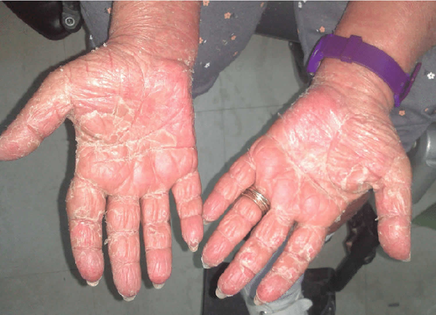
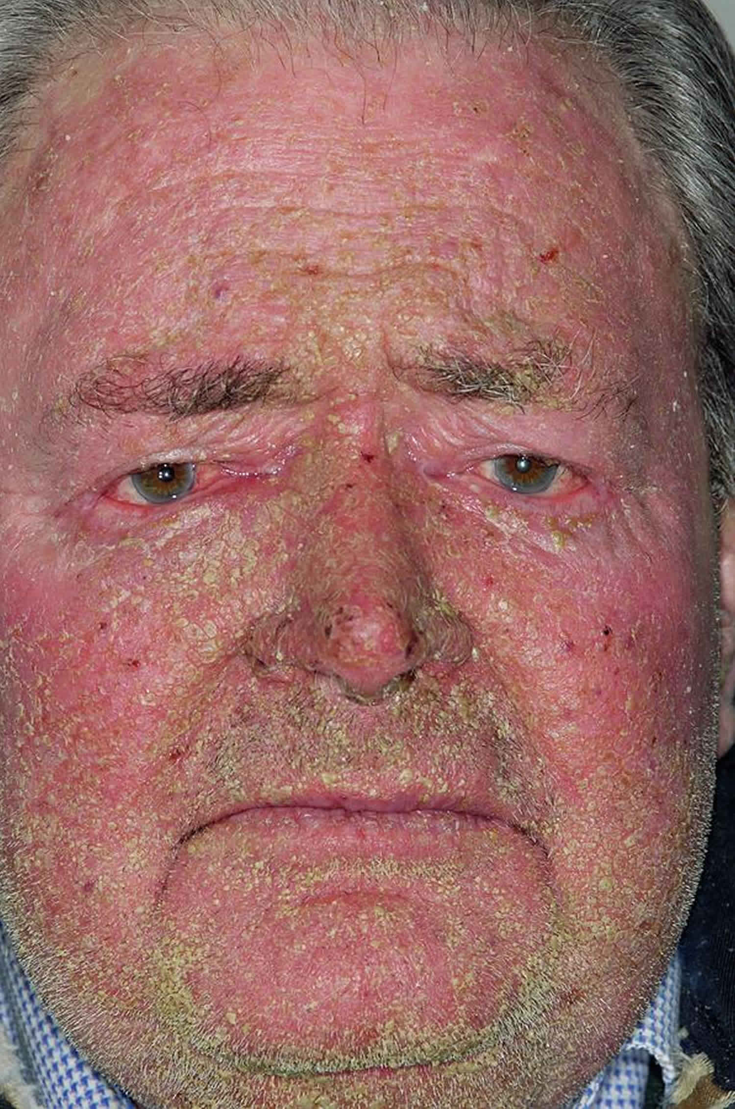
Figure 2. Exfoliative dermatitis face
Exfoliative dermatitis causes
The most common causes of exfoliative dermatitis are preexisting dermatoses, drug reactions, malignancies and other miscellaneous or idiopathic disorders. Exfoliative dermatitis may occur in severe cases of many common skin conditions, such as eczema and psoriasis. It is sometimes caused by an allergy to medicines, chemicals that come in contact with the skin, or by body-wide disease.
Sometimes the cause is unknown.
Primary dermatologic disorders
Dermatologic disorders occasionally present as exfoliative dermatitis. The most common of these are psoriasis, atopic dermatitis, seborrheic dermatitis, contact dermatitis and pityriasis rubra pilaris. Other dermatoses associated with erythroderma are listed below.
Dermatoses associated with exfoliative dermatitis 2:
- Atopic dermatitis
- Candidiasis
- Contact dermatitis
- Dermatophytosis
- Ichthyosis
- Lichen planus
- Mastocytosis
- Nummular eczema
- Pemphigus
- Photosensitive eczema
- Pityriasis rubra pilaris
- Prurigo
- Psoriasis
- Reiter’s syndrome
- Scabies
- Seborrheic dermatitis
- Staphylococcus scalded skin syndrome
- Stasis with autoeczematization
- Subacute cutaneous lupus erythematosus
- Vitamin deficiency
Medications
Since the earliest descriptions of exfoliative dermatitis, medications have been known to be important causative agents. Hence, the apparent increase in cases of exfoliative dermatitis may be related to the introduction of many new drugs. Drug eruptions that initially present as morbilliform, lichenoid or urticarial rashes may progress to generalized exfoliative dermatitis. Antiepileptic medications, antihypertensive medications, antibiotics, calcium channel blockers and a variety of topical agents 1 can cause exfoliative dermatitis, but theoretically, any drug may cause exfoliative dermatitis.
A pseudolymphoma reaction with fever, arthralgias, lymphadenopathy, hepatosplenomegaly, anemia and erythroderma may develop as a result of hypersensitivity to dapsone or antiepileptic drugs. If cutaneous pathology also mimics cutaneous T-cell lymphoma, it can be very difficult to differentiate a drug-induced skin condition from exfoliative dermatitis associated with a malignancy 1.
Drugs associated with exfoliative dermatitis 2:
- Acetaminophen
- Actinomycin D (Cosmegan)
- Allopurinol (Zyloprim)
- Aminoglycosides
- Aminophylline
- Amiodarone (Cordarone)
- Arsenic
- Aztreonam (Azactam)
- Barbiturates
- Calcium channel blockers
- Captopril (Capoten)
- Carbamazepine (Tegretol)
- Cephalosporins
- Chinese herbs
- Chloroquine (Aralen)
- Chlorothiazide (Diuril)
- Chlorpromazine (Thorazine)
- Chlorpropamide (Diabinese)
- Cimetidine (Tagamet)
- Cisplatin (Platinol)
- Clofazimine (Lamprene)
- Clotrimazole (Lotrimin)
- Codeine
- Cyclobenzaprine (Flexeril)
- Dapsone
- Dimercaprol (BAL in Oil)
- Ethylenediamines
- Gold
- Hydantoins
- Hydroxychloroquine (Plaquenil)
- Interleukin-2 (Proleukin)
- Interferon alfa (Roferon-A, Intron A, Alferon N)
- Interferon beta (Avonex, Betaseron)
- Iodine (Pima syrup)
- Isoniazid (Laniazid, Nydrazid; also in Rifamate, Rimactane)
- Isosorbide dinitrate (Isordil, Sorbitrate)
- Isotretinoin (Accutane)
- Lithium (Eskalith, Lithobid)
- Mefloquine (Larium)
- Mephyntoin (Mesantoin)
- Mercurials
- Mercury
- Mexilitene (Mexitil)
- Minocycline (Dynacin, Minocin, Vectrin)
- Mitomycin-C (Mutamycin)
- Neomycin (Neosporin)
- Nitrofurantoin (Furadantin, Macrodantin)
- Omeprazole (Prilosec)
- Para-amino salicylic acid (Sodium P.A.S.)
- Penicillins
- Phenolphthalein (Agoral, Alophen, Modane)
- Phenothiazines
- Phenobarbital (Donnatal, Bellatal)
- Phenytoin (Dilantin)
- Quinacrine
- Quinidine (Quinidex)
- Ranitidine (Zantac)
- Rifampin (Rifadin, Rimactane; also in Rifamate)
- Streptomycin
- Sulfadiazine
- Sulfonamides
- Sulfonylureas
- Terbutaline (Brethine, Bricanyl)
- Tetrachloroethylene
- Tetracyclines
- Thalidomide (Synovir)
- Thiazide diuretics
- Trimethoprim (Trimpex; also in Bactrim, Septra)
- Tolbutamide (Orinase)
- Vancomycin (Vancocin)
Cancers
Cancers are a major cause of exfoliative dermatitis. Reticuloendothelial neoplasms, as well as internal visceral malignancies, can produce erythroderma, with the former being the more predominant cause.
The cutaneous T-cell lymphomas are the lymphomas most commonly associated with exfoliative dermatitis. The most notable member of this group is mycosis fungoides. Studies indicate that mycosis fungoides may cause 25 to 40 percent of all cases of malignancy-related erythroderma.6,7 The erythroderma may arise as a progression from a previous cutaneous T-cell lymphoma lesion or appear simultaneously with the cutaneous T-cell lymphoma, or it may precede the appearance of the cutaneous T-cell lymphoma lesion. When it precedes cutaneous T-cell lymphoma lesions, exfoliative dermatitis becomes the presenting sign of the underlying malignancy.
The time interval between the appearance of exfoliative dermatitis and the appearance of cutaneous T-cell lymphoma lesions can vary from months to years or even decades. Sézary syndrome, the leukemic variant of mycosis fungoides, is also associated with exfoliative dermatitis. The erythrodermic form of mycosis fungoides and the Sézary syndrome may also be difficult to distinguish from benign erythroderma. Immunophenotypic studies with the use of advanced antibody panels may be useful in the differential diagnosis of these two forms.10 Reticulum cell sarcoma is another form of cutaneous T-cell lymphoma that may cause exfoliative dermatitis.
Acute and chronic leukemia may also cause exfoliative dermatitis. The relative risk of leukemia inducing erythroderma is highly variable, ranging from 11 to 50 percent 3.
Internal (visceral) malignancies cause about 1 percent of all cases of exfoliative dermatitis 3. Frequently, exfoliative dermatitis is the presenting sign of the malignancy. Patients with carcinoma of the colon, lung, prostate and thyroid have presented with erythroderma. More recently, carcinomas of the fallopian tube 4, larynx 5 and esophagus 6 have been reported as causes of exfoliative dermatitis. Insidious development of the erythroderma, progressive debilitation of the patient, absence of previous skin disease and resistance to standard therapy are features that may suggest an underlying malignancy 3.
Other associated disorders
Exfoliative dermatitis is also associated with disorders that cannot easily be classified into groups. Exfoliative dermatitis has been reported in association with hepatitis, acquired immunodeficiency syndrome, congenital immunodeficiency syndrome (Omenn’s syndrome) and graft-versus-host disease 7.
In reviews of erythroderma, a significant percentage of patients (about 25 percent) do not receive a specific etiologic diagnosis. Some of these patients undergo spontaneous resolution. Other cases are ultimately classifiable as another dermatosis. A significant number of these patients eventually progress to cutaneous T-cell lymphoma 8.
Exfoliative dermatitis pathogenesis
Exfoliative dermatitis is the result of a dramatic increase in the epidermal turnover rate. In patients with this disorder, the mitotic rate and the absolute number of germinative skin cells are higher than normal. Moreover, the time necessary for cells to mature and travel through the epidermis is decreased. This compressed maturation process results in an overall greater loss of epidermal material, which is manifested clinically as severe scaling and shedding. Normal epidermis undergoes some exfoliation every day, but the scales that are lost contain little, if any, important viable material, such as nucleic acids, soluble proteins and amino acids.4 In exfoliative dermatitis, however, protein and folate losses may be high 9.
The pathogenesis of exfoliative dermatitis is a matter of debate. In recent years, clinicians have come to believe that this condition is secondary to a complicated interaction of cytokines and cellular adhesion molecules. Interleukin (IL)-1, IL-2, IL-8, intercellular adhesion molecule 1 (ICAM-1), tumor necrosis factor and interferon gamma are the cytokines that may have roles in the pathogenensis of exfoliative dermatitis 1.
Exfoliative dermatitis prevention
Risk for exfoliative dermatitis may be reduced by following the provider’s instructions on skin care for psoriasis or other skin conditions.
Exfoliative dermatitis symptoms
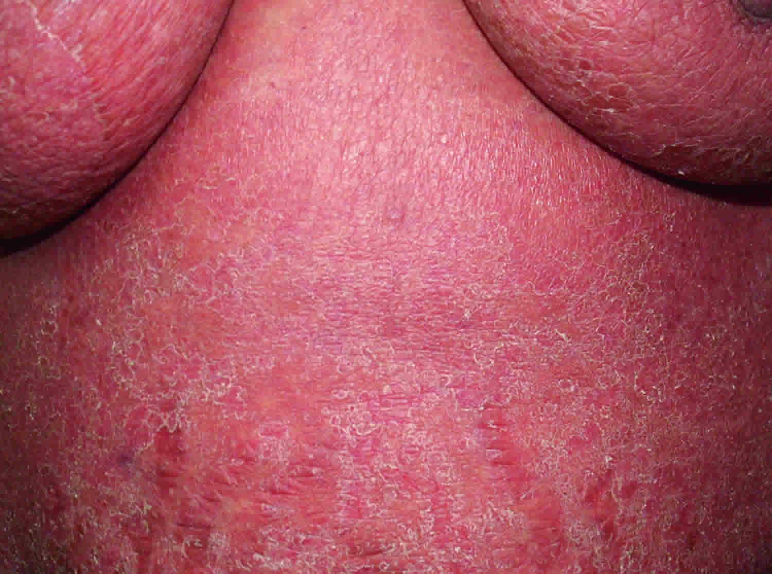
Exfoliative dermatitis symptoms include pruritus, malaise, and chills. Clinically, the first stage of exfoliative dermatitis is erythema (redness), often beginning as single or multiple pruritic patches, involving especially the head, trunk and genital region. These patches tend to spread to involve all or nearly all of the body, after a matter of days or weeks, most of the skin surface is covered with an erythematous, pruritic eruption. Usually, but not always, the palms of the hands, the soles of the feet and the mucous membranes are spared. In some studies, the nose and paranasal area are spared. This has been called the “nose sign” 10.
Extensive epidermal sloughing leads to abnormal thermoregulation, nutritional deficiencies because of extensive protein losses, increased metabolic rate with a hypercatabolic state, and hypovolemia due to transdermal fluid losses.
Once the erythema is well established, scaling inevitably follows. The scales may be small or large, superficial or deep. Acute processes usually favor large scales, whereas chronic processes produce smaller ones. The exfoliative process also may involve the scalp, with 25 percent of patients developing alopecia 11. Nails can often become dystrophic, particularly in patients with preexisting psoriasis 11.
The most frequently noted symptoms in patients with exfoliative dermatitis include malaise, pruritis and a chilly sensation. Both hyperthermia and hypothermia are reported. Other clinical findings include lymphadenopathy, hepatomegaly, splenomegaly, edema of the foot or ankle 11 and gynecomastia 12.
The scaling that occurs in exfoliative dermatitis can have severe metabolic consequences, depending on the intensity and the duration of the scaling. Since cutaneous function as a multiprotective barrier is so disrupted in exfoliative dermatitis, the body loses heat, water, protein and electrolytes, and renders itself much more vulnerable to infection. Exfoliative dermatitis is also a risk factor for epidemic spread of methicillin-resistant Staphylococcus aureus.6,20
Heat loss is another major concern that accompanies a defective skin barrier in patients with exfoliative dermatitis. Loss of normal vasoconstrictive function in the dermis, decreased sensitivity to the shivering reflex and extra cooling that comes from evaporation of the fluids leaking out of the weeping skin lesions all result in thermoregulatory dysfunction that can cause hypothermia or hyperthermia 13. The basal metabolic rate also is increased in patients with exfoliative dermatitis. A catabolic state thus ensues, which is often responsible for significant weight loss.
Each of these physiologic disruptions is potentially life-threatening. Hypothermia can result in ventricular flutter, decreased heart rate and hypotension. Increased peripheral blood flow can result in high-output cardiac failure. Hypervolemia can also occur in patients with exfoliative dermatitis, contributing to the likelihood of cardiac failure 14.
Exfoliative dermatitis symptoms may include any of the following:
- Redness and scaling with weeping or crusting that cover over 80% to 90% of the body
- Skin is itchy or painful and often has an odor
- Swelling of the arms or legs
- Fast heart beat
- Loss of fluids leading to dehydration
- Loss of temperature regulation by the body
There may be secondary infections of the skin.
Exfoliative dermatitis possible complications
Exfoliative dermatitis complications may include:
- Secondary infections that can lead to sepsis (bodywide inflammatory response)
- Fluid loss that can result in dehydration and an imbalance of minerals (electrolytes) in the body
- Heart failure
Exfoliative dermatitis diagnosis
Diagnosis is by history and examination. Your health care provider will perform an examination and ask about your symptoms. The skin will be carefully checked, often with an instrument called a dermascope. Most of the time, the cause can be identified after the exam.
Laboratory evaluation of patients with erythroderma is generally not very helpful in determining a specific diagnosis. Typical laboratory values include mild anemia, leukocytosis, eosinophilia, elevated erythrocyte sedimentation rate, abnormal serum protein electrophoresis with a polyclonal elevation in the gamma globulin region, and elevated IgE levels 8.
Blood counts and bone marrow studies may reveal an underlying leukemia. Analysis for circulating Sézary cells may be helpful, but only if the cells are identified in unequivocally large numbers.
If needed, the following tests may be ordered:
- Biopsy of the skin
- Allergy testing
- Other tests to find the cause of erythroderma
Preexisting skin disease may underlie the extensive erythema and suggest a cause. Biopsy is often nonspecific but is indicated when mycosis fungoides is suspected. Blood tests may reveal hypoproteinemia, hypocalcemia, and iron deficiency; however, these findings are not diagnostic.
Exfoliative dermatitis treatment
Since exfoliative dermatitis can quickly lead to serious complications, your doctor will start treatment right away. This usually involves strong doses of cortisone medicines to reduce inflammation. Supportive care consists of correction of dehydration, correction of electrolyte abnormalities and nutritional deficiencies, and comprehensive wound care and dressings to prevent bacterial superinfection. Other measures include bed rest, lukewarm soaks or baths, bland emollients and oral antihistamines 15. Because drug eruptions and contact dermatitis cannot be ruled out by history alone, all drugs should be stopped if possible or changed. Skin care is with emollients and colloidal oatmeal baths. Weak topical corticosteroids (eg, 1 to 2.5% hydrocortisone ointment) may be used. Corticosteroids (prednisone 40 to 60 mg po once/day for 10 days, then tapered) are used for severe disease.
Other treatments may include:
- Medicines to treat the underlying cause of erythroderma, such as psoriasis
- Antibiotics for any infection
- Dressings applied to the skin
- Ultraviolet light
In patients with chronic idiopathic erythroderma, emollients and topical steroids may be effective. Other patients may warrant PUVA (psoralen plus ultraviolet A) phototherapy, systemic steroids (if psoriasis has been ruled out), retinoids (for exfoliative dermatitis secondary to psoriasis and pityriasis rubra pilaris), or immunosuppressive agents such as methotrexate (Rheumatrex) and azathioprine (Imuran) 16.
When used as adjunctive therapy, behavior modification designed to eliminate persistent scratching has been successful in reducing the rate of excoriation and increasing the rate of healing 17.
No uniformity of opinion exists concerning the best treatment for cutaneous T-cell lymphoma. Options include use of PUVA light therapy, total-body electron beam irradiation, topical nitrogen mustard, systemic chemotherapy and extracorporeal photopheresis. Consultation with an oncologist who is well-versed in treatment of cutaneous T-cell lymphoma is advisable once the disease progresses to the tumor stage.
Exfoliative dermatitis prognosis
Exfoliative dermatitis may be life threatening; hospitalization is often necessary. Even though exfoliative dermatitis is a complex disorder involving many factors, the underlying disease is usually the key determinant of the course and prognosis. Drug-induced exfoliative dermatitis is usually short-lived once the inciting medication is withdrawn and appropriate therapy is administered. Cases related to drug reactions have the shortest duration, lasting 2 to 6 weeks after the drug is withdrawn.
Patients with underlying skin disorders may respond much more slowly to therapy, but clearing almost always occurs eventually. The clinical course of patients with malignancies depends on the type of malignancy and the response to appropriate therapy. Patients who have exfoliative dermatitis of unknown cause tend to have an unpredictable course, usually replete with multiple remissions and exacerbations 11.
In patients who develop complications (i.e., infection, fluid and electrolyte abnormalities, cardiac failure), the rate of mortality is often high. The most common causes of death in patients with exfoliative dermatitis are pneumonia, septicemia and heart failure 2.
Complications of exfoliative dermatitis are not widely reported. However, patchy, diffuse areas of postinflammatory hyperpigmentation and hypopigmentation may occur, especially in patients with darker skin 18. One case of posterythrodermic generalized vitiligo beginning six weeks after the onset of exfoliative dermatitis has been reported 19. Residual eruptive nevi and keloid formation are rare sequelae. Mild to severe alopecia and transient or permanent nail dystrophy also may be encountered.
References- Wilson DC, Jester JD, King LE Jr. Erythroderma and exfoliative dermatitis. Clin Dermatol. 1993;11:67–72.
- Exfoliative Dermatitis. Am Fam Physician. 1999 Feb 1;59(3):625-630. https://www.aafp.org/afp/1999/0201/p625.html
- Rosen T, Chappell R, Drucker C. Exfoliative dermatitis: presenting sign of internal malignancy. South Med J. 1979;72:652–3.
- Axelrod JH, Herbold DR, Freel JH, Palmer SM. Exfoliative dermatitis: presenting sign of fallopian tube carcinoma. Obstet Gynecol. 1988;71(6 Pt 2):1045–7
- Faure M, Bertrand C, Mauduit G, Souteyrand P, Thivolet J. Paraneoplastic erythroderma: apropos of a case. Dermatologica. 1985;170:147–51.
- Deffer TA, Overton-Keary PP, Goette DK. Erythroderma secondary to esophageal carcinoma [Letter]. J Am Acad Dermatol. 1985;13(2 Pt 1):311–2.
- Brooks EG, Wirt DP, Klimpel GR, Vaidya S, Goldblum RM. In vivo and in vitro suppression of T-cell receptor alpha/beta CD4-CD8-T lymphocytes by cyclosporine A. Clin Immunol Immunopathol. 1993;67(3 Pt 1):224–31.
- Thestrup-Pedersen K, Halkier-Sorensen L, Sogaard H, Zachariae H. The red man syndrome. Exfoliative dermatitis of unknown etiology: a description and follow-up of 38 patients. J Am Acad Dermatol. 1988;18:1307–12.
- Hild DH. Folate losses from the skin in exfoliative dermatitis. Arch Intern Med. 1969;123:51–7.
- Agarwal S, Khullar R, Kalla G, Malhotra YK. Nose sign of exfoliative dermatitis: a possible mechanism [Letter]. Arch Dermatol. 1992;128:704.
- Freedberg IM. Exfoliative dermatitis. In: Fitzpatrick TB, Eisen AZ, Wolff K, Freedberg IM, Austen KF. Dermatology in general medicine. 4th ed. New York: McGraw Hill, 1993:527–30.
- Shuster S, Brown JB. Gynecomastia and urinary estrogens in patients with generalized skin disease. Lancet. 1962;1:1358.
- Nicolis GD, Helwig EB. Exfoliative dermatitis. A clinicopathologic study of 135 cases. Arch Dermatol. 1973;108:788–97.
- Grice KA, Bettley FR. Skin water loss and accidental hypothermia in psoriasis, ichthyosis, and erythroderma. Br Med J. 1967;4:195–8.
- Mogavera HS. Exfoliative dermatitis. In: Provost TT, Farmer ER, eds. Current therapy in dermatology. 2d ed. Philadelphia: Decker, 1988:20–1.
- Shelley WB, Shelley ED. Erythroderma. In: Shelley WB, Shelley ED, eds. Advanced dermatologic therapy. Philadelphia: Saunders, 1987:185–9.
- Cataldo MF, Varni JW, Russo DC, Estes SA. Behavior therapy techniques in treatment of exfoliative dermatitis. Arch Dermatol. 1980;116:919–22.
- Gibson LE, Perry HO. Papulosquamous eruptions and exfoliative dermatitis. In: Moschella SL, Hurley HJ, eds. Dermatology. 3d ed. Philadelphia: Saunders, 1992:607–51.
- Torres JE, Sanchez JL. Disseminated pyogenic granuloma developing after an exfoliative dermatitis. J Am Acad Dermatol. 1995;32(2 Pt 1):280–2.


