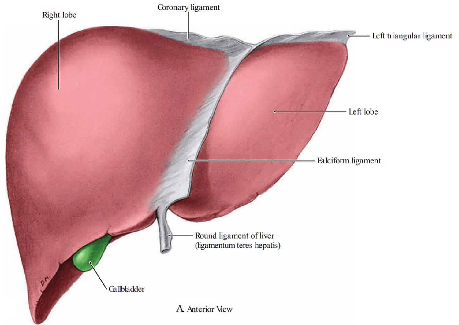
Falciform ligament
The falciform ligament is the thin, sickle-shaped, fibrous structure that connects the anterior part of the liver to the anterior abdominal wall and diaphragm. The falciform ligament can be seen drooping from the liver hilum when looking inside the abdomen during surgery. The falciform ligament attaches to the liver between the right and left lobes as well as attaching to the inferior diaphragmatic surface 1. The falciform ligament splits the liver into anatomic left and right along the anterior aspect 2. The free, inferior border of the falciform ligament contains the paraumbilical veins and the round ligament of the liver (ligamentum teres hepatis, which is the remnant of the embryonic umbilical vein) which courses along a fissure situated between the inferior surface of the right and left lobes. Paraumbilical veins become prominent and more patent during portal hypertension when the portal vein, draining the gastrointestinal system, becomes engorged with blood which is unable to fully enter the liver. If the portal hypertension is severe enough, the paraumbilical veins will form a caput medusa of engorged blood vessels surrounding the umbilicus. The presence of a caput medusa is a stigmata of portal hypertension and severe liver dysfunction as seen in end-stage liver disease.
Falciform ligament of liver is a remnant of the ventral mesentery of the fetus. The falciform ligament derives from the ventral mesentery, which is a part of the embryological foregut and forms a connection between the ventral abdominal wall and the liver. The umbilical vein, which is within the umbilical cord, carries oxygen-rich blood from the placenta to the fetal liver. Once the child is born, the umbilical vein degenerates because maternal-fetal circulation is no longer needed to sustain fetal life as the placenta is no longer intact. After birth, the umbilical vein remnant forms the round ligament of the liver, which in adults, contains paraumbilical veins.
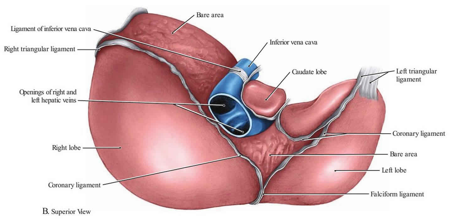
The falciform ligament serves as an anatomical landmark during abdominal surgery. The hepatic veins, which are hidden from view on the bare surface of the liver and drain into the inferior vena cava, are located immediately inferior to the falciform ligament. Additionally, the round ligament located in the free margin of the falciform ligament enters the umbilicus and serves as a significant landmark for the inner anterior abdominal wall. It also acts as an important landmark during gallbladder surgeries.
In patients with portal hypertension, the falciform ligament becomes recanalized with blood vessels. The venous congestion that occurs as a result of portal hypertension causes blood to shunt towards the anterior abdominal wall. Once blood starts pooling in the falciform ligament, periumbilical bruising will be evident. With increasing portal hypertension, caput medusae will present as a clinical feature resulting from engorged and distended superficial epigastric and periumbilical veins on the anterior surface of the abdomen. When one visualizes this feature, a diagnosis of portal hypertension should be suspected. Rarely, cysts may develop on the falciform ligament which may cause vague upper abdominal pain. A CT scan is the best means for assessment of falciform ligament pathology.
The falciform ligament is a cause of acute abdomen in select patients under various circumstances 3. For instance, there have been case reports of falciform ligament necrosis 4. Also, hematomas, abscesses, and lipomas have been documented 5. By far, the most common issues that can occur with the falciform ligament include cysts, tumors, and engorgement due to portal hypertension from conditions such as cirrhosis, Budd-Chiari, and malignancy. Congenital defects can occur within the falciform ligament during the process of development that may predispose a person to internal hernias 6.
Figure 1. Falciform ligament
Falciform ligament function
The falciform ligament is a double-layered extension of parietal peritoneum that sweeps off the anterior abdominal wall to divide the liver into the asymmetric left and right lobes. When looking at the falciform ligament from a coronal view, the most distal aspect of the falciform ligament ends in a free edge containing the round ligament of the liver and the paraumbilical veins. The proximal portion will diverge to merge with the right and left coronary ligaments which surround the bare liver area which superimposes against the inferior diaphragmatic surface. The round ligament of the liver is the fetal remnant of the umbilical vein, which once traveled from the placenta to the fetal liver to deliver oxygenated blood 7.
References- Garbar V, Newton BW. Anatomy, Abdomen and Pelvis, Falciform Ligament. [Updated 2019 Mar 6]. In: StatPearls [Internet]. Treasure Island (FL): StatPearls Publishing; 2019 Jan-. Available from: https://www.ncbi.nlm.nih.gov/books/NBK539858
- Vernon H, Kasi A. Anatomy, Abdomen and Pelvis, Liver. [Updated 2019 Jan 16]. In: StatPearls [Internet]. Treasure Island (FL): StatPearls Publishing; 2019 Jan-.Available from: https://www.ncbi.nlm.nih.gov/books/NBK500014
- Priola AM, Priola SM, Cataldi A, Marci V, Fava C. Acute abdomen as an unusual presentation of hepatic PEComa. A case report. Tumori. 2009 Jan-Feb;95(1):123-8.
- Sørensen J, Møller AM, Håkansson T. [Acute abdomen caused by necrosis of the falciform ligament of the liver]. Ugeskr. Laeg. 1983 Feb 21;145(8):583.
- Sari S, Ersöz F, Güneş ME, Paşaoğlu E, Arikan S. Hematoma of the falciform ligament: a rare cause of acute abdomen. Turk J Gastroenterol. 2011;22(2):213-5.
- Sourtzis S, Canizares C, Thibeau JF, Philippart P, Damry N. An unusual case of herniation of small bowel through an iatrogenic defect of the falciform ligament. Eur Radiol. 2002 Mar;12(3):531-3.
- Vernon H, Kasi A. Anatomy, Abdomen and Pelvis, Liver. [Updated 2019 Jan 16]. In: StatPearls [Internet]. Treasure Island (FL): StatPearls Publishing; 2019 Jan-. Available from: https://www.ncbi.nlm.nih.gov/books/NBK500014


