Gastric outlet obstruction
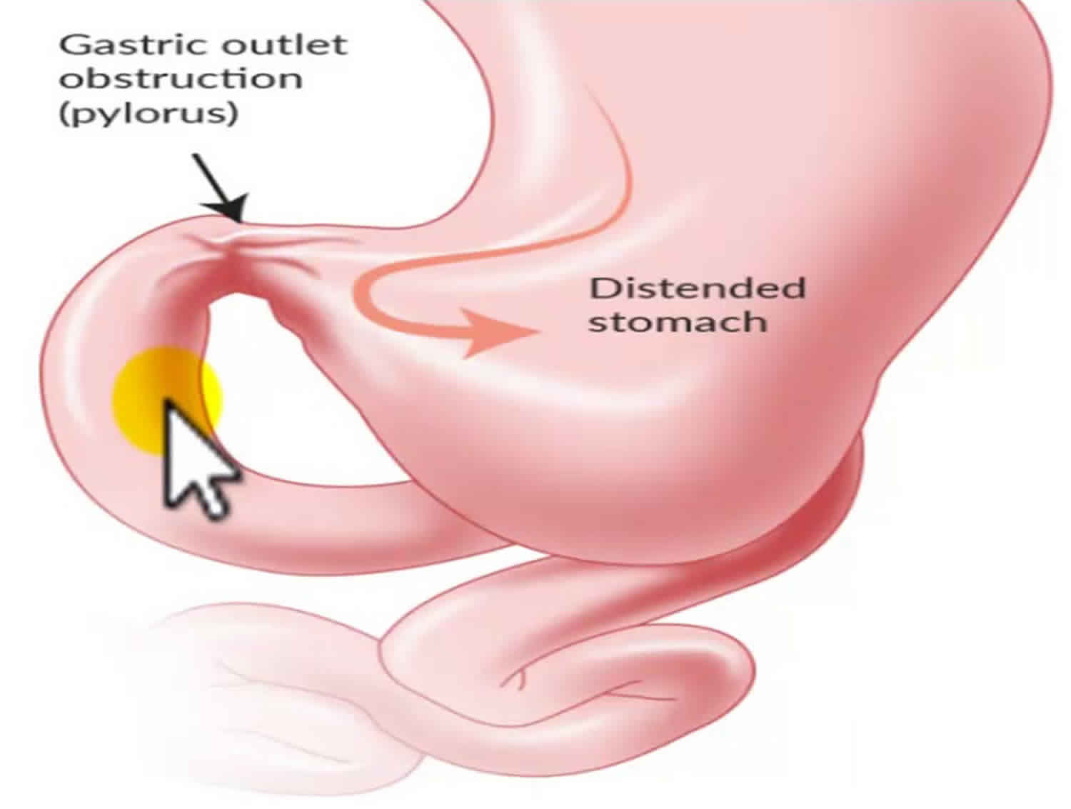
Gastric outlet obstruction also known as pyloric obstruction is not a single entity; gastric outlet obstruction is the clinical and pathophysiological consequence of any disease process that produces a mechanical impediment to gastric emptying 1. The stomach is divided into four portions: cardia, body, antrum, and pylorus. Inflammation, scarring, or infiltration of the antrum and pylorus are associated with the development of gastric outlet obstruction 1. The duodenum begins immediately beyond the pylorus and mostly is a retroperitoneal structure, wrapping around the head of the pancreas. The duodenum classically is divided into four portions. It is intimately related to the gallbladder, liver, and pancreas; therefore, a malignant process of any adjacent structure may cause outlet obstruction due to extrinsic compression. Clinical entities that can result in gastric outlet obstruction generally are categorized into two well-defined groups of causes: benign and malignant. This classification facilitates discussion of management and treatment. In the past, when peptic ulcer disease was more prevalent, benign causes were the most common; however, one review showed that only 37% of patients with gastric outlet obstruction have benign disease and the remaining patients have obstruction secondary to cancer 2. Malignant gastric outlet obstruction is a common condition that results from locally advanced malignancies in the upper gastrointestinal tract, such as pancreatic, gastric, and other carcinomas 3.
The incidence of gastric outlet obstruction has been reported to be less than 5% in patients with peptic ulcer disease, which is the leading benign cause of the problem. Five percent to 8% of ulcer-related complications result in an estimated 2000 operations per year in the United States 4.
The incidence of gastric outlet obstruction in patients with peripancreatic malignancy, the most common malignant etiology, has been reported as 15-20%.
Gastric outlet obstruction can be a diagnostic and treatment dilemma. Despite medical advances in the acid suppression mechanism, the incidence of gastric outlet obstruction remains a prevalent clinical problem in benign peptic ulcer disease. Also, an increase in the number of cases of gastric outlet obstruction seems to be noted secondary to malignancy; this is possibly due to improvements in cancer therapy, which allow patients to live long enough to develop this complication.
As part of the initial workup, exclude the possibility of functional nonmechanical causes of obstruction, such as diabetic gastroparesis. Once a mechanical obstruction is confirmed, differentiate between benign and malignant processes because definitive treatment is based on recognition of the specific underlying cause.
Two types of procedures for malignant gastric outlet obstruction, namely, gastrojejunostomy with laparotomy or a laparoscopic approach and endoscopic stenting, are currently available. Although numerous previous reports have clarified the benefits and drawbacks of each procedure, whether gastrojejunostomy or endoscopic stenting should be used in patients with gastric outlet obstruction that results from gastric cancer who may have a longer life expectancy than patients with other malignancies has not been determined.
Gastric outlet obstruction causes
The major benign causes of gastric outlet obstruction are peptic ulcer disease, gastric polyps, ingestion of caustics, pyloric stenosis, congenital duodenal webs, gallstone obstruction (Bouveret syndrome), pancreatic pseudocysts, and bezoars.
Peptic ulcer disease manifests in approximately 5% of all patients with gastric outlet obstruction. Ulcers within the pyloric channel and first portion of the duodenum usually are responsible for outlet obstruction. Obstruction can occur in an acute setting secondary to acute inflammation and edema or, more commonly, in a chronic setting secondary to scarring and fibrosis. Helicobacter pylori has been implicated as a frequent associated finding in patients with gastric outlet obstruction, but its exact incidence has not been defined precisely.
Within the pediatric population, pyloric stenosis constitutes the most important cause of gastric outlet obstruction. Pyloric stenosis occurs in 1 per 750 births. It is more common in boys than in girls and also is more common in first-born children. Pyloric stenosis is the result of gradual hypertrophy of the circular smooth muscle of the pylorus.
Pancreatic cancer is the most common malignancy causing gastric outlet obstruction. Outlet obstruction may occur in 10-20% of patients with pancreatic carcinoma. Other tumors that may obstruct the gastric outlet include ampullary cancer, duodenal cancer, cholangiocarcinomas, and gastric cancer. Metastases to the gastric outlet also may be caused by other primary tumors.
Gastric outlet obstruction symptoms
Nausea and vomiting are the cardinal symptoms of gastric outlet obstruction. Vomiting usually is described as nonbilious, and it characteristically contains undigested food particles. In the early stages of obstruction, vomiting may be intermittent and usually occurs within 1 hour of a meal.
Patients with gastric outlet obstruction resulting from a duodenal ulcer or incomplete obstruction typically present with symptoms of gastric retention, including early satiety, bloating or epigastric fullness, indigestion, anorexia, nausea, vomiting, epigastric pain, and weight loss. They are frequently malnourished and dehydrated and have a metabolic insufficiency. Weight loss is frequent when the condition approaches chronicity and is most significant in patients with malignant disease.
Abdominal pain is not frequent and usually relates to the underlying cause (eg, peptic ulcer disease or pancreatic cancer).
Physical examination often demonstrates the presence of chronic dehydration and malnutrition. A dilated stomach may be appreciated as a tympanitic mass in the epigastric area and/or left upper quadrant.
Dehydration and electrolyte abnormalities can be demonstrated by routine laboratory examinations. Increases in blood urea nitrogen (BUN) and creatinine are late features of dehydration.
Prolonged vomiting causes loss of hydrochloric acid and produces an increase of bicarbonate in the plasma to compensate for the lost chloride and sodium. The result is a hypokalemic hypochloremic metabolic alkalosis. Alkalosis shifts the intracellular potassium to the extracellular compartment, and the serum positive potassium is increased factitiously. With continued vomiting, the renal excretion of potassium increases in order to preserve sodium. The adrenocortical response to hypovolemia intensifies the exchange of potassium for sodium at the distal tubule, with subsequent aggravation of the hypokalemia.
Gastric outlet obstruction diagnosis
A physical exam may reveal:
- Signs of dehydration, such as dry skin and mouth, less tearing when crying, and dry diapers
- Swollen belly
- Olive-shaped mass when feeling the upper belly, which is the abnormal pylorus in pyloric stenosis
Obtain a complete blood count (CBC). Check the hemoglobin and hematocrit to rule out the possibility of anemia. Obtain an electrolyte panel. As noted previously, identifying and correcting electrolyte abnormalities that tend to occur is essential. Liver function tests may be helpful, particularly when a malignant etiology is suspected. A test for Helicobacter pylori is helpful when the diagnosis of peptic ulcer disease is suspected.
Imaging studies
Plain abdominal radiography, contrast upper gastrointestinal (GI) studies (Gastrografin or barium), and computed tomography (CT) with oral contrast are helpful. Plain radiographs, including the obstruction series (ie, supine abdomen, upright abdomen, chest posteroanterior), can demonstrate the presence of gastric dilatation and may be helpful in distinguishing the differential diagnosis.
Upper endoscopy
Upper endoscopy can help visualize the gastric outlet and may provide a tissue diagnosis when the obstruction is intraluminal.
The sodium chloride load test is a traditional clinical nonimaging study that may be helpful. The traditional sodium chloride load test is performed by infusing 750 mL of sodium chloride solution into the stomach via a nasogastric tube. A diagnosis of gastric outlet obstruction (gastric outlet obstruction) is made if more than 400 mL remains in the stomach after 30 minutes.
Nuclear gastric emptying studies measure the passage of orally administered radionuclide over time. Unfortunately, both the nuclear test and the saline load test may produce abnormal results in functional states.
Barium upper GI studies are very helpful because they can delineate the gastric silhouette and demonstrate the site of obstruction. An enlarged stomach with a narrowing of the pyloric channel or first portion of the duodenum helps differentiate gastric outlet obstruction from gastroparesis.
The specific cause may be identified as an ulcer mass or intrinsic tumor.
In the presence of peptic ulcer disease, perform endoscopic biopsy to rule out the presence of malignancy. In the case of peripancreatic malignancy, CT-guided biopsy may be helpful in establishing a preoperative diagnosis. Needle-guided biopsy also may be helpful in establishing the presence of metastatic disease. This knowledge may impact the magnitude of the procedure planned to alleviate the gastric outlet obstruction. Histologic findings relate to the individual underlying cause.
Gastric outlet obstruction treatment
Gastric outlet obstruction due to benign ulcer disease may be treated medically if results of imaging studies or endoscopy determine that acute inflammation and edema are the principal causes of the outlet obstruction (as opposed to scarring and fibrosis, which may be fixed).
If medical therapy conducted for a reasonable period fails to alleviate the gastric outlet obstruction, then surgical intervention becomes appropriate. Typically, if resolution or improvement is not seen within 48-72 hours, surgical intervention is necessary. The choice of surgical procedure depends upon the patient’s particular circumstances; however, vagotomy and antrectomy should be considered the criterion standard against which the efficacy of other procedures is measured.
In cases of malignant obstruction, weigh the extent of surgical intervention for the relief of gastric outlet obstruction against the malignancy’s type and extent, as well as the patient’s anticipated long-term prognosis. As a guiding principle, undertake major tumor resections in the absence of metastatic disease in a patient who can withstand such a procedure from a nutritional standpoint. In patients with largely metastatic disease, determine the degree of surgical intervention for palliation in light of the patient’s realistic prognosis and personal wishes.
Contraindications for surgery relate to the underlying medical condition. Most patients benefit from an initial period of gastric decompression, hydration, and correction of electrolyte imbalances. In patients who are severely malnourished, postponing surgical intervention until the nutritional status has been optimized may be wise. In selective cases, some patients may benefit from total parenteral nutrition (TPN) or distal tube feeding (eg, placed via a percutaneous jejunostomy).
One of the relative contraindications for surgery is the presence of advanced malignancy; in these cases, in which life expectancy may be limited to a few months, palliation via endoscopically placed stents should be considered.
Overall, every patient with gastric outlet obstruction deserves evaluation by a surgeon. Even if the patient has unresectable disease, palliative surgical measures may improve the quality of life.
Medical therapy
Initial management of gastric outlet obstruction should be the same regardless of the primary cause. After a diagnosis is made, admit patients for hydration and correction of electrolyte abnormalities. Remembering that the metabolic alkalosis of gastric outlet obstruction responds to the administration of chloride is important; therefore, sodium chloride solution should be the initial intravenous (IV) fluid of choice. Potassium deficits are corrected after repletion of volume status and after replacement of chloride.
Place a nasogastric tube to decompress the stomach. Occasionally, a large tube is required because the undigested food blocks tubes with small diameters.
When acute peptic ulcer disease has been identified as a primary cause of gastric outlet obstruction, focus treatment on the reduction of acid production. Histamine-2 (H2) blockers and proton pump inhibitors are the mainstay of treatment.
Treat H pylori infection, when identified, according to current recommendations. Although most patients improve temporarily with treatment, scarring and fibrosis may worsen over time. Pneumatic balloon dilatation of a chronic, benign stricture can be performed via endoscopy. Patients who are candidates for balloon dilatation are likely to present with recurrent gastric outlet obstruction.
Published series using this technique report success rates of over 76% after multiple dilatations 5, though the rate of failure and recurrent obstruction is higher in patients treated with balloon dilatation who have not also been treated for Helicobacter pylori infection 6. Patients who are negative for Helicobacter pylori do not respond favorably to balloon dilatation and should be considered for surgical treatment early in the process 7.
Further treatment is tailored to the underlying cause; this is where the distinction between benign and malignant disease becomes important.
Surgical therapy
Perform standard preoperative evaluation in these patients. Correct fluid and electrolyte abnormalities prior to surgery. Perform gastric decompression by NG tube and suction and alert the anesthesiologist to the potential risk for aspiration upon induction.
Perform a preoperative nutritional evaluation and initiate appropriate nutritional therapy (ie, TPN or enteral feedings via a percutaneous jejunostomy placed distal to the obstruction) as soon as possible. Maximizing preoperative nutrition can greatly reduce or eliminate postoperative complications related to delayed healing.
Management of benign disease
More than 75% of patients presenting with gastric outlet obstruction eventually require surgical intervention 8. Surgical intervention usually provides definitive treatment of gastric outlet obstruction, but it may result in its own comorbid consequences. Operative management should offer relief of obstruction and correction of the acid problem.
The most common surgical procedures performed for gastric outlet obstruction related to peptic ulcer disease are vagotomy and antrectomy, vagotomy and pyloroplasty, truncal vagotomy and gastrojejunostomy, pyloroplasty, and laparoscopic variants 9 of the aforementioned procedures. The video below demonstrates robotic-assisted pyloroplasty.
Vagotomy and antrectomy with Billroth II reconstruction (gastrojejunostomy) seem to offer the best results. Vagotomy and pyloroplasty and pyloroplasty alone, although used with some success, can be technically difficult to perform due to scarring at the gastric outlet. A combination of balloon dilatation and highly selective vagotomy has been described, but it is associated with gastroparesis and a high recurrence rate.
Placement of a jejunostomy tube at the time of surgery should be considered. This provides temporary feeding access in already malnourished patients. Also, in chronically dilated partial obstructions, the stomach may be slow to recover a normal rate of emptying.
The role of the laparoscopic approach in the treatment of gastric outlet obstruction is under investigation and may represent a valid form of therapy with low morbidity. The experience of several international centers has been published. One group in China performed laparoscopic truncal vagotomy and gastrojejunostomy for gastric outlet obstruction related to peptic ulcer disease, with nearly complete resolution of symptomatology. The investigators reported no conversions to open procedure or mortalities. Twenty-seven percent of patients did experience transiently delayed gastric emptying, which resolved with conservative measures 10.
Kim et al 11 also reported good results from the use of laparoscopic truncal vagotomy with gastrojejunostomy, including shorter operating times and hospital stays in comparison with the open procedure.
Hall et al 12 performed a double-blind, multicenter, randomized, controlled trial comparing patient recovery following laparoscopic pyloromyotomy to that after open pyloromyotomy in infants with pyloric stenosis. The investigators found that among the 87 infants who underwent the laparoscopic procedure, the median (interquartile range) postoperative time needed to achieve full enteral feeding was 18.5 hours, compared with 23.9 hours in the 93 infants who underwent open pyloromyotomy. The median postoperative length of stay in the laparoscopic and open pyloromyotomy groups were 33.6 hours and 43.8 hours, respectively.
The study also found that the incidence of postoperative vomiting was similar in the open and laparoscopic groups, as was the frequency of intraoperative and postoperative complications. The authors suggested that open and laparoscopic pyloromyotomy are safe means of treating infantile pyloric stenosis. Because of its apparent advantages, however, they recommended that in centers with suitable laparoscopic experience, the laparoscopic form of the procedure be used.
Management of malignant disease
The management of gastric outlet obstruction secondary to malignancy is controversial. Of patients with periampullary cancer, 30-50% present with nausea and vomiting at the time of diagnosis 13. Most of these tumors are unresectable (approximately 40% of gastric cancers and 80-90% of periampullary cancers) 14. When tumors are found to be unresectable, 13-20% of patients eventually develop gastric outlet obstruction before they succumb to their disease. The 1-year survival rate is poor.
Gastrojejunostomy remains the surgical treatment of choice for gastric outlet obstruction secondary to malignancy. Although surgeons traditionally have preferred an antecolic anastomosis to prevent further obstruction by advancing tumor growth, a publication evaluating the retrocolic anastomosis in this setting challenges conventional wisdom 15. Results demonstrate that a retrocolic anastomosis may be associated with decreased incidences of delayed gastric emptying (6% vs 17%) and late gastric outlet obstruction (2% vs 9%).
Other groups have illustrated that partial stomach-partitioning gastrojejunostomy decreases the rates of delayed gastric emptying as compared with traditional gastrojejunostomy 16. Feeding jejunostomy should again be considered to combat malnutrition and slow recovery of gastric emptying.
Internationally, studies are under way using laparoscopic gastrojejunostomy instead of the open procedure. In the United States, critics cite a nearly 20% conversion rate and a delay in the return of gut function as reasons to not perform the procedure laparoscopically. Comparisons of laparoscopic gastrointestinal anastomosis versus the open procedure have revealed less morbidity and mortality, shorter hospital stays, fewer blood transfusions, and faster gastrointestinal transit recovery time 17.
Researchers at Johns Hopkins Hospital 18 have attempted endoscopic transgastric approaches to create a gastrojejunostomy in a porcine model. As natural orifice transluminal surgery gains more widespread interest, these novel approaches may become more popular.
Chopita et al 19 reported on the use of magnetic endoscopic gastroenteric anastomosis in 15 patients with malignant gastroenteric obstruction. The procedure had an 86.7% success rate, with the authors noting the additional benefits of shorter duration of hospital stay and good quality of life in patients. Although still experimental, this procedure may one day be a surgical option.
JH et al 20 reported that gastrojejunostomy was preferable to metal stent placement in providing palliation of gastric outlet obstruction caused by unresectable or metastatic cancer in patients with a good performance status.
Self-expandable metallic stents also have been used for the treatment of gastric outlet obstruction in a malignant setting 21. Metallic stents have previously been used successfully to treat stenosis of such areas as the blood vessels, bile duct, esophagus, and trachea. With the development of newer stents and delivery systems, metallic stents may have a role in the nonsurgical treatment of gastroduodenal obstruction. Stents may allow the physician to avoid complicated surgical procedures.
Currently, only the Wallstent has FDA approval for palliation in malignant gastroduodenal obstruction. Significant complications include the following: malposition, misdeployment, tumor ingrowth or overgrowth, migration, bleeding, and perforation 22.
A review of 19 studies published in 2004 quoted clinical success rates of 80-90% 23. Subsequent multicenter trials using the enteral Wallstent in 176 patients with malignant gastric outlet obstruction resulted in 89% of patients tolerating oral intake for a median of 219 days postprocedure. Of the 84% in whom the stent was successful after the initial procedure, 22% required restenting to tolerate an oral diet. In addition, as other studies have demonstrated, chemotherapy was independently associated with an increased tolerance in oral intake 24.
One proposed solution uses covered metallic stents that have a lower incidence of tumor ingrowth. A 60% rate of tumor ingrowth in uncovered stents versus a 10% rate of tumor ingrowth in covered stents has been reported. Furthermore, with the double stent technique, that is, simultaneous placement of both covered stents and uncovered stents, lower early restenosis rates have been achieved. A stent patency of 21.5 days for uncovered stents versus 150 days for double stents has been achieved 25.
Of the 62 patients studied by Maetani et al 26, half received uncovered stents and half covered stents. The authors found no statistical differency in patency, but the triple-covered stent resulted in less frequent dysfunction four weeks after stenting.
Several retrospective studies have been performed to compare the results of stenting withm those of surgical intervention. Survival rates are equivalent; however, costs, length of stay, and number of subsequent procedures are all decreased following stenting 27. In addition, a delay of gastric emptying and morbidity decrease with the use of metallic stents 28. These promising results suggest that stents may eventually replace surgery as palliative intervention for unresectable periampullary malignancies.
A 2011 study from the Netherlands discusses the use of a D-Weave Niti-S nitinol stent specifically for the duodenum. A key outcome in this palliative procedure was a significant improvement in global health status and median 82-day survival. The authors report a marvelous technical and robust clinical success rate with patency up to 190 days, and a 25% complication rate 29.
In a 2014 study designed to compare the outcomes of endoscopic enteral stent insertion for malignant gastric outlet obstruction in older (≥65 years; n = 26) versus younger (n = 56) patients, Mansoor and Zeb 30 reported comparable rates of technical success (100% in older patients vs 97% in younger patients) and clinical success in the two groups. The complication rates were also comparable (27% in older patients vs 23% in younger patients).
The role of prophylactic gastrojejunostomy in cases of malignant gastric outlet obstruction is a question that remains to be answered. Some surgeons argue that prophylactic gastrojejunostomy may increase postoperative morbidity, primarily due to delayed gastric emptying.
This issue was addressed in a study by Lillemoe and Cameron 15, in which 87 patients with unresectable periampullary cancer were randomized to receive or not receive a prophylactic gastrojejunostomy. Although there were no significant differences in morbidity, length of hospital stay, and survival rates, the prophylactic gastrojejunostomy group had a 0% (0/44) incidence of gastric outlet obstruction versus 19% (8/43) in the other group. The authors concluded that prophylactic gastrojejunostomy significantly decreased the incidence of late gastric outlet obstruction and should be performed routinely when a patient is undergoing surgical palliation for periampullary cancer. Comparable data are supported by other series.
Gastric outlet obstruction treatment complications
Although the risk is small, patients undergoing endoscopic treatment with either balloon dilatation or stenting are at risk for perforation. Several literature reports exist regarding migration of the stents and reocclusion requiring further intervention.
Operative complications in patients undergoing surgery for gastric outlet obstruction often are related to the nutritional status of the patients. Commencing nutritional support upon recognition of the presence of gastric outlet obstruction is important. If surgery is anticipated, delaying the surgery or any intervention until TPN has been instituted for at least 1 week is often prudent.
Acute intervention may be technically difficult because of significant gastric dilatation and gastric wall edema. This circumstance may increase the rate of anastomotic leak. On occasion, delaying surgical intervention for several days while the stomach is decompressed by nasogastric suction may be prudent.
Alert patients undergoing gastric resection for benign or malignant disease to the possibility of well-known postgastrectomy syndromes, such as dumping, alkaline gastritis, and afferent loop syndrome. Severe symptoms may be present in 1-2% of patients.
Long-term monitoring
Closely monitor patients after surgery and upon discharge 31. After relief of gastric outlet obstruction, patients may continue to experience gastric dysmotility and may require medication to stimulate gastric emptying and motility.
In patients with malignancy, the potential for progressive and recurrent disease always remains. These patients should be monitored by a surgeon or an oncologist.
Closely monitor patients whose treatment consisted of balloon dilatation; most of these patients require subsequent dilatations to achieve satisfactory results.
References- Gastric outlet obstruction. https://emedicine.medscape.com/article/190621-overview
- Andersson A, Bergdahl L. Carcinoid tumors of the appendix in children. A report of 25 cases. Acta Chir Scand. 1977. 143(3):173-5.
- Treatment of gastric outlet obstruction that results from unresectable gastric cancer: Current evidence. World J Gastrointest Endosc. 2016 Feb 10; 8(3): 165–172. doi: 10.4253/wjge.v8.i3.165 https://www.ncbi.nlm.nih.gov/pmc/articles/PMC4734975
- Gibson JB, Behrman SW, Fabian TC, Britt LG. Gastric outlet obstruction resulting from peptic ulcer disease requiring surgical intervention is infrequently associated with Helicobacter pylori infection. J Am Coll Surg. 2000 Jul. 191(1):32-7.
- Lam YH, Lau JY, Fung TM, et al. Endoscopic balloon dilation for benign gastric outlet obstruction with or without Helicobacter pylori infection. Gastrointest Endosc. 2004 Aug. 60(2):229-33.
- Taskin V, Gurer I, Ozyilkan E, Sare M, Hilmioglu F. Effect of Helicobacter pylori eradication on peptic ulcer disease complicated with outlet obstruction. Helicobacter. 2000 Mar. 5(1):38-40.
- Gouma DJ, van Geenen R, van Gulik T, de Wit LT, Obertop H. Surgical palliative treatment in bilio-pancreatic malignancy. Ann Oncol. 1999. 10 Suppl 4:269-72.
- Doberneck RC, Berndt GA. Delayed gastric emptying after palliative gastrojejunostomy for carcinoma of the pancreas. Arch Surg. 1987 Jul. 122(7):827-9.
- Abdel-Salam WN, Katri KM, Bessa SS, et al. Laparoscopic-assisted truncal vagotomy and gastro-jejunostomy: trial of simplification. J Laparoendosc Adv Surg Tech A. 2009 Apr. 19(2):125-7.
- Siu WT, Tang CN, Law BK, Chau CH, Yau KK, Yang GP, et al. Vagotomy and gastrojejunostomy for benign gastric outlet obstruction. J Laparoendosc Adv Surg Tech A. 2004 Oct. 14(5):266-9.
- Kim SM, Song J, Oh SJ, et al. Comparison of laparoscopic truncal vagotomy with gastrojejunostomy and open surgery in peptic pyloric stenosis. Surg Endosc. 2009 Jun. 23(6):1326-30.
- Hall NJ, Pacilli M, Eaton S, et al. Recovery after open versus laparoscopic pyloromyotomy for pyloric stenosis: a double-blind multicentre randomised controlled trial. Lancet. 2009 Jan 31. 373(9661):390-8.
- Khullar SK, DiSario JA. Gastric outlet obstruction. Gastrointest Endosc Clin N Am. 1996 Jul. 6(3):585-603.
- Lillemoe KD, Sauter PK, Pitt HA, Yeo CJ, Cameron JL. Current status of surgical palliation of periampullary carcinoma. Surg Gynecol Obstet. 1993 Jan. 176(1):1-10.
- Lillemoe KD, Cameron JL, Hardacre JM, Sohn TA, Sauter PK, Coleman J, et al. Is prophylactic gastrojejunostomy indicated for unresectable periampullary cancer? A prospective randomized trial. Ann Surg. 1999 Sep. 230(3):322-8; discussion 328-30.
- Arciero CA, Joseph N, Watson JC, Hoffman JP. Partial stomach-partitioning gastrojejunostomy for malignant duodenal obstruction. Am J Surg. 2006 Mar. 191(3):428-32.
- Alam TA, Baines M, Parker MC. The management of gastric outlet obstruction secondary to inoperable cancer. Surg Endosc. 2003 Feb. 17(2):320-3.
- Kantsevoy SV, Jagannath SB, Niiyama H, Chung SS, Cotton PB, Gostout CJ, et al. Endoscopic gastrojejunostomy with survival in a porcine model. Gastrointest Endosc. 2005 Aug. 62(2):287-92.
- Chopita N, Vaillaverde A, Cope C, et al. Endoscopic gastroenteric anastomosis using magnets. Endoscopy. 2005 Apr. 37(4):313-7.
- No JH, Kim SW, Lim CH, Kim JS, Cho YK, Park JM, et al. Long-term outcome of palliative therapy for gastric outlet obstruction caused by unresectable gastric cancer in patients with good performance status: endoscopic stenting versus surgery. Gastrointest Endosc. 2013 Jul. 78 (1):55-62.
- Chopita N, Landoni N, Ross A, Villaverde A. Malignant gastroenteric obstruction: therapeutic options. Gastrointest Endosc Clin N Am. 2007 Jul. 17(3):533-44, vi-vii.
- Adler DG, Merwat SN. Endoscopic approaches for palliation of luminal gastrointestinal obstruction. Gastroenterol Clin North Am. 2006 Mar. 35(1):65-82, viii.
- Baron TH. Surgical versus endoscopic palliation of malignant gastric outlet obstruction: big incision, little incision, or no incision?. Gastroenterology. 2004 Oct. 127(4):1268-9.
- Telford JJ, Carr-Locke DL, Baron TH, Tringali A, Parsons WG, Gabbrielli A, et al. Palliation of patients with malignant gastric outlet obstruction with the enteral Wallstent: outcomes from a multicenter study. Gastrointest Endosc. 2004 Dec. 60(6):916-20.
- Song GA, Kang DH, Kim TO, Heo J, Kim GH, Cho M, et al. Endoscopic stenting in patients with recurrent malignant obstruction after gastric surgery: uncovered versus simultaneously deployed uncovered and covered (double) self-expandable metal stents. Gastrointest Endosc. 2007 May. 65(6):782-7.
- Maetani I, Mizumoto Y, Shigoka H, Omuta S, Saito M, Tokuhisa J, et al. Placement of a triple-layered covered versus uncovered metallic stent for palliation of malignant gastric outlet obstruction: a multicenter randomized trial. Dig Endosc. 2014 Mar. 26 (2):192-9.
- Del Piano M, Ballare M, Montino F, Todesco A, Orsello M, Magnani C, et al. Endoscopy or surgery for malignant GI outlet obstruction?. Gastrointest Endosc. 2005 Mar. 61(3):421-6.
- Wong YT, Brams DM, Munson L, Sanders L, Heiss F, Chase M, et al. Gastric outlet obstruction secondary to pancreatic cancer: surgical vs endoscopic palliation. Surg Endosc. 2002 Feb. 16(2):310-2.
- van Hooft JE, van Montfoort ML, Jeurnink SM, et al. Safety and efficacy of a new non-foreshortening nitinol stent in malignant gastric outlet obstruction (DUONITI study): a prospective, multicenter study. Endoscopy. 2011 Aug. 43(8):671-5.
- Mansoor H, Zeb F. Enteral stents are safe and effective to relieve malignant gastric outlet obstruction in the elderly. J Gastrointest Cancer. 2015 Mar. 46 (1):42-7.
- Huang YL, Lee HC, Yeung CY, et al. Sonogram before and after pyloromyotomy: the pyloric ratio in infantile hypertrophic pyloric stenosis. Pediatr Neonatol. 2009 Jun. 50(3):117-20.


