Hemihyperplasia
Hemihyperplasia also called hemihypertrophy, is a condition in which there is excessive (hyper) growth (trophy) of only one side (hemi) of one or more body parts 1. The overgrowth may be limited to a portion of the body, such as a leg or arm, or it may involve several different areas of the body, including the arms, face (causing asymmetry of the nose, eyes or cheeks), tongue, jaw, teeth and ears. There may be associated asymmetric hypertrophy of internal organs. All tissue types can be affected, including the bones, skin, muscle, fat and nerves that are connected to the area of overgrowth. Hemihyperplasia may not be apparent at birth, but becomes most noticeable as the child grows.
Hemihyperplasia can be an isolated hemihyperplasia (occur by itself) or be part of well-defined genetic syndromes such as in the case of Beckwith-Wiedemann syndrome, Proteus syndrome, Russell-Silver syndrome, Klippel-Trenaunay-Weber syndrome, McCune-Albright syndrome and Sotos syndrome 2. Isolated hemihyperplasia is usually sporadic, but a number of familial occurrences have been described. In most cases, the cause of isolated hemihypertrophy is unknown. In cases where hemihyperplasia is part of a genetic syndrome, the cause depends on the specific syndrome.
The reported incidence of hemihyperplasia is thought to occur in around one in 86,000 live births, but this number may change as there is more agreement on a definition and more people looking for it.
Rowe 3 proposed a classification system for hemihyperplasia, based on anatomic site of involvement. According to this classification, complex hemihyperplasia is defined as involvement of half of the body (at least one arm and one leg), simple hemihyperplasia is the involvement of a single limb and hemifacial hyperplasia is the involvement of one side of the face. The degree of asymmetry is variable, and mild cases are easily overlooked 4. The prevalence of isolated hemihyperplasia is difficult to establish accurately because many cases may be so mild as not to come to medical attention. The prevalence for hemihyperplasia was reported as approximately 1 in 86,000 5.
Hemihyperplasia can be diagnosed at birth or appear later in childhood, and can follow an irregular growth pattern. At times new growth may be excessive, while at other times it may be modest.
Hemihyperplasia treatment may include surgery to correct the differences in the affected body part(s) 6.
The risk of tumor development in isolated hemihyperplasia is approximately one in 20, or around 5%. Because most of the cancers occur in the abdomen, the recommendation has been made (by the participants of the First International Conference on Molecular and Clinical Genetics of Childhood Renal Tumors–among others) that children with hemihypertrophy receive a screening abdominal ultrasound every 3 months until age 7 and, at minimum, a careful physical examination every 6 months until growth is completed. There is currently inadequate indication to screen children above 6 years of age 7.
Figure 1. Hemihyperplasia face
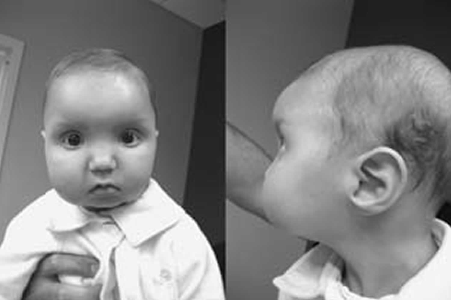
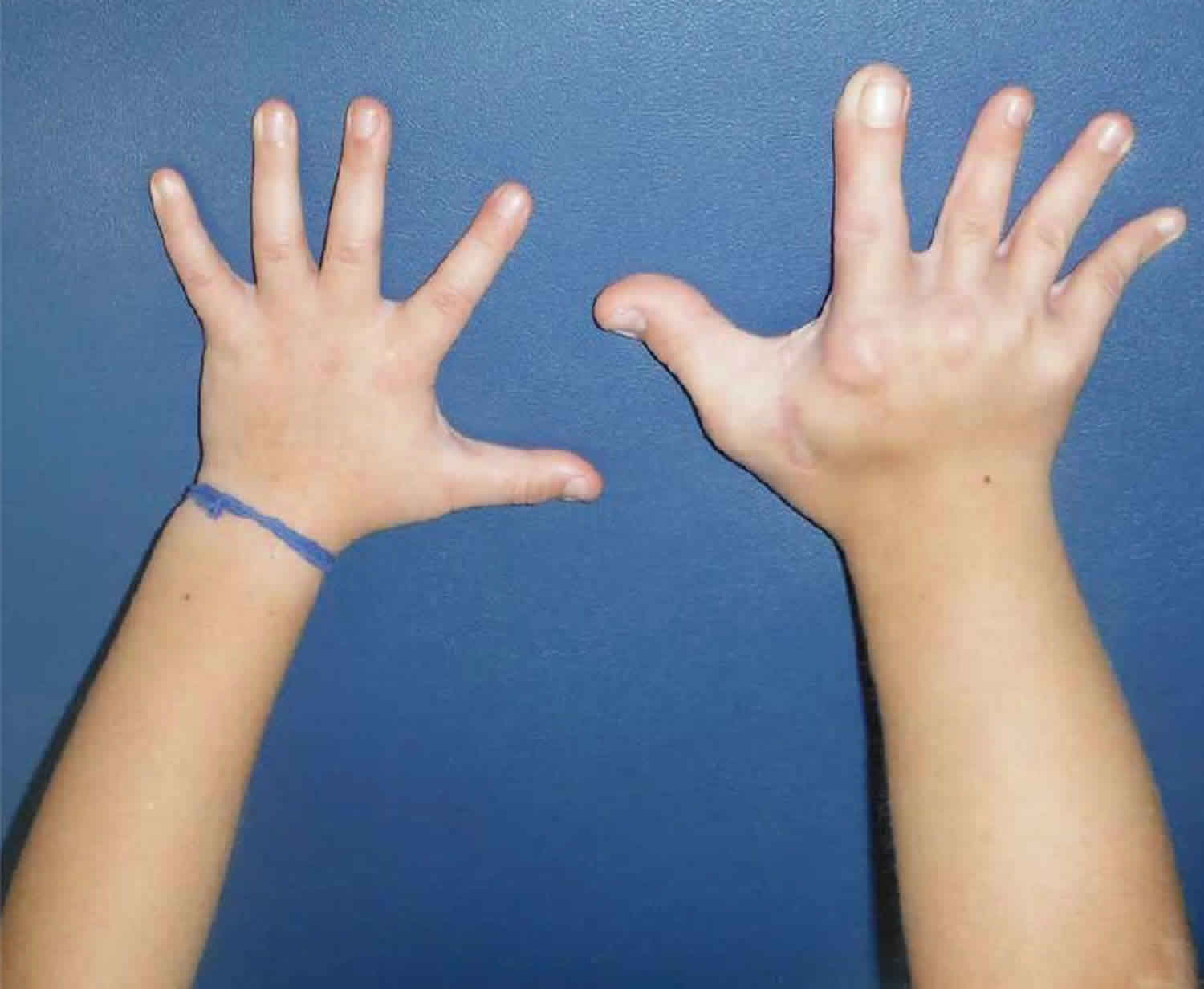
Figure 2. Hemihyperplasia involving the upper and lower right extremities
Footnote: A child with hemihyperplasia involving the upper and lower right extremities. The leg length discrepancy can be noted by the pelvic tilt.
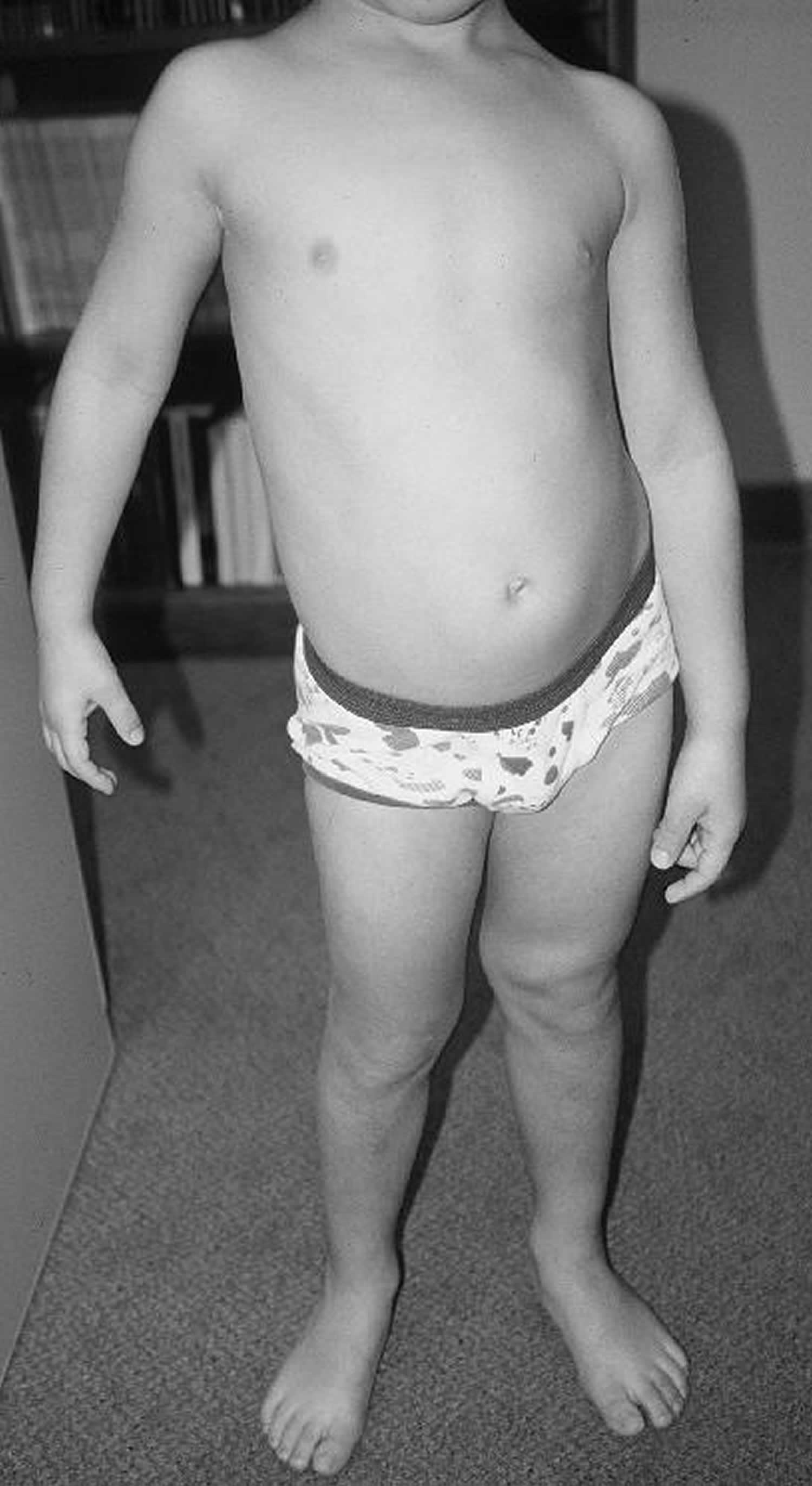
[Source 9 ]Figure 3. Isolated hemihyperplasia


Footnote: Enlargement of the left hand and forearm (arrows) and enlargement of the left foot, leg, and thigh (arrows).
[Source 10 ]Hemihyperplasia causes
In most cases, hemihyperplasia is isolated, meaning it occurs without signs of other problems. There is no single cause for hemihyperplasia, but there are genetic causes that can be signs of a medical condition. The cause varies and is looked at on an individual basis.
In some cases, hemihyperplasia can be a sign of a medical condition such as:
- Beckwith-Wiedemann syndrome
- Kippel-Trenaunay syndrome
- Proteus syndrome
- Neurofibromatosis type 1
- Russel-Silver syndrome
- Sotos syndrome
Beckwith-Wiedemann syndrome is the most common cause of hemihypertrophy or hemihyperplasia. The condition was named after American pediatric pathologist John Bruce Beckwith in 1963, and German pediatrician Hans-Rudolf Wiedemann in 1964, reported the syndrome independently 11.
The cause of Beckwith-Wiedemann syndrome is complex. About 80-90% of patients have a known molecular aberration that affects the regulation of a group of imprinted genes implicated in cell cycle progression and somatic growth control located in the chromosome 11p15.5 12. Genomic imprinting is an epigenetic-regulated process by which only one copy of a gene is expressed depending on the sex of the parent carrying the allele. Two imprinting control regions (IC1 and IC2) regulate the gene expression from the 11p15 region. Normally, methylation (silencing of gene expression) occurs in the paternal allele at IC1 and the maternal allele at IC2. In an individual with Beckwith-Wiedemann syndrome, the molecular defects more commonly described are 13:
- Loss of methylation at IC2 on the maternal allele in 50 to 60% of cases
- Paternal uniparental isodisomy of 11p15 in 20 to 25% of cases
- The gain of methylation at IC1 on the maternal allele in 5 to 10% of cases
- Autosomal dominant maternal point mutations in CDKN1C (regulated by IC2) in 5% of sporadic cases and 40% of familial cases
- Chromosomal rearrangements (duplications, translocations, deletions or inversions) in less than 1% of cases
- The unknown molecular defect in 10 to 15% of cases
Hemihyperplasia signs and symptoms
Hemihyperplasia is the enlargement of one part or side of the body causing asymmetry. Children with hemihyperplasia may not show any symptoms other than a subtle difference between the two sides of the face. As overgrowth progresses, a greater difference may be seen and the overgrowth may lead to difficulty with eating, chewing seeing and breathing. Appearance-related concerns may arise as the disease progresses.
Hemihypertrophy is often linked with mild mental retardation, genito-urinary anomalies, and an oncogenic potential (Wilms’ tumor) 14. Wilms’ tumor accounts for most renal neoplasms in childhood, and occurs with roughly equal incidence in both genders and all races, with a yearly incidence of 7.8 per million children younger than 15 years 15. An imperative feature of Wilms’ tumor is the association with congenital anomalies, the most common being genitourinary anomalies (4.4%), and hemihypertrophy (29%) 15. The risk of tumor development in isolated hemihyperplasia is approximately one in 20, or around 5%. The best follow-up plan is to follow the patients until the age of 7 years; these children should have abdominal ultrasound scans at 3 monthly intervals. With daily caretaker abdominal examination at the discretion of the doctor or parent. There is currently inadequate indication to screen children above 7 years of age 7.
Hemihyperplasia diagnosis
Hemihyperplasia is diagnosed with clinical examination and supplemented with radiologic studies such as a CT scan or MRI. A genetic counselor and a genetics doctor will meet with you in the clinic. They will ask about your family’s medical history, examine your child and make recommendations.
Labs and/or radiology studies may be done.
Screening recommendations for isolated hemihyperplasia or Beckwith-Wiedemann syndrome:
- A blood test called Alpha-Fetoprotein (AFP) tumor marker every 3 months until age 4.
- An abdominal ultrasound every 3 months until age 8.
- A kidney ultrasound every year from age 8 until mid-adolescence.
Although most people do not get tumors, screening tests are done to find tumors early when treatment is most effective and less invasive.
Hemihyperplasia treatment
There is no cure for hemihyperplasia and treatment depends on the cause of your child’s hemihyperplasia. Your child may be recommended to see an orthopedics provider for treatment of abnormal limb size.
Treatment of hemihyperplasia addresses both functional and appearance-related purposes. Procedures performed include suction-assisted lipectomy, excision of excessive skin and subcutaneous tissue, and contouring or reducing facial bones.
The goal of surgery is to preserve as much nerve and muscle function as possible. Surgery can occur on an outpatient basis, or if it is more extensive it will require hospitalization for a one to two day period.
Incisions can vary around the face depending on the area that is hyperplastic but, in general, are hidden in creases, folds, or intraorally. A moderate amount of soft tissue swelling can occur following surgery and eating may be compromised short term following discharge. Swelling will improve over time and any numbness or the like as a consequence of the surgery will generally improve.
References- Heilstedt HA, Bacino CA. A case of familial isolated hemihyperplasia. BMC Med Genet. 2004;5:1. Published 2004 Feb 2. doi:10.1186/1471-2350-5-1 https://www.ncbi.nlm.nih.gov/pmc/articles/PMC373451
- Ballock RT, Wiesner GL, Myers MT, et al. Hemihypertrophy. Concepts and controversies. J Bone Joint Surg Am. 1997;79(11):1731–8.
- Rowe NH. Hemifacial hypertrophy: Review of the literature and addition of four cases. Oral Surg. 1962;15:572–6.
- Hoyme HE, Seaver LH, Jones KL, et al. Isolated hemihyperplasia (hemihypertrophy): report of a prospective multicenter study of the incidence of neoplasia and review. Am J Med Genet. 1998;79(4):274–8.
- Ringrose RE, Jabbour JT, Keele DK. Hemihypertrophy. Pediatrics. 1965;36(3):434–48.
- Shuman C, Beckwith JB, Weksberg R. Beckwith-Wiedemann Syndrome. 2000 Mar 3 [Updated 2016 Aug 11]. In: Adam MP, Ardinger HH, Pagon RA, et al., editors. GeneReviews® [Internet]. Seattle (WA): University of Washington, Seattle; 1993-2020. Available from: https://www.ncbi.nlm.nih.gov/books/NBK1394
- Giani I, Lapi E, Pezzati M. [Idiopathic congenital hemihypertrophy: a report on 6 cases] Pediatr Med Chir 1991; 13: 83-89. Italian.
- Mikhail, Fady & Sathienkijkanchai, Achara & Robin, Nathaniel & Prucka, Sandra & Biggerstaff, Julie & Komorowski, Jan & Andersson, Robin & Bruder, Carl & Piotrowski, Arkadiusz & Díaz de Ståhl, Teresita & Dumanski, Jan & Carroll, Andrew. (2007). Overlapping phenotype of Wolf-Hirschhorn and Beckwith-Wiedemann syndromes in a girl with der(4)t(4;11)(pter;pter). American journal of medical genetics. Part A. 143A. 1760-6. 10.1002/ajmg.a.31821
- A case of familial isolated hemihyperplasia. BMC Med Genet. 2004; 5:1. doi: 10.1186/1471-2350-5-1 https://www.ncbi.nlm.nih.gov/pmc/articles/PMC373451
- Mohanna, Mabrook & Sallam, Abdulkhalig. (2014). Idiopathic hemihypertrophy. Saudi medical journal. 35. 403-5.
- Cohen MM. Beckwith-Wiedemann syndrome: historical, clinicopathological, and etiopathogenetic perspectives. Pediatr. Dev. Pathol. 2005 May-Jun;8(3):287-304.
- Borjas Mendoza PA, Mendez MD. Beckwith Wiedemann Syndrome. [Updated 2020 Jun 27]. In: StatPearls [Internet]. Treasure Island (FL): StatPearls Publishing; 2020 Jan-. Available from: https://www.ncbi.nlm.nih.gov/books/NBK558993
- Wang KH, Kupa J, Duffy KA, Kalish JM. Diagnosis and Management of Beckwith-Wiedemann Syndrome. Front Pediatr. 2020;7:562. Published 2020 Jan 21. doi:10.3389/fped.2019.00562
- Tan TY, Amor DJ. Tumor surveillance in Beckwith-Wiedemann syndrome and hemihyperplasia: a critical review of the evidence and suggested guidelines for local practice. J Paediatr Child Health 2006; 42: 486-490.
- Behrman RE, Kliegman RM, Jenson HB. Nelson Textbook of Pediatrics. 16th ed. Philadelphia (PA): W.B. Saunders Company; 2000. p. 1554, 1736.


