Hypomelanosis of Ito
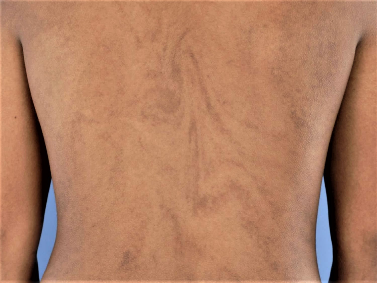
Hypomelanosis of Ito also called incontinentia pigmenti achromians, is a very rare birth defect that causes streaked, whirled, or mottled patches of light-colored (hypopigmented) skin 1. Hypomelanosis of Ito is part of a rare genetic neurocutaneous syndrome 2. These skin changes often develop within the first two years of life. Other symptoms may include varying degrees of learning disability, seizures, increased body hair, scoliosis, and strabismus.
Hypomelanosis of Ito represents the third most frequent neurocutaneous disease, after neurofibromatosis type 1 and tuberous sclerosis 2. Hypomelanosis of Ito is characterized by linear nevoid hypopigmentation along the lines of Blashko located on the limbs and the trunk. This cutaneous pattern appears at birth or in early childhood and usually becomes evident in infancy. The phenotype of hypomelanosis of Ito would be the result of a mosaic dermal abnormality of monogenic or chromosomal origin. Hypomelanosis of Ito is a rare neurocutaneous disease with multisystemic involvement. The most common defects involve the central nervous and musculoskeletal systems. Hypomelanosis of Ito is currently a descriptive rather than definitive diagnosis. Blaschkoid or mosaic hypomelanosis is a better descriptive term.
Hypomelanosis of Ito presentation is variable and behavioral problems and seizures are common 3. Fifty percent have an IQ <80 4. Males are often infertile 4. X-chromosomal abnormalities, primarily mosaicism, have been reported 4. Neuroimaging occasionally shows abnormalities contralateral to the skin lesion 4. Differential diagnosis includes vitiligo, incontinentia pigmenti, tuberous sclerosis, and other phakomatoses.
Hypomelanosis of Ito is characterized by:
- Streaky, patchy, whorl-like, or linear hypopigmented macules occurring on any part of the body
- Palms, scalp and soles of the feet are usually not affected
- Swirling patterns around the trunk and linear patterns down the legs and arms are referred to as Blaschko’s lines
- Lesions first appear as small 0.5-1 cm hypopigmented or white macules that merge to form larger patches
- Macules cover more than two dermatomes and are often on both sides of the body
- Patches are not symmetrical
While the exact cause is not known, hypomelanosis of Ito syndrome is strongly linked to its genetics and many patients have chormosomal abnormalities. The disease may be caused by abnormal nerve termination in the involved areas of the skin. Girls tend to be affected more commonly than boys. Treatment depends on the problems that are presented 1.
Currently there is not a cure for hypomelanosis of Ito. There is no treatment for the skin patches. Cosmetics or clothing may be used to cover the patches. Seizures, scoliosis, and other problems are treated as needed. Therapies are aimed at treating the symptoms in the child (e.g., seizures, scoliosis, strabismus). Children with this condition often receive their care from a multidisciplinary team of healthcare providers, including a pediatric ophthalmologist, neurologist, orthopedic specialist and others as needed. The overall prognosis of the child will depend on the severity of the associated symptoms.
Figure 1. Hypomelanosis of Ito
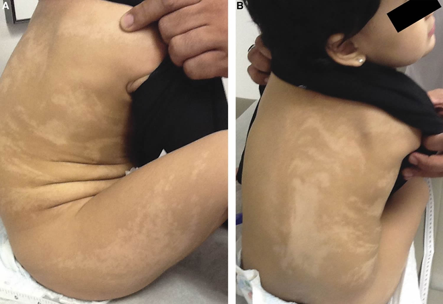
Footnote: A 2-year-old girl presented with developmental delay. Examination revealed height, weight, and head circumference above the 95th percentile, appendicular hypotonia, right hemibody hypopigmented lesions following the lines of Blaschko, and right hypopigmented iris (Figure, A and B). EEG showed disorganized background. Genetic microarray was normal. Photograph showing unilateral hemibody hypopigmented whorled lesions following the lines of Blaschko pathognomonic for hypomelanosis of Ito (incontinentia pigmenti achromians)
[Source 5 ]What causes hypomelanosis of Ito?
The cause of hypomelanosis of Ito remains unknown but scientists believe it is a problem with genes. The familial occurrence was described as suggesting an underlying chromosomal abnormality. Several models of inheritance have been proposed: X-linked, autosomal-dominant, and recessive genes transmission. However, to date, there is no evidence for genetic transmission. Most cases are sporadic. Chromosomal mosaicism and chromosomal translocations were reported 6.
Some cases have been associated with an underlying chromosomal abnormality. The skin patterning may reflect “mosaicism.” In mosaicism the person has some cells with normal chromsomes, and some with the chromosomal or gene abnormality 7. Mosaicism often leads to 2 cell lineages, which results in areas of hypopigmented (light areas of skint) and hyperpigmented skin (darker areas of skin). X-chromosome alterations are also found in hypomelanosis of Ito, and recent studies show that X-chromosome inactivation, activation, and mosaicism as the main causes of these differences in the skin. In less than 3% of the patients there is a family history of hypomelanosis of Ito–type skin lesions. Although hypomelanosis of Ito syndrome is most commonly a de novo occurrence (without any other cases in the family), familial cases appear to be transmitted as an autosomal dominant trait. About 10% of the patients report a family history of seizures or epilepsy 8.
People with specific questions about genetic risks or genetic testing for themselves or family members should speak with a genetics professional.
Resources for locating a genetics professional in your community are available online:
- The National Society of Genetic Counselors (https://www.findageneticcounselor.com/) offers a searchable directory of genetic counselors in the United States and Canada. You can search by location, name, area of practice/specialization, and/or ZIP Code.
- The American Board of Genetic Counseling (https://www.abgc.net/about-genetic-counseling/find-a-certified-counselor/) provides a searchable directory of certified genetic counselors worldwide. You can search by practice area, name, organization, or location.
- The Canadian Association of Genetic Counselors (https://www.cagc-accg.ca/index.php?page=225) has a searchable directory of genetic counselors in Canada. You can search by name, distance from an address, province, or services.
- The American College of Medical Genetics and Genomics (http://www.acmg.net/ACMG/Genetic_Services_Directory_Search.aspx) has a searchable database of medical genetics clinic services in the United States.
Hypomelanosis of Ito symptoms
Skin symptoms are most often visible by the time a child is about 2 years old. Hypomelanosis of Ito is present at birth, and patients usually undergo examination in their first or second year of life. Approximately 75% of patients with hypomelanosis of Ito seek care by the time they are aged 2 years. One fourth of patients seek care between ages 2 and 5 years. Skin lesions may become more pigmented over time and blend well with normally pigmented skin.
Hypomelanosis of Ito is characterized by streaked, whirled, or mottled patches of light-colored skin along the Blaschko lines. Other associated symptoms vary. The following symptoms have been described in cases of hypomelanosis of Ito reported in the medical literature.
Other symptoms develop as the child grows, and may include:
- Central nervous system (brain and spinal cord) defects
- Chest that caves in (pectus excavatum)
- Chest that caves out (pectus carinatum)
- Crossed eyes (strabismus)
- Finger anomalies
- Hearing problems
- Increased body hair (hirsutism)
- Large head size
- Learning difficulties
- Low muscle tone
- Mental retardation
- Motor retardation
- Nystagmus (sudden involuntary eye movements)
- Retinal degeneration
- Scoliosis
- Seizures
- Skeletal defects (e.g., short stature, facial and limb asymmetry)
- Small head size
- Streaked, whorled or mottled patches of skin on the arms, legs, and middle of the body
- Intellectual disability, including autism spectrum and learning disability
- Mouth or tooth abnormalities
Hypomelanosis of Ito complications
Problems that may result from hypomelanosis of Ito include:
- Discomfort and walking problems due to scoliosis
- Emotional distress, related to the physical appearance
- Intellectual disability
- Injury from seizures
Hypomelanosis of Ito diagnosis
The diagnosis of hypomelanosis of Ito is clinical. Ultraviolet light (Wood lamp) examination of the skin can enhance the hypopigmentation and help to confirm the diagnosis 9. Besides any patient with hypopigmentation along the lines of Blashko should be evaluated for neurological, musculoskeletal, cardiac, genitourinary, and ophthalmological abnormalities although computed tomography (CT) and magnetic resonance imaging (MRI) should be performed only in cases of neurologic symptoms. Skeletal radiography should be completed in all cases. In the case of patients with seizure disorders, electromyography (EEG) should be performed.
Tests that may be done include any of the following:
- CT or MRI scan of the head for a child with seizures and nervous system symptoms
- X-rays for a child with skeletal problems
- EEG to measure electrical activity of the brain in a child with seizures
- Genetic testing
Diagnostic criteria have been established for hypomelanosis of Ito 10:
Major criteria:
- Non-hereditary cutaneous hypopigmented linear streaks or patches involving more than two body segments, appearing at birth or in the first months
- One or more neurological or musculoskeletal manifestations.
Minor criteria
- Chromosomal anomalies
- Two or more congenital malformation, excluding nervous and musculoskeletal systems.
The diagnosis is retained with one major or minor criterion or two minor standards.
Hypomelanosis of Ito treatment
Treatment of hypomelanosis of Ito is symptom directed, targeting the specific clinical manifestations. Skin lesions do not require special treatment. Camouflage make-up may be employed for cosmetic concerns. Special education and anticonvulsants are often necessary. Close patient follow-up is needed to monitor for complications. Genetic counseling should be part of the management scheme.
Associated diseases, including the following, require appropriate specialty care:
- Seizures
- Mental retardation
- Hearing abnormalities
- Tooth deformities
- Visual problems
- Orthopedic problems
Diagnosis is important to guide genetic counseling.
Hypomelanosis of Ito prognosis
The overall prognosis of a child with hypomelanosis of Ito will depend on the type and severity of symptoms that develop. The life expectancy is normal for symptoms that only involve the skin changes. In most cases, skin color eventually turns to normal. Death is rare.
References- Hypomelanosis of Ito. https://medlineplus.gov/ency/article/001461.htm
- Chamli A, Litaiem N. Hypomelanosis of Ito. [Updated 2019 Apr 10]. In: StatPearls [Internet]. Treasure Island (FL): StatPearls Publishing; 2019 Jan-. Available from: https://www.ncbi.nlm.nih.gov/books/NBK538268
- Assogba K, Ferlazzo E, Striano P, et al. Heterogeneous seizure manifestations in hypomelanosis of Ito: report of four new cases and review of the literature. Neurol Sci 2010;31:9–16.
- Hsieh DT, Sarnat HB. Clinical summary: hypomelanosis of Ito. In: Medlink Neurology [online].
- Teaching NeuroImages: Hypomelanosis of Ito. Aunali S. Khaku, Vishnumurthy S. Hedna, Anuranjita Nayak. Neurology Mar 2013, 80 (12) e130; DOI: 10.1212/WNL.0b013e318288697b
- Glover MT, Brett EM, Atherton DJ. Hypomelanosis of Ito: spectrum of the disease. J. Pediatr. 1989 Jul;115(1):75-80.
- Ito hypomelanosis. https://www.orpha.net/consor/cgi-bin/OC_Exp.php?lng=EN&Expert=435
- Hypomelanosis of Ito. https://emedicine.medscape.com/article/1068339-overview
- Ardinger HH, Bell WE. Hypomelanosis of Ito. Wood’s light and magnetic resonance imaging as diagnostic measures. Arch. Neurol. 1986 Aug;43(8):848-50.
- Ruiz-Maldonado R, Toussaint S, Tamayo L, Laterza A, del Castillo V. Hypomelanosis of Ito: diagnostic criteria and report of 41 cases. Pediatr Dermatol. 1992 Mar;9(1):1-10.


