What is intravenous pyelogram
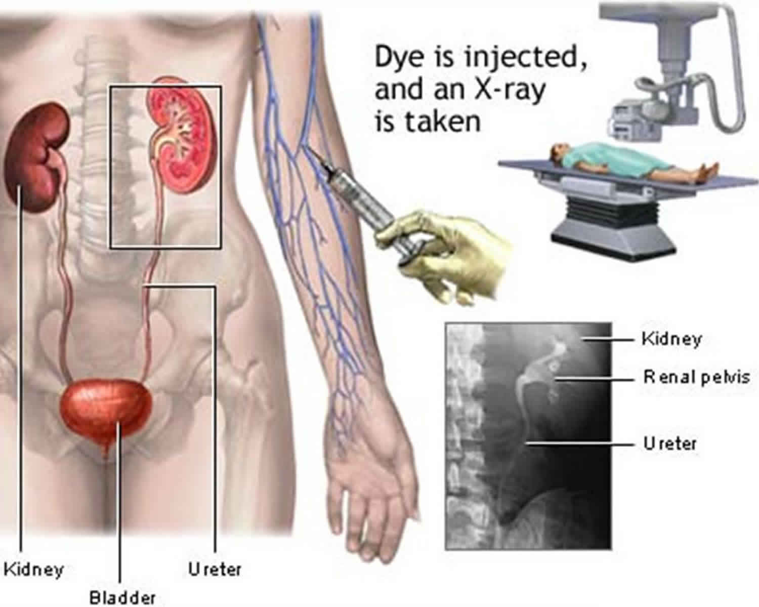
Intravenous pyelogram (IVP) also referred as intravenous urography (IVU) or excretory urography (EU), is a x-ray exam that uses an injection of iodine-containing contrast material to evaluate your kidneys, ureters and urinary bladder and help diagnose blood in the urine or pain in your side or lower back. When the iodine-containing contrast material is injected into a vein in the patient’s arm, it travels through the blood stream and collects in the kidneys and urinary tract, turning these areas bright white on the x-ray images. An IVP allows the radiologist to view and assess the anatomy and function of the kidneys, ureters and the bladder. An intravenous pyelogram (IVP) may provide enough information to allow your doctor to treat you with medication and avoid surgery. Today, however, intravenous pyelogram has been largely replaced by CT urography.
Inform your doctor if there’s a possibility you are pregnant and discuss any recent illnesses, medical conditions, medications you’re taking and allergies, especially to iodine-based contrast materials. Your doctor may instruct you to take a mild laxative the evening before the exam and to not eat or drink anything after midnight. Wear loose, comfortable clothing and leave jewelry at home. You may be asked to wear a gown.
Intravenous pyelogram indications
An intravenous pyelogram examination helps the radiologist assess abnormalities in the urinary system, as well as how quickly and efficiently the patient’s system is able to handle fluid waste.
The exam is used to help diagnose symptoms such as blood in the urine or pain in the side or lower back.
The IVP exam can enable the radiologist to detect problems within the urinary tract resulting from:
- kidney stones
- enlarged prostate
- tumors in the kidney, ureters or urinary bladder
- scarring from urinary tract infection
- surgery on the urinary tract
- congenital anomalies of the urinary tract
- check for normal function of kidneys
- check for anatomical variants or congenital anomalies (e.g. horse-shoe kidney)
- check the course of the ureters
- detect and localize a ureteric obstruction (urolithiasis)
- assess for synchronous upper tract disease in those with bladder transitional cell carcinoma (TCC)
Intravenous pyelogram preparation
Your doctor will give you detailed instructions on how to prepare for your intravenous pyelogram study.
You will likely be instructed not to eat or drink after midnight on the night before your exam. You may also be asked to take a mild laxative (in either pill or liquid form) the evening before the procedure.
You should inform your physician of any medications being taken and if there are any allergies, especially to iodinated contrast materials. Also inform your doctor about recent illnesses or other medical conditions.
You will be asked to remove some of your clothes and to wear a gown during the exam. You may also be asked to remove jewelry, removable dental appliances, eye-glasses and any metal objects or clothing that might interfere with the x-ray images.
Women should always inform their physician and x-ray technologist if there is any possibility that they are pregnant. Many imaging tests are not performed during pregnancy so as not to expose the fetus to radiation. If an x-ray is necessary, precautions will be taken to minimize radiation exposure to the baby.
How is the intravenous pyelogram procedure performed?
Intravenous pyelogram examination is usually done on an outpatient basis.
You will lie on the table and still x-ray images are taken. The contrast material is then injected, usually in a vein in your arm, followed by additional still images. The number of images taken depends on the reason for the examination and your anatomy.
You must hold very still and may be asked to keep from breathing for a few seconds while the x-ray picture is taken to reduce the possibility of a blurred image. The technologist will walk behind a wall or into the next room to activate the x-ray machine.
As the contrast material is processed by the kidneys, a series of images is taken to determine the actual size of the kidneys and to image the urinary tract in action as it begins to empty. The technologist may apply a compression band around the body to better visualize the urinary structures.
When the examination is complete, you may be asked to wait until the radiologist determines that all the necessary images have been obtained.
An IVP study is usually completed within an hour. However, because some kidneys function at a slower rate, the exam may last up to four hours.
What will I experience during and after the intravenous pyelogram procedure?
The intravenous pyelogram is usually a relatively comfortable procedure.
You will feel a minor sting as the contrast material is injected into your arm through a small needle. Some patients experience a flush of warmth, a mild itching sensation and a metallic taste in their mouth as it begins to circulate throughout their body. These common side effects usually disappear within a minute or two and are harmless. Rarely, some patients will experience an allergic reaction. Itching that persists or is accompanied by hives, can be easily treated with medication. In very rare cases, a patient may become short of breath or experience swelling in the throat or other parts of the body. These can be indications of a more serious reaction to the contrast material that should be treated promptly. Tell the radiologist immediately if you experience these symptoms as he/she is well prepared to treat this.
During the imaging process, you may be asked to turn from side to side and to hold several different positions to enable the radiologist to capture views from several angles. Near the end of the exam, you may be asked to empty your bladder so that an additional x-ray can be taken of your urinary bladder after it empties.
The contrast material used for IVP studies will not discolor your urine or cause any discomfort when you urinate.
What are the benefits vs. risks of intravenous pyelogram?
Benefits of intravenous pyelogram
- Imaging of the urinary tract with intravenous pyelogram is a minimally invasive procedure.
intravenous pyelogram images provide valuable, detailed information to assist physicians in diagnosing and treating urinary tract conditions from kidney stones to cancer. - An intravenous pyelogram can often provide enough information about kidney stones and urinary tract obstructions to direct treatment with medication and avoid more invasive surgical procedures.
- No radiation remains in a patient’s body after an x-ray examination.
- X-rays usually have no side effects in the typical diagnostic range for this exam.
Risks of intravenous pyelogram
- There is always a slight chance of cancer from excessive exposure to radiation. However, the benefit of an accurate diagnosis far outweighs the risk.
- The effective radiation dose for this procedure varies. Exposures are generally in the 65-75 kV range, mA of 600-1000, with exposure of <0.1 sec. Higher kV ranges reduce contrast of the renal parenchyma.
- Contrast materials used in intravenous pyelogram studies can cause adverse allergic reactions in some people, sometimes requiring medical treatment.
- Women should always inform their physician or x-ray technologist if there is any possibility that they are pregnant.
What are the limitations of intravenous pyelogram exams?
An intravenous pyelogram shows details of the inside of the urinary tract including the kidneys, ureters and bladder. Computed tomography (CT) or magnetic resonance imaging (MRI) may add valuable information about the functioning tissue of the kidneys and surrounding structures nearby the kidneys, ureters and bladder. Small urinary tract tumors and stones are more easily identified on these examinations.
Intravenous pyelogram exams are not usually indicated for pregnant women.
The uses for intravenous pyelogram in infants and children are limited. Other tests, including ultrasound, can be used in most cases to evaluate the kidneys and bladder. In general, intravenous pyelograms are rarely done in pediatric patients.


