Lentigo maligna
Lentigo maligna also known as Hutchinson’s melanotic freckle, is an early stage of melanoma. Lentigo maligna is a type of melanoma called ‘in situ’ melanoma 1. ‘In situ’ means that the cancer cells have not had the opportunity to spread anywhere else in the body. In lentigo maligna the cancer cells are confined to the upper layer of the skin (epidermis). When the cancer cells spread deeper into the skin (to dermis) it is called lentigo maligna melanoma. Lentigo maligna occurs most commonly in sun damaged areas such as the face and neck, particularly the nose and cheek, in fair skinned people over the age of 60. Lentigo maligna is a slow growing condition which can take years to develop. It grows slowly in diameter over 5 to 20 years or longer 2. Melanoma is a potentially lethal disease and lentigo maligna should be diagnosed and excised as soon as possible.
Lentigo maligna has a lower rate of transformation to invasive melanoma than the other forms of melanoma in situ (under 5% overall). However, the risk of invasive melanoma is greater in larger lesions, with up to 50% of those with diameter of greater than 4 cm being reported to have an invasive focus.
Transformation into lentigo maligna melanoma can be suggested by 3:
- Development of a nodule
- Increasing number of colors
- Ulceration or bleeding
Unlike superficial spreading melanoma, lentigo maligna is not related to the number of melanocytic nevi (moles) or atypical nevi.
Lentigo maligna can be cured with surgery. However, if the whole area is not removed completely with the appropriate surgery, some may develop into an invasive melanoma. It is therefore important to have it removed with a rim of normal skin (an adequate surgical margin). There are also preventative measures which can be taken that will further lower the risk of recurrence in the future.
Surgical excision
- Lentigo maligna is usually removed by surgical excision with an appropriate margin around the lesion for the best outcome. More specialized surgery or other investigations may be needed in some cases.
Non-surgical removal
- In cases where surgical excision cannot be performed (lesion is too large, surgery will be too deforming or contraindicated), other treatment methods include radiotherapy, cryotherapy and imiquimod cream. Then close monitoring of the lesion is required.
Figure 1. Skin anatomy

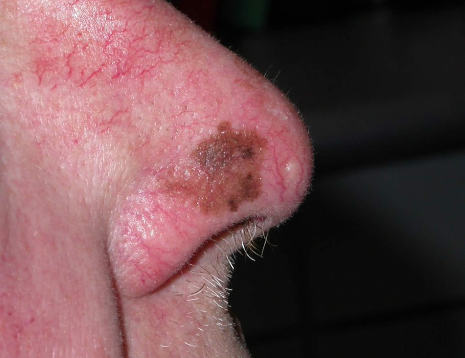
Figure 2. Lentigo maligna on the nose
While it may seem more convenient to shave off or cut out a mole yourself, there are 3 very good reasons a dermatologist should remove it:
- Skin cancer: If the mole contains skin cancer, some of the cancer cells can stay in the skin and even spread.
- Scarring: You can disfigure your skin causing a scar.
- Infection: A dermatologist uses sterile equipment to prevent infection.
Lentigo maligna causes
The cause of lentigo maligna is sun exposure or solarium use. Total UV exposure is important – lesions tend to arise in later middle age and elderly patients and are more common in outdoor workers. Lentigo maligna is a proliferation of malignant pigment cells (melanocytes) along the basal layer of the epidermis and within the hair follicle. What triggers the cells to become malignant is unknown but genetic mutations may start within primitive stem cells. Solar damage results in a degree of immune tolerance, allowing abnormal cells to grow unchecked.
Factors that predispose a person to developing lentigo maligna or associated condition include:
- chronic sun damaged/solar-induced skin damage
- fair skin complexion
- male gender
- a personal history of non melanoma skin cancer and precancerous lesions
- older individuals (those between 60 to 80 years are most commonly affected).
Thus lentigo maligna is more common in outdoor workers, in older people and in association with solar damage and keratinocyte skin cancer (basal cell carcinoma, squamous cell carcinoma). Although often occurring in those with very fair skin (skin phototype 1 and 2), it may also occur in those who tan quite easily (phototype 3). It is rare in brown or black skin (phototype 4-6).
Lentigo maligna is more common in males than females. The majority of patients with lentigo maligna are older than 40 years, and the peak age of diagnosis is be between 60 and 80 years.
Lentigo maligna symptoms
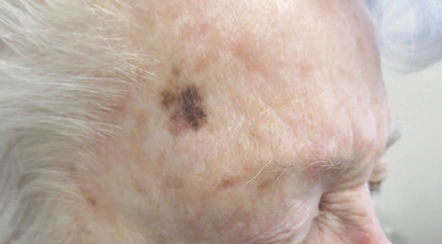
Lentigo maligna presents as a slowly growing or changing patch of discolored skin. Lentigo maligna commonly looks like a freckle, age spot, sun spot or brown patch that slowly changes shape and grows in size. The spot may be large in size, irregularly shaped with a smooth surface, and of multiple shades of brown and sometimes other colors.
At first, it often resembles common freckles or brown marks (lentigines). It becomes more distinctive and atypical in time, often growing to several centimetres over several years or even decades. Like other flat forms of melanoma, it can be recognised by the ABCDE rule: Asymmetry, Border irregularity, Color variation, large Diameter and Evolving.
The characteristics of lentigo maligna include:
- Large size: >6 mm and often several centimeters in diameter at diagnosis
- Irregular shape
- Variable pigmentation – colors may include light brown or tan, dark brown, pink, red or white
- Smooth surface.
Thickening of part of the lesion, increasing number of colors, ulceration or bleeding can be markers that the lesion is changing into a lentigo maligna melanoma.
Invasive melanoma is reported to arise within lentigo maligna in 3-10% of cases 2. It may be difficult to determine whether this has occurred just from the appearance, but the following features are very suspicious:
- Thickening of part of the lesion
- Increasing number of colors, especially blue or black
- Ulceration or bleeding
- Itching or stinging
Lentigo maligna diagnosis
It is vitally important to diagnose lentigo maligna and lentigo maligna melanoma accurately. Lentigo maligna is diagnosed clinically by a dermatologist, sometimes with the help of a dermatoscope (a tool used to magnify and look closely at skin moles) and in some centres, by confocal microscopy. The lesion may be difficult to distinguish from a benign, non-cancerous lesion. If the lesion is suspect, the doctor will biopsy or remove it to confirm the diagnosis. New techniques are being evaluated to help identify the margin of lentigo maligna prior to excision biopsy.
Other tests are not necessary in the majority of patients but those with invasive melanoma that is more than 1 mm thick may be advised to have imaging studies, lymph node biopsy and blood tests.
New tests are being developed to determine specific genetic mutations with lentigo maligna melanoma, which may inform future targeted therapy.
Diagnostic excision biopsy of lesion suspicious of melanoma
If a skin lesion is clinically suspicious of lentigo maligna, it is best cut out (excision biopsy) with a 2–3 mm margin. Partial biopsy is less accurate than complete excision biopsy, as a single small biopsy could miss a malignant focus. However sometimes the lesion is very large, and before performing significant surgery, a partial biopsy is arranged to confirm the diagnosis. The doctor should remove a long ellipse of skin, take biopsies from multiple sites or carefully shave a representative area for histology.
The pathological diagnosis of melanoma and its precursors can be very difficult. Some lesions clinically typical of lentigo maligna are reported to show junctional melanocytic proliferation alone (with or without atypia), others have the criteria to diagnose in situ melanoma, and a few show invasive cancer.
The histological features of lentigo maligna include a predominantly junctional confluent proliferation of melanocytes and extension along adnexal structures. Solar elastosis (degeneration of elastic tissue within dermis) is typically prominent. Immunostains eg SAC R21 may improve accuracy of diagnosis in borderline cases.
Pathology report in melanoma
The pathologist’s report should include a macroscopic description of the specimen and melanoma (the naked eye view), and a microscopic description. The following features should be reported if there is invasive melanoma.
- Diagnosis of primary melanoma
- Breslow thickness to the nearest 0.1 mm
- Clark level of invasion
- Margins of excision i.e. the normal tissue around the tumor
- Mitotic rate – a measure of how fast the cells are proliferating
- Whether or not there is ulceration
The report may also include comments about the cell type and its growth pattern, invasion of blood vessels or nerves, inflammatory response, regression and whether there is associated in-situ disease.
What is Breslow thickness?
The Breslow thickness is reported for invasive melanomas. It is measured vertically in millimeters from the top of the granular layer (or base of superficial ulceration) to the deepest point of tumor involvement. It is a strong predictor of outcome; the thicker the melanoma, the more likely it is to metastasize (spread).
What is the Clark level of invasion?
The Clark level indicates the anatomic plane of invasion. The deeper the Clark level, the greater the risk of metastasis (secondary spread). It is useful in predicting outcome in thin tumors, and less useful for thicker ones in comparison to the value of the Breslow thickness.
Table 1. Clark level
| Level | Characteristics |
|---|---|
| Level 1 | In situ melanoma |
| Level 2 | Melanoma has invaded papillary dermis |
| Level 3 | Melanoma has filled papillary dermis |
| Level 4 | Melanoma has invaded reticular dermis |
| Level 5 | Melanoma has invaded subcutaneous tissue |
Lentigo maligna treatment
There are a few ways of treating lentigo maligna. In most cases, lentigo maligna should undergo surgical excision. This means cutting it out and repairing the defect by simply closing the wound and stitching it up, creating a flap or by skin grafting.
Lentigo maligna has an unusually high risk of recurrence (up to 20%). For this reason, it is usually removed with a margin of healthy tissue, which can be difficult to achieve on facial skin. The ideal margin for all forms of melanoma in situ is 5-10mm, depending on how well defined are the edges of the lesion. Recommended margins for invasive melanoma are based on its thickness: it is 1 cm if less than 1 mm and 1-2 cm if the melanoma is over 1 mm in depth. If the margin and extent of the lentigo maligna is unclear, mapped serial excision may be recommended (margin-controlled, Mohs micrographic surgery or ‘slow Mohs’), in an attempt to remove all the malignant cells and to spare healthy skin.
Other treatments for lentigo maligna may be considered if it is difficult to remove the lesion surgically, or surgery will be very deforming, or there is a major contraindication to surgery:
- Radiotherapy – superficial X-rays or electrons
- Cryotherapy – undertaken by an expert
- Imiquimod cream – not yet fully evaluated.
However, these treatments do not offer as high cure rates as complete surgical excision.
Because the risk of invasive melanoma is small, one option in very elderly patients with large lentigo maligna is to simply photograph and watch the lesion carefully, biopsying any areas suspicious of invasive disease because of clinical or dermoscopic change.
Lentigo maligna prognosis
Lentigo maligna has a good prognosis when detected early and treated appropriately. However, a small percentage of lesions may be undetected and progress to become an invasive melanoma which is potentially life-threatening.
Regular follow-up and full skin examinations are need with the treating doctor to review past treatment sites and monitor areas of suspicion.
References- Lentigo maligna. https://www.bad.org.uk/for-the-public/patient-information-leaflets/lentigo-maligna
- Lentigo maligna and lentigo maligna melanoma. https://dermnetnz.org/topics/lentigo-maligna-and-lentigo-maligna-melanoma
- Melanoma: lentigo maligna melanoma (including lentigo maligna). http://www.pcds.org.uk/clinical-guidance/lentigo-maligna-melanoma-and-lentigo-maligna


