What is lentigo
Lentigo is a pigmented flat or slightly raised lesion with a clearly defined edge. Unlike an ephelis (freckle), lentigo does not fade in the winter months. There are several kinds of lentigo.
The name lentigo originally referred to its appearance resembling a small lentil. The plural of lentigo is lentigines, although “lentigos” is also in common use.
Lentigines can affect males and females of all ages and races. Solar lentigines are especially prevalent in fair skinned adults. Lentigines associated with syndromes are present at birth or arise during childhood.
Lentigo causes
Common forms of lentigo are due to exposure to ultraviolet (UV) radiation:
- Sun damage including sunburn
- Indoor tanning
- Phototherapy, especially photochemotherapy (PUVA)
Ionising radiation, eg radiation therapy, can also cause lentigines.
Several familial syndromes associated with widespread lentigines originate from mutations in Ras-MAP kinase, mTOR signalling and PTEN pathways.
What are the clinical features of lentigines?
Lentigines have been classified into several different types depending on what they look like, where they appear on the body, causative factors, and whether they are associated to other diseases or conditions.
Lentigines may be solitary or more often, multiple. Most lentigines are smaller than 5 mm in diameter.
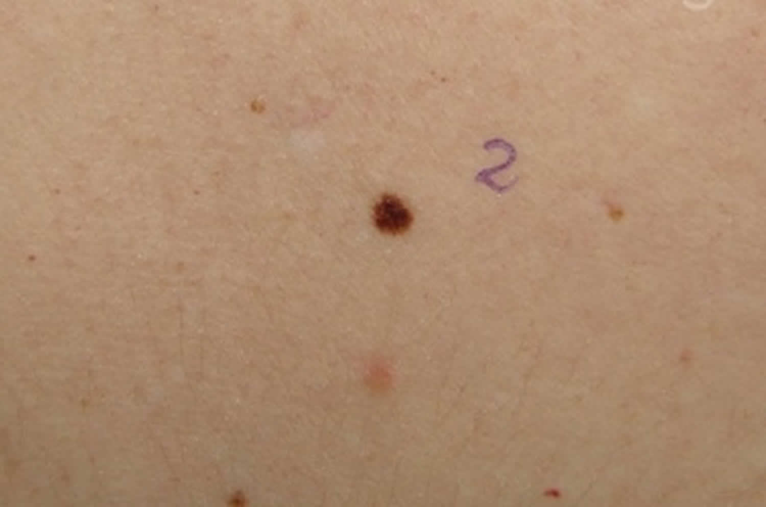
Lentigo simplex
Lentigo simplex is the most common form of lentigo. A single lesion or multiple lesions (lentigines) may be present at birth or more commonly first develop in early childhood. Lentigo simplex is not induced by sun exposure, and it is not associated with any medical diseases or conditions. It is also referred to as simple lentigo and juvenile lentigo.
Lentigo simplex is:
- A precursor to junctional nevus
- Arises during childhood and early adult life
- Found on trunk and limbs
- Small brown round or oval macule or thin plaque
- Jagged or smooth edge
- May have a dry surface
- May disappear in time
Figure 1. Lentigo simplex
What causes lentigo simplex?
The cause of lentigo simplex is unknown. Multiple lentigines can occur without associated conditions, in which case the condition is referred to as lentigines profusa or generalized lentigines. When multiple lentigines occur with associated abnormalities, the condition forms its own disease entity. These include Peutz-Jeghers syndrome, Xeroderma pigmentosum, LAMB syndrome, LEOPARD syndrome (Multiple Lentigines Syndrome) and Carney’s complex.
Clinical features of lentigo simplex
Lentigo simplex lesions may occur anywhere on the skin or mucous membranes. Only a few lesions may develop initially, but over time up until adult life they may become more numerous. Rarely, lesions may erupt suddenly or occur in vast numbers. Lesions appear as:
- Round or oval shaped macule (flat spot or patch) 3-15mm in diameter
- Margin or edge of the macule can be jagged or smooth
- A single even colour ranging from light brown to black
- Lesions are symptomless, i.e.: not painful or itchy
Lentigo simplex lesions are distinguished from freckles by their darker colour, comparative sparseness and scattered distribution. They also do not darken or increase in number on sun exposure, as do freckles.
Lentigo simplex treatment
Lentigo simplex lesions are benign (non-cancerous) lesions that cause no harm. However, their appearance is sometimes similar to melanomas or other cancerous lesions so they need to be examined carefully. Also, the presence or development of multiple lentigines may indicate the presence of associated abnormalities. Sometimes these defects may not show up clinically for many years, so a patient with lentigines should be followed up for long periods.
In some patients, the lesions may fade or disappear spontaneously over the course of years. If necessary, lentigines can be removed permanently through the use of chemical peels, cryotherapy, laser treatments or simple surgical excision.
Actinic lentigo
Actinic lentigo also called solar lentigo, is a harmless patch of darkened skin. Actinic lentigo results from exposure to ultraviolet (UV) radiation, which causes local proliferation of melanocytes and accumulation of melanin within the skin cells (keratinocytes). Actinic lentigos or lentigines are very common, especially in people over the age of 40 years. Sometimes they are also known as an “old age spot” or “senile lentigines”.
Actinic lentigo is:
- A precursor to seborrhoeic keratosis
- Found on chronically sun exposed sites such as hands, face, lower legs
- May also follow sunburn to shoulders
- Yellow, light or dark brown regular or irregular macule or thin plaque
- May have a dry surface
- Often has moth-eaten outline
- Can slowly enlarge to several centimeters in diameter
- May disappear, often through the process known as lichenoid keratosis
- When atypical in appearance, may be difficult to distinguish from melanoma in situ
Seborrheic keratoses may arise within actinic lentigos. This results in localized thickening and change in texture within the actinic lentigo.
Solar lentigines may become inflamed, when they are called lichenoid keratoses or lichen-planus like keratoses (due to the pattern of inflammation seen on histopathology). Lichenoid keratoses gradually disappear.
Figure 2. Actinic lentigo
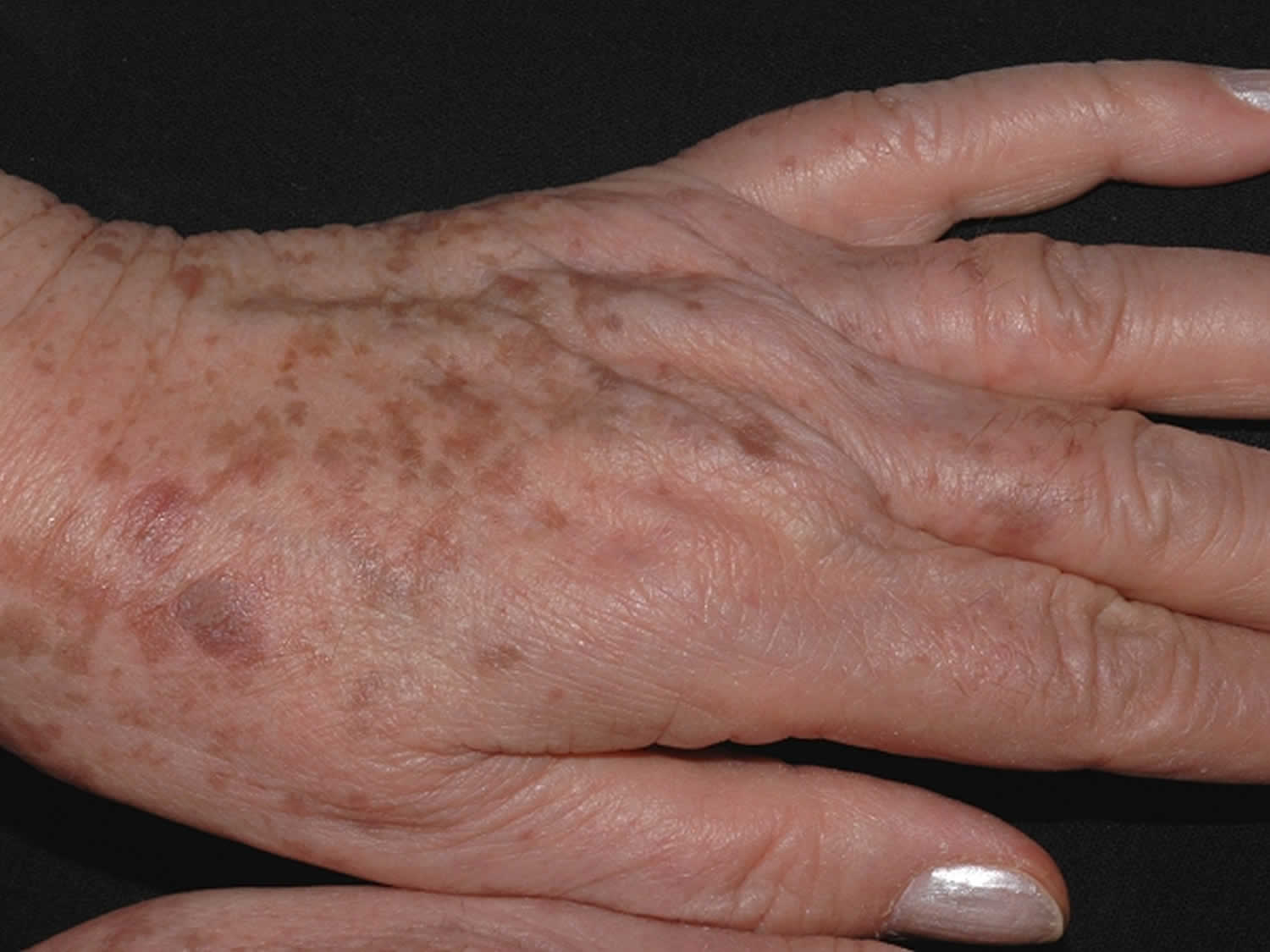
What does actinic lentigo look like?
Actinic lentigo is a flat, well-circumscribed patch. It can be round, oval or irregular in shape. Color varies from skin-colored, tan to dark brown or black, and size varies from a few millimeters to several centimeters in diameter. They can be slightly scaly.
Solar lentigines are found as groups of similar lesions on sun-exposed sites, particularly the face or the back of hands. Actinic lentigos occur in light and dark skin, but tend to be more numerous in fair-skinned individuals.
How is actinic lentigo diagnosed?
Actinic lentigo is often diagnosed on its clinical appearance. On occasion, it can be difficult to differentiate an irregular actinic lentigo from melanoma, a potentially dangerous form of skin cancer, and the term atypical actinic lentigo may be used.
Examination using dermatoscopy can clarify the diagnosis. If there is still diagnostic doubt, a skin biopsy may be performed for histological examination.
Actinic lentigo treatment
If left untreated, actinic lentigo will most likely persist indefinitely. Cryotherapy and laser surgery can destroy them, but treatment may leave a temporary or permanent white or dark mark.
Bleaching agents such as hydroquinone are not effective.
Preventative measures include minimizing sun exposure and use of sunscreens, but this needs to start early in life.
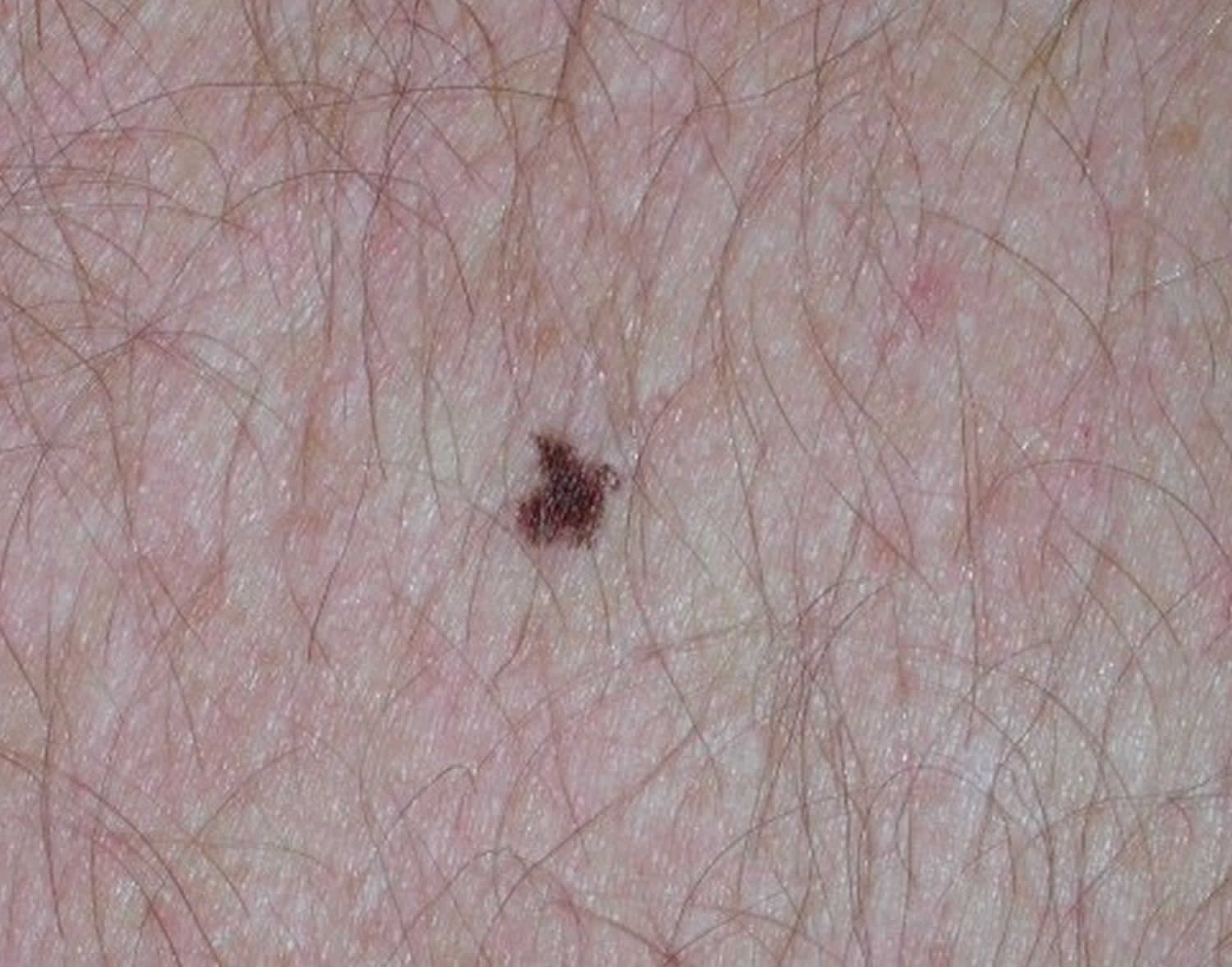
Ink spot lentigo
The characteristic features of ink spot lentigo (reticulated lentigo) allow one to make the clinical diagnosis of a benign lentigo 1.
- Also known as reticulated lentigo or reticulated melanotic macule 2
- Few in number compared to actinic lentigos or solar lentigines
- Follows sunburn in very fair skinned individuals
- Dark brown to black irregular ink spot-like macule
Histopathologically, ink spot lentigo (reticulated lentigo) was mainly characterized by a sharply circumscribed hyperpigmentation of the lower epidermis with accentuation at the tips of elongated and clubbed rete ridges. In addition, a normal or slightly increased melanocyte number in the basal layer and an infiltrate of melanophages in the upper dermis was noted. Ink spot lentigo (reticulated lentigo) shows histopathologic features similar to those of melanotic macules on volar skin and mucous membranes. Because of its characteristic clinical, dermatoscopic and histopathologic features ink spot lentigo (reticulated lentigo) can be regarded as a distinctive clinicopathologic entity.
Figure 3. Ink spot lentigo
Generalized lentigines
- Found on any exposed or covered site from early childhood
- Small macules may merge to form larger patches
- Not associated with a syndrome
- Also called lentigines profusa, multiple lentigines
Lentigo maligna
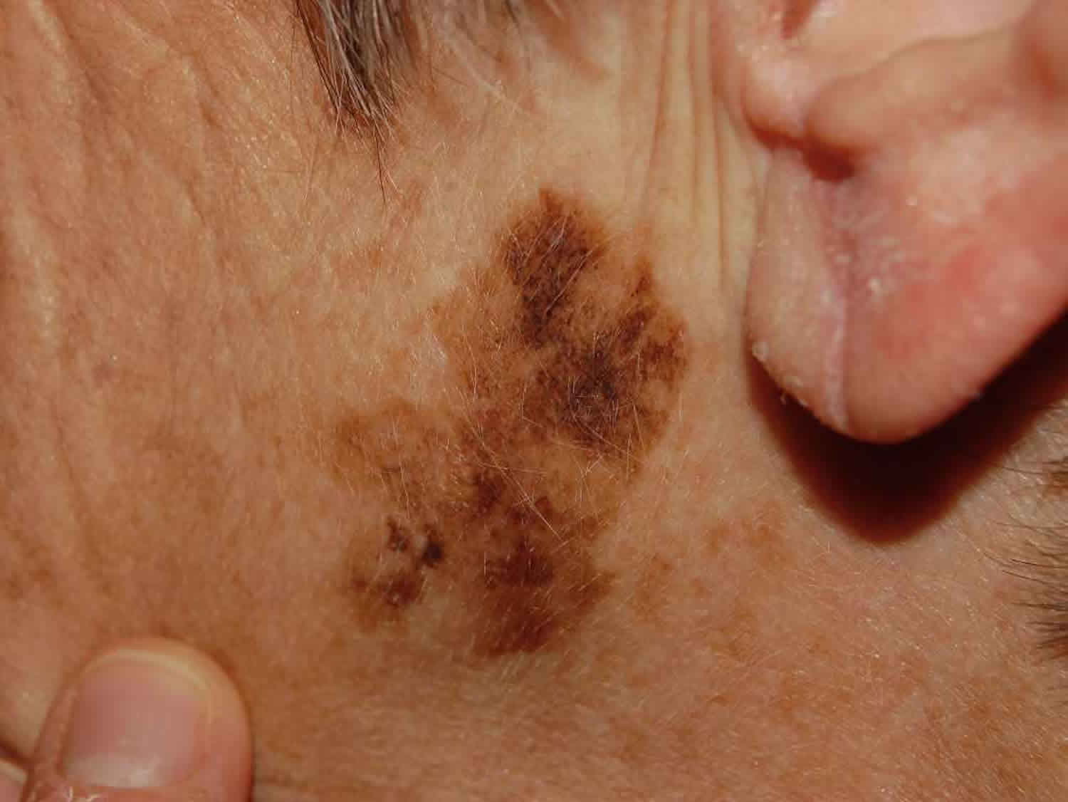
Lentigo maligna is a precursor to lentigo maligna melanoma, a potentially serious form of skin cancer. Lentigo maligna is also known as Hutchinson melanotic freckle.
Lentigo maligna is an early form of melanoma in which the malignant cells are confined to the tissue of origin, the epidermis, hence it is often reported as ‘in situ’ melanoma. It occurs in sun damaged skin so is generally found on the face or neck, particularly the nose and cheek. It grows slowly in diameter over 5 to 20 years or longer.
Lentigo maligna melanoma is diagnosed when the malignant melanoma cells have invaded into the dermis and deeper layers of skin. Lentigo maligna has a lower rate of transformation to invasive melanoma than the other forms of melanoma in situ (under 5% overall). However, the risk of invasive melanoma is greater in larger lesions, with up to 50% of those with diameter of greater than 4 cm being reported to have an invasive focus.
Figure 4. Lentigo maligna
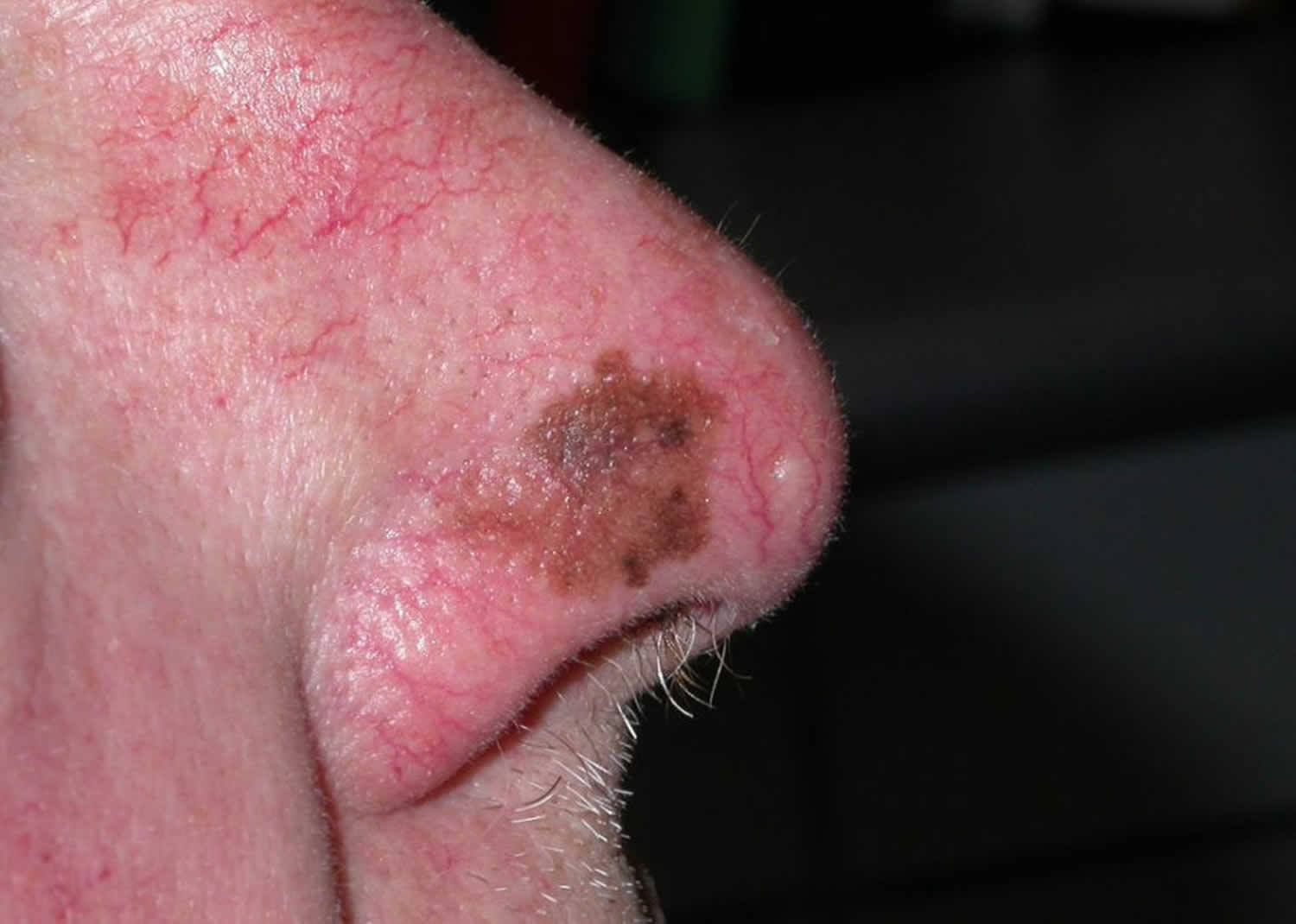
What does lentigo maligna look like?
Lentigo maligna presents as a slowly growing or changing patch of discoloured skin. At first, it often resembles common freckles or brown marks (lentigines). It becomes more distinctive and atypical in time, often growing to several centimetres over several years or even decades. Like other flat forms of melanoma, it can be recognised by the ABCDE rule: Asymmetry, Border irregularity, Colour variation, large Diameter and Evolving.
The characteristics of lentigo maligna include:
- Large size: >6 mm and often several centimetres in diameter at diagnosis
- Irregular shape
- Variable pigmentation – colours may include light brown or tan, dark brown, pink, red or white
- Smooth surface.
Invasive melanoma is reported to arise within lentigo maligna in 3-10% of cases. It may be difficult to determine whether this has occurred just from the appearance, but the following features are very suspicious.
- Thickening of part of the lesion
- Increasing number of colors, especially blue or black
- Ulceration or bleeding
- Itching or stinging
What is the cause of lentigo maligna?
Lentigo maligna is a proliferation of malignant pigment cells (melanocytes) along the basal layer of the epidermis and within the hair follicle. What triggers the cells to become malignant is unknown but genetic mutations may start within primitive stem cells.
Solar damage results in a degree of immune tolerance, allowing abnormal cells to grow unchecked.
What tests should be done if I have lentigo maligna?
It is essential to diagnose lentigo maligna and lentigo maligna melanoma accurately. Clinical diagnosis is aided by dermoscopy and in some centres, by confocal microscopy. New techniques are being evaluated to help identify the margin of lentigo maligna prior to excision biopsy.
Other tests are not necessary in the majority of patients but those with invasive melanoma that is more than 1 mm thick may be advised to have imaging studies, lymph node biopsy and blood tests.
New tests are being developed to determine specific genetic mutations with lentigo maligna melanoma, which may inform future targeted therapy.
Lentigo maligna treatment
In most cases, lentigo maligna should undergo surgical excision. This means cutting it out and repairing the defect by simply closing the wound and stitching it up, creating a flap or by skin grafting.
Lentigo maligna has an unusually high risk of recurrence (up to 20%). For this reason, it is usually removed with a margin of healthy tissue, which can be difficult to achieve on facial skin. The ideal margin for all forms of melanoma in situ is 5-10mm, depending on how well defined are the edges of the lesion. Recommended margins for invasive melanoma are based on its thickness: it is 1 cm if less than 1 mm and 1-2 cm if the melanoma is over 1 mm in depth. If the margin and extent of the lentigo maligna is unclear, mapped serial excision may be recommended (margin-controlled, Mohs micrographic surgery or ‘slow Mohs’), in an attempt to remove all the malignant cells and to spare healthy skin.
Other treatments for lentigo maligna may be considered if it is difficult to remove the lesion surgically, or surgery will be very deforming, or there is a major contraindication to surgery:
- Radiotherapy – superficial X-rays or electrons
- Cryotherapy – undertaken by an expert
- Imiquimod cream – not yet fully evaluated.
However, these treatments do not offer as high cure rates as complete surgical excision.
Because the risk of invasive melanoma is small, one option in very elderly patients with large lentigo maligna is to simply photograph and watch the lesion carefully, biopsying any areas suspicious of invasive disease because of clinical or dermoscopic change.
Lentigo melanoma
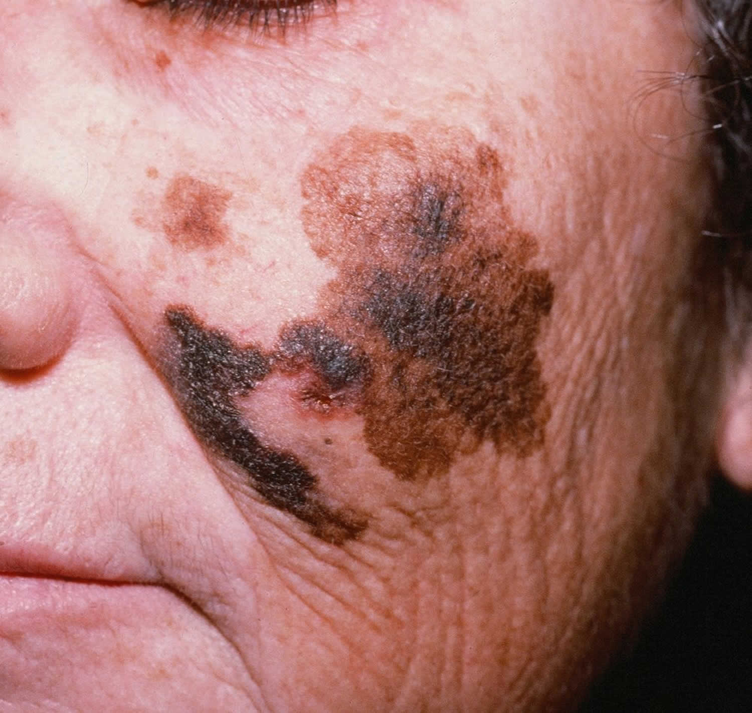
Lentigo melanoma also called lentigo maligna melanoma is the first presentation of melanoma in which the cancerous cells are limited to the tissue of origin, the epidermis 3. Therefore, lentigo melanoma (lentigo maligna melanoma) is often reported as “in situ’” melanoma. Lentigo maligna melanoma accounts for about 10% of melanomas and typically occurs on chronically sun exposed skin, most commonly on the neck or face, especially around the cheeks and nose and occasionally on the back, the forearms, or the lower legs. Lentigo maligna melanoma is most common among the elderly. Lentigo maligna melanoma grows from a few millimeters to centimeters within 5 to 20 years or sometimes even longer. As a palpable lesion, the pigment of lentigo maligna melanoma is usually dark brown and a few millimeters large. It is the slowest growing and the least common type of melanoma. Although it is a slow-growing malignancy, the downside is that lentigo maligna melanoma can merge with other tumors like melanocytic nevus when developing. If detected early and treated, the prognosis is very good.
The incidence of lentigo maligna is highest in Hawaii and Australia with males being affected more often than females. Majority of the patients with the disease are older than 40 years, and the peak age of diagnosis is between 60 and 80 years. A study reported a 52% increase in the occurrence of lentigo maligna melanoma among people of age within 45 to 64 within 1990 and 2000 in the United States. The risk of the formation of the tumors is higher in fair-skinned people with a history of melanoma skin cancer and markers of actinic skin damage (solar lentigines and actinic keratoses).
Figure 5. Lentigo maligna melanoma
Lentigo melanoma causes
Lentigo maligna is the preinvasive form of melanoma while lentigo maligna melanoma occurs when atypical melanocytes invade the dermal layer of the skin, thereby making the cancerous cells prone to metastasis. Prolonged damage to the skin causes it as a result of cumulative exposure to ultraviolet rays.
About 10% to15% of lentigo maligna melanoma patients have a family member with the disease. First-degree relatives have 8 to 12-times the risk of having lentigo maligna melanoma than individuals who do not have a familial history of lentigo melanoma. Organ transplant and pills that weakens the immune system of an individual can also cause the formation of the cancerous cells. Undue exposure to ultraviolet radiation and a history of severe sunburn can also provoke the development of lentigo maligna melanoma, especially in adolescents and adults. A skin disorder called dysplastic nevus syndrome is also known to increase the risks of lentigo maligna melanoma. Xeroderma pigmentosum, a defect in the protein responsible for the repair of the DNA damaged by ultra-violet rays has also been discovered to be an underlying cause of lentigo maligna melanoma.
Lentigo maligna melanoma risk factors
Risk factors for development of lentigo maligna include the following: a history of sunburns, a history of nonmelanoma skin cancers, advanced age, lighter skin types, and tendency to form solar lentigines. Although lentigo maligna occurs on chronically sun-damaged skin, it is thought that intermittent sunburns, rather than cumulative sun exposure, are a risk factor for lentigo maligna 4.
Studies have shown that lentigo maligna has a different genetic make-up than other types of melanoma. Unlike the other types of melanoma, a genetic propensity to form atypical nevi is not seen in lentigo maligna. In lentigo maligna, there is a higher incidence of p53 mutations compared with BRAF mutations. BRAF may not play a significant role in lentigo maligna as it does with other types of melanoma 5.
Lentigo melanoma clinical features
Lentigo maligna melanoma manifests as an irregular patch of discolored skin. Initially, it often resembles brown marks (lentigines) or common freckles, but along the line, it becomes more noticeable and begins to expand across the skin. Like other forms of melanoma, it can be understood by the ABCDE rule of malignancy: asymmetry, border irregularity, color variation, large diameter, and evolving of a mole.
It is not possible to diagnose lentigo maligna melanoma with the naked eye. Patients most often come to the hospital with complaints of a large diameter irregular lesion or nodule on their skin. Melanoma patients need a thorough and complete physical examination, with particular attention to the lymph nodes and skin. Hence, an excisional biopsy should be conducted to diagnose lentigo maligna melanoma.
Lentigo melanoma diagnosis
Accurate diagnosis of lentigo maligna melanoma is critical. Clinical investigation is aided by dermoscopy and in some centers, by confocal microscopy. Dermoscopy helps in differentiating lentigo maligna from other skin lesions while confocal fluorescence microscopy is used to view the lesion in 3 dimensions. New techniques are being tested and evaluated to help classify the extent of lentigo maligna melanoma before excision biopsy. The gold standard for diagnosing lentigo maligna melanoma is a full-thickness skin biopsy and dermatopathological analysis of the biopsy specimen. Other tests are not mandatory in the majority of cases, but those with invasive melanoma deeper than 1-mm thick should have imaging studies and a lymph node biopsy as part of their workup.
Lentigo melanoma staging
Lentigo maligna melanoma is staged to determine the degree of severity, presence or absence of metastasis and to prescribe the best treatment plan.
The staging system most often used for melanoma is the American Joint Committee on Cancer (AJCC) TNM system, which is based on 3 key pieces of information:
- The extent of the tumor (T): How deep has the cancer grown into the skin? Is the cancer ulcerated?
- Tumor thickness: The thickness of the melanoma is called the Breslow measurement. In general, melanomas less than 1 millimeter (mm) thick (about 1/25 of an inch) have a very small chance of spreading. As the melanoma becomes thicker, it has a greater chance of spreading.
- Ulceration: Ulceration is a breakdown of the skin over the melanoma. Melanomas that are ulcerated tend to have a worse outlook.
- The spread to nearby lymph nodes (N): Has the cancer spread to nearby lymph nodes?
- The spread (metastasis) to distant sites (M): Has the cancer spread to distant lymph nodes or distant organs such as the lungs or brain?
Numbers or letters after T, N, and M provide more details about each of these factors. Higher numbers mean the cancer is more advanced. Once a person’s T, N, and M categories have been determined, this information is combined in a process called stage grouping to assign an overall stage.
The staging system in the table below uses the pathologic stage (also called the surgical stage). It is determined by examining tissue removed during an operation. Sometimes, if surgery is not possible right away or at all, the cancer will be given a clinical stage instead. This is based on the results of a physical exam, biopsy, and imaging tests. The clinical stage will be used to help plan treatment. Sometimes, though, the cancer has spread further than the clinical stage estimates, and may not predict the patient’s outlook as accurately as a pathologic stage.
There are both clinical and pathologic staging systems for melanoma. Since most cancers are staged with the pathologic stage, we have included that staging system below. If your cancer has been clinically staged, it is best to talk to your doctor about your specific stage.
The table below is a simplified version of the TNM system. It is based on the most recent AJCC system, effective January 2018. It’s important to know that melanoma cancer staging can be complex. If you have any questions about the stage of your cancer or what it means, please ask your doctor to explain it to you in a way you understand.
Table 1. American Joint Committee on Cancer (AJCC) Melanoma Skin Cancer Stages
| AJCC Stage | Melanoma Stage description | |
| 0 | The cancer is confined to the epidermis, the outermost skin layer. It has not spread to nearby lymph nodes or distant sites. This stage is also known as melanoma in situ. | |
| I
| The cancer is no more than 2mm (2/25 of an inch) thick and might or might not be ulcerated. It has not spread to nearby lymph nodes or to distant sites. | |
| II | The cancer is at least 1.01 mm and may be thicker than 4.0 mm. It might or might not be ulcerated. It has not spread to nearby lymph nodes (N0) or to distant sites (M0).
| |
| IIIA | The cancer is no more than 2.0 mm thick. It might or might not be ulcerated. It has spread to 3 or less lymph node(s), but it is so small that it is only seen under the microscope. It has not spread to distant sites. | |
| IIIB
| There is no sign of the primary cancer AND:
It has not spread to distant sites. | |
| OR | ||
The cancer is no more than 4.0 mm thick. It might or might not be ulcerated AND:
It has not spread to distant sites. | ||
| IIIC | There is no sign of the primary cancer AND:
It has not spread to distant sites. | |
| OR | ||
The cancer is no more than 4.0 mm thick. It might or might not be ulcerated AND:
It has not spread to distant sites. | ||
| OR | ||
The cancer is between 2.1 and 4.0mm OR thicker than 4.0 mm. It might or might not be ulcerated AND:
It has not spread to distant sites. | ||
| OR | ||
The cancer is thicker than 4.0 mm and is ulcerated AND:
It has not spread to distant sites. | ||
| IIID | The cancer is thicker than 4.0 mm and is ulcerated AND:
It has not spread to distant sites. | |
| IV | The cancer can be any thickness and might or might not be ulcerated. It might or might not have spread to nearby lymph nodes. It has spread to distant lymph nodes or organs such as the lungs, liver or brain. | |
Lentigo melanoma treatment
Lentigo melanoma treatment depends on the thickness of the invasive component of the lentigo maligna melanoma, and it is similar to other melanomas of the same thickness and stage.
Lentigo maligna melanoma should be completely removed surgically. If possible, there should be a 1 cm margin of normal skin around the tumor, but the margin may depend on the site of the lesion and how close it is to important structures like the mouth, eye or nose. If the local lymph nodes are enlarged due to melanoma, they should also be completely removed, which entails a major surgical procedure under general anesthetic.
However, for patients who cannot have surgery, several conservative therapies can be applied. These therapies include cryotherapy, chemotherapy, and immune therapy with topical imiquimod. Occasionally radiotherapy is applied to destroy melanoma cells, though studies have shown mixed feelings concerning the effectiveness of this therapy option because it may miss focal lentigo maligna melanoma. Non-surgical therapies have been documented to have a recurrence rate of 20% to 100% in 5 years. This shows that surgical resection is the best treatment modality.
Lentigo melanoma prognosis
The prognosis of lentigo maligna melanoma is fair unless it has metastasized (spread). A patient’s prognosis depends significantly on the time, overall wellness of the patient, diagnosis, and depth of the lesion.
The SEER (Surveillance, Epidemiology, and End Results) database tracks 5-year relative survival rates for melanoma skin cancer in the United States, based on how far the cancer has spread. The SEER database, however, does not group cancers by AJCC TNM stages (stage 1, stage 2, stage 3, etc.). Instead, it groups cancers into localized, regional, and distant stages:
- Localized: There is no sign that the cancer has spread beyond the skin where it started. This would include AJCC stage I and stage II cancers.
- Regional: The cancer has spread beyond the skin where it started to nearby structures or lymph nodes. This would include mainly stage III cancers in the AJCC system.
- Distant: The cancer has spread to distant parts of the body, such as the lungs, liver, or skin on other parts of the body. This would include stage IV cancers.
Table 2. 5-year relative survival rates for melanoma skin cancer (Based on people diagnosed with melanoma between 2008 and 2014)
| SEER stage | 5-year relative survival rate |
| Localized | 98% |
| Regional | 64% |
| Distant | 23% |
| All SEER stages combined | 92% |
Footnote: SEER = Surveillance, Epidemiology, and End Results
- These numbers apply only to the stage of the cancer when it is first diagnosed. They do not apply later on if the cancer grows, spreads, or comes back after treatment.
- These numbers don’t take everything into account. Survival rates are grouped based on how far the cancer has spread, but your age, overall health, how well the cancer responds to treatment, and other factors can also affect your outlook. For example, younger people tend to have a better outlook than older people, regardless of the stage. And people who have weakened immune systems, such as those who have had organ transplants or who are infected with HIV, are at greater risk of dying from their melanoma.
- People now being diagnosed with melanoma may have a better outlook than these numbers show. Treatments improve over time, and these numbers are based on people who were diagnosed and treated at least five years earlier.
How can lentigines be prevented?
Lentigines associated with exposure ultraviolet radiation can be prevented by very careful sun protection. Clothing is more successful at preventing new lentigines than are sunscreens.
Lentigo diagnosis
Lentigines are usually diagnosed clinically by their typical appearance. Concern regarding possibility of melanoma may lead to:
- Dermatoscopy
- Diagnostic excision biopsy
Histopathology of a lentigo shows:
- Thickened epidermis
- An increased number of melanocytes along the basal layer of epidermis
- Unlike junctional melanocytic naevus, there are no nests of melanocytes
- Increased melanin pigment within the keratinocytes
- Additional features depending on type of lentigo
In contrast, an ephelis (freckle) shows sun-induced increased melanin within the keratinocytes, without an increase in number of cells.
Lentigo treatment
Most lentigines are left alone. Attempts to lighten them may not be successful. The following approaches are used:
- SPF 50+ broad-spectrum sunscreen
- Hydroquinone bleaching cream
- Alpha hydroxy acids
- Vitamin C
- Retinoids
- Azelaic acid
- Chemical peels
Individual lesions can be permanently removed using:
- Cryotherapy
- Intense pulsed light
- Pigment lasers.
Lentigines prognosis
Lentigines usually persist. They may increase in number with age and sun exposure. Some in sun-protected sites may fade and disappear.
References- Bolognia JL. Reticulated Black Solar Lentigo (`Ink Spot’ Lentigo). Arch Dermatol. 1992;128(7):934–940. doi:10.1001/archderm.1992.01680170066008
- [Reticular lentigo]. Hautarzt. 1997 Mar;48(3):181-5. https://www.ncbi.nlm.nih.gov/pubmed/?term=9182089
- Charifa A, Chen CSJ. Cancer, Melanoma, Lentigo Maligna. [Updated 2018 Oct 27]. In: StatPearls [Internet]. Treasure Island (FL): StatPearls Publishing; 2019 Jan-. Available from: https://www.ncbi.nlm.nih.gov/books/NBK482163
- Gaudy-Marqueste C, Madjlessi N, Guillot B, Avril MF, Grob JJ. Risk factors in elderly people for lentigo maligna compared with other melanomas: a double case-control study. Arch Dermatol. 2009;145(4):418–23. http://dx.doi.org/10.1001/archdermatol.2009.1
- Purdue MP, From L, Kahn HJ, Armstrong BK, Kricker A, Gallagher RP, McLaughlin JR, Klar NS, Marrett LD. Etiologic factors associated with p53 immunostaining in cutaneousmalignant melanoma. Int J Cancer. 2005;117(3):486–93


