Linea alba
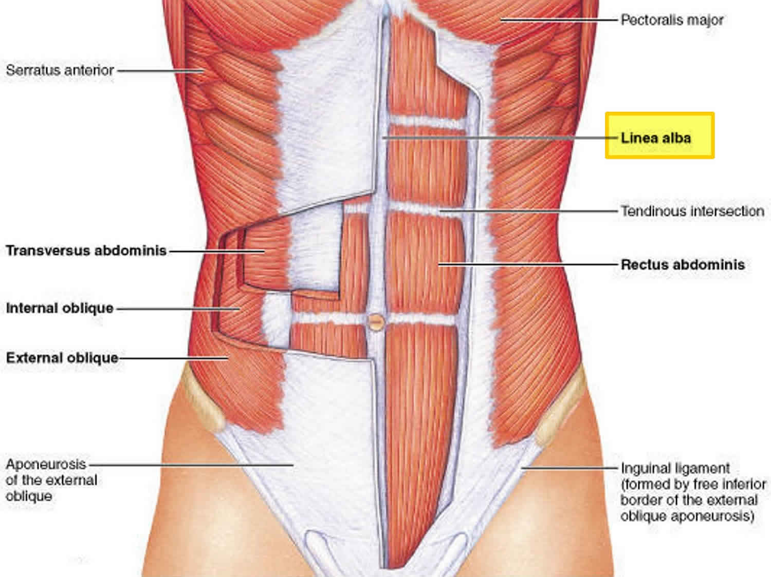
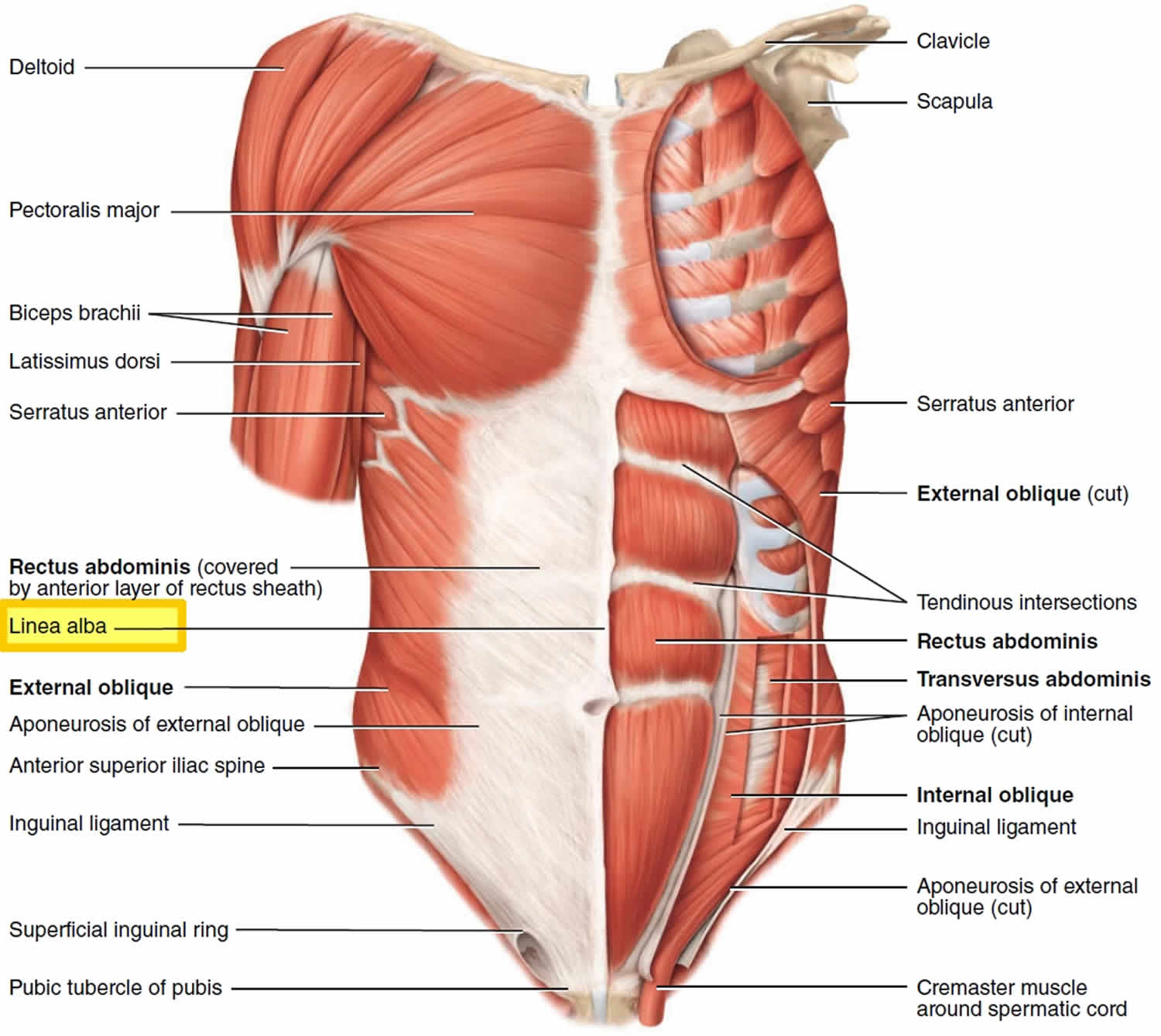
Linea alba is Latin for “white line”, is a single midline tough fibrous band line in the anterior abdominal wall formed by the median fusion of the layers of the rectus sheath medial to the bilateral rectus abdominis muscles. Linea alba attaches to the xiphoid process of the sternum and the pubic symphysis. The umbilicus passes through the linea alba.
In muscular individuals its presence can be seen on the skin, forming the depression between the left and right halves of a “six pack”.
Because linea alba consists of mostly connective tissue, and does not contain any primary nerves or blood vessels, a median incision through the linea alba is a common surgical approach (laparotomy incision) 1.
In the latter stages of pregnancy, the linea alba stretches to increase the distance between the rectus abdominis muscles. Paraumbilical herniae can occur through the linea alba.
Figure 1. Linea alba anatomy
Linea alba function
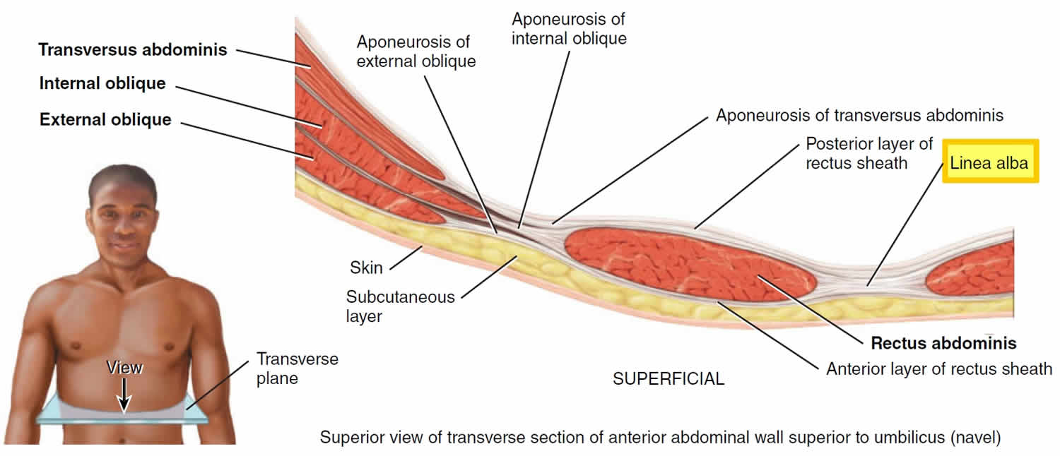
It is formed by the fusion of the aponeuroses (sheathlike tendons) of the external oblique, internal oblique, and transversus abdominis muscles form the rectus sheaths, which enclose the rectus abdominis muscles. The sheaths meet at the midline to form the linea alba. As linea alba is prolongation of rectus sheaths and more generally, fascias of lateral muscles, its structure is known to follow fiber orientation of lateral abdominal muscles.
Linea alba is the most common site for laparotomy incision 1 and it plays a key role in abdominal wall biomechanics during and after closure of incisions or hernia defects 2.
References- World Health Organization (2003), Surgical Care at the District Hospital (SCDH) manual-Chap.6 Laparotomy and abdominal trauma.
- van’t Riet, M., Steyerberg, E. W., Nellensteyn, J., Bonjer, H. J. & Jeekel, J. (2002), ‘Meta-analysis of techniques for closure of midline abdominal incisions’, British Journal of Surgery 89(11), 1350–1356.


