What is lung abscess
Lung abscess is a pus-filled cavity in the lung surrounded by inflamed tissue and caused by an infection. Lung abscess is defined as necrosis of the pulmonary tissue and formation of cavities containing necrotic debris or fluid caused by microbial infection 1. The formation of multiple small (<2 cm) abscesses is occasionally referred to as necrotizing pneumonia or lung gangrene 1. Both lung abscess and necrotizing pneumonia are manifestations of a similar pathologic process. Failure to recognize and treat lung abscess is associated with poor clinical outcome. Lung abscesses are often complicated to manage and difficult to treat and, in some cases, may be life-threatening.
A lung abscess is usually caused by bacteria that normally live in the mouth and are inhaled into the lungs. Lung abscess is most commonly caused by aspiration of oral secretions by patients who have impaired consciousness. Lung abscesses likely occur more commonly in elderly patients because of the increased incidence of periodontal disease and the increased prevalence of dysphagia and aspiration. However, a case series from an urban center with high prevalence of alcoholism reported a mean age of 41 years 2.
Lung abscess symptoms include fatigue, loss of appetite, night sweats, fever, weight loss, and a persistent cough that brings up sputum.
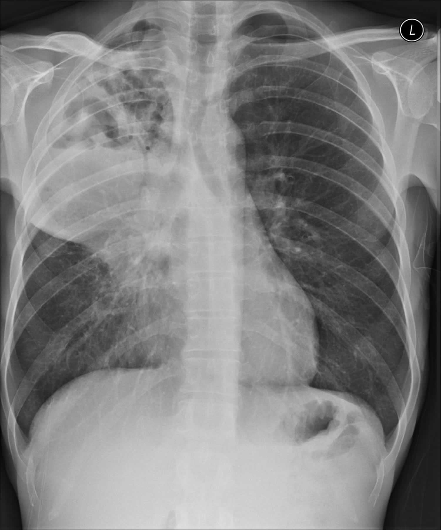
Diagnosis is usually determined with a chest x-ray.
People usually need to take antibiotics for several weeks before a lung abscess clears up. Lung abscess treatment usually is with clindamycin or combination beta-lactam/beta-lactamase inhibitors.
As a result of the widespread availability of antibiotics, the incidence of lung abscesses has dramatically reduced. Similarly, mortality has reduced. The elderly, immunocompromised, malnourished, debilitated, and those who do not have access to antibiotics are particularly susceptible and have the worst prognosis 3. Particularly due to an increased number of immunocompromised (secondary to HIV/AIDS and iatrogenic immunosuppression) the rate has once more increased 4.
Empyema vs Lung abscess
Empyemas are purulent inflammatory collections within a body cavity. Contrast this with abscesses, which arise within parenchymal tissue, rather than occupying a pre-existing anatomical space.
Colloquially, the standalone term empyema is used to refer to thoracic empyemas but there are various different types of empyema which are described by the organ affected:
- gallbladder empyema
- subdural empyema
- thoracic empyema
- joint empyema (of any synovial joint)
- empyema cystitis (urinary bladder)
Like abscesses, as the vascular supply to an empyema is poor, antimicrobial treatment on its own is usually insufficient to treat the underlying infection and drainage of the collection is usually required. This is often done percutaneously with ultrasound guidance although open surgical drainage can be necessary in complex cases.
Causes of lung abscess
Most lung abscesses develop after aspiration of oral secretions by patients with gingivitis or poor oral hygiene. Typically, patients have altered consciousness as a result of alcohol intoxication, illicit drugs, anesthesia, sedatives, or opioids. Older patients and those unable to handle their oral secretions, often because of neurologic disease, are also at risk. Lung abscesses can also develop secondary to endobronchial obstruction (eg, due to bronchial carcinoma) or to immunosuppression (eg, due to HIV/AIDS or after transplantation and use of immunosuppressive drugs).
Patients at the highest risk for developing lung abscess have the following risk factors:
- Periodontal disease
- Seizure disorder
- Alcohol abuse
- Dysphagia
Other patients at high risk for developing lung abscess include individuals with an inability to protect their airways from massive aspiration because of a diminished gag or cough reflex, caused by a state of impaired consciousness (eg, from alcohol or other CNS depressants, general anesthesia, or encephalopathy).
Infrequently, the following infectious causes of pneumonia may progress to parenchymal necrosis and lung abscess formation:
- Pseudomonas aeruginosa
- Klebsiella pneumoniae
- Staphylococcus aureus (may result in multiple abscesses)
- Streptococcus pneumoniae
- Nocardia species
- Actinomyces species
- Fungal species
An abscess may develop as an infectious complication of a preexisting bulla or lung cyst.
An abscess may develop secondary to carcinoma of the bronchus. The bronchial obstruction causes postobstructive pneumonia, which may lead to abscess formation. Underlying lung cancer in edentulous patients with lung abscesses should also be considered.
A less common cause of lung abscess is necrotizing pneumonia that may develop from hematogenous seeding of the lungs due to suppurative thromboembolism (eg, septic embolism due to IV drug use) or right-sided endocarditis. In contrast to aspiration and obstruction, these conditions typically cause multiple rather than isolated lung abscesses.
Pathogens
The most common pathogens of lung abscesses due to aspiration are anaerobic bacteria, but about half of all cases involve both anaerobic and aerobic organisms (see Infectious Causes of Cavitary Lung Lesions). The most common anaerobic pathogens are Peptostreptococcus, Fusobacterium, Prevotella, and Bacteroides. The most common aerobic pathogens are streptococci and staphylococci—sometimes methicillin-resistant Staphylococcus aureus (MRSA). Occasionally, cases are due to gram-negative bacteria, especially Klebsiella. Immunocompromised patients with lung abscess are most commonly infected with Pseudomonas aeruginosa and other gram-negative bacilli but also may have infection with Nocardia, Mycobacteria sp, or fungi. Rare cases of pulmonary gangrene or fulminant pneumonia with sepsis have been reported with pathogens such as methicillin-resistant Staphylococcus aureus (MRSA), Pneumococcus, and Klebsiella. Some patients, especially those from developing countries, are at risk of abscess due to Mycobacterium tuberculosis, and rare cases are due to amebic infection (eg, with Entamoeba histolytica), paragonimiasis, or infection with Burkholderia pseudomallei.
Introduction of these pathogens into the lungs first causes inflammation, which, over a week or two, leads to tissue necrosis and then abscess formation. The abscess usually ruptures into a bronchus, and its contents are expectorated, leaving an air- and fluid-filled cavity. In about one third of cases, direct or indirect extension (via bronchopleural fistula) into the pleural cavity results in empyema.
Lung abscess symptoms
Lung abscess symptoms depend on whether the abscess is caused by anaerobic or other bacterial infection. A lung abscess may be asymptomatic in a small proportion of patients in the early stages. Typical lung abscess symptoms are described below.
Symptoms of lung abscess due to anaerobic bacteria or mixed anaerobic and aerobic bacteria are usually chronic (e.g., occurring over weeks or months) and include productive cough, fever, night sweats, and weight loss. Patients may also present with hemoptysis and pleuritic chest pain. Sputum may be purulent or blood-streaked and classically smells or tastes foul.
Symptoms of lung abscess due to aerobic bacteria develop more acutely and resemble bacterial pneumonia. Abscesses due to organisms other than anaerobes (e.g., Mycobacteria,Nocardia) lack putrid respiratory secretions and may be more likely to occur in nondependent lung regions.
Signs of lung abscess, when present, are nonspecific and resemble those of pneumonia: decreased breath sounds indicating consolidation or effusion, temperature ≥ 100.4 °F (38° C), crackles over the affected area, egophony, and dullness to percussion in the presence of effusion. Patients typically have signs of periodontal disease and a history of a predisposing cause of aspiration, such as dysphagia or a condition causing impaired consciousness.
Lung abscess complications
Complications of lung abscess include the following:
- Rupture into pleural space causing empyema
- Pleural fibrosis
- Trapped lung
- Respiratory failure
- Bronchopleural fistula
- Pleural cutaneous fistula
In a patient with coexisting empyema and lung abscess, draining the empyema while continuing prolonged antibiotic therapy is often necessary.
Lung abscess diagnosis
Lung abscess is suspected based on history in a patient who is aspiration-prone due to altered consciousness or dysphagia and is confirmed by chest x-ray showing cavitation.
Diagnostic tests
- Chest x-ray
- Sometimes chest CT scan
- Sputum cultures (unless anaerobic infection is very likely), including for fungi and mycobacteria
- Bronchoscopy as needed to exclude cancer, detect unusual pathogens such as fungi or mycobacteria, and in immunocompromised patients
- Culture of any pleural fluid
In an anaerobic infection due to aspiration, chest x-ray classically shows consolidation with a single cavity containing an air-fluid level in portions of the lung that would be dependent when the patient is recumbent (eg, the posterior segments of the upper lobes or the superior or lateral basal segments of the lower lobes). This pattern helps distinguish anaerobic abscess from other causes of cavitary pulmonary disease, because diffuse or embolic pulmonary disease often causes multiple cavitations, and TB typically involves the apices.
CT is not routinely needed (eg, if cavitation is clear on chest x-ray in a patient who has risk factors for lung abscess). However, CT may be useful when cavitation is suggested but not clearly seen on the chest x-ray, when an underlying pulmonary mass obstructing the drainage of a lung segment is suspected, or when abscess needs to be differentiated from empyema or bulla with an air-fluid level.
Bronchial carcinoma can lead to obstruction that causes pneumonia and abscess formation. Bronchial carcinoma should be suspected in patients who do not respond to antimicrobial treatment or have atypical findings such as a cavitary lesion and no fever. Bronchoscopy is sometimes done to exclude cancer or the presence of a foreign body or to detect unusual pathogens, such as fungi or mycobacteria. Bronchoscopy is done if patients are immunocompromised.
Procedures
Diagnostic material uncontaminated by bacteria colonizing the upper airway may be obtained for anaerobic culture from the following:
- Blood culture
- Pleural fluid (if empyema present)
- Transtracheal aspirate
- CT-guided transthoracic needle aspirate
- Surgical specimens
- Fiberoptic bronchoscopy with protected brush
- Bronchoalveolar lavage with quantitative cultures
Expectorated sputum and other methods of sampling the upper airway do not yield useful results for anaerobic culture because the oral cavity is extensively colonized with anaerobes. Blood cultures are infrequently positive in patients with lung abscess. If the empyema is due to an anaerobic organism, the fluid is foul and can suggest the diagnosis even though cultures may not grow an organism, given the difficulties that occur in specimen transport (eg, aerobic exposure, need for anaerobic culture media).
The other modalities listed are invasive, costly, and require laboratory expertise. Bronchoscopy using a protected brush to obtain a specimen uncontaminated by the upper airway or quantitative culture of organisms from the bronchoalveolar lavage fluid has been advocated to establish bacteriologic diagnosis of lung abscesses. However, care must to taken with manipulation of a brush to ensure that the abscess is not punctured, allowing spillage of the contents of the abscess cavity into the airways. The experience with these techniques in diagnosis of anaerobic lung infections is limited and the diagnostic yield is uncertain. Perhaps most importantly, cultures obtained by any of these methods are unlikely to be positive after the initiation of antibiotics 5.
Flexible fiberoptic bronchoscopy is performed to exclude bronchogenic carcinoma whenever bronchial obstruction is suspected 6.
Cultures
Anaerobic bacteria are rarely identifiable on culture because uncontaminated specimens are difficult to obtain and because most laboratories do not culture anaerobes well or often. If sputum is putrid, then anaerobic infection is assumed to be the cause. However, if empyema is present, pleural fluid provides a good source for anaerobic culture.
When clinical findings make anaerobic infection less likely, aerobic, fungal, or mycobacterial infection should be suspected, and attempts should be made to identify a pathogen. Cultures of sputum, bronchoscopic aspirates, or both are helpful.
Lung abscess treatment
Lung abscesses are usually managed with prolonged antibiotics and physiotherapy with postural drainage 7. Bronchoscopy may be beneficial in establishing bronchial patency to improve drainage 8.
- IV antibiotics or, for less seriously affected patients, oral antibiotics
- Percutaneous or surgical drainage of any abscess that does not respond to antibiotics or of any empyema.
- An accompanying empyema must be drained.
Treatment is with antibiotics. Clindamycin 600 mg IV q 6 to 8 h is usually the drug of choice because it has excellent activity against streptococci and anaerobic organisms. The primary alternative is a combination β-lactam/β-lactamase inhibitor (eg, ampicillin/sulbactam 1 to 2 g IV every 6 hours). Other alternatives include a carbapenem (eg, imipenem/cilastatin 500 mg IV every 6 hours) or combination therapy with metronidazole 500 mg every 8 hours plus penicillin 2 million units IV every 6 hours. Less seriously ill patients may be given oral antibiotics such as clindamycin 300 mg orally every 6 hours or amoxicillin/clavulanate 875/125 mg orally every 12 hours. IV regimens can be converted to oral ones when the patient fever subsides. For very serious infections involving MSRA, the best treatment is vancomycin or linezolid.
Optimal duration of treatment is unknown, but common practice is to treat until the chest x-ray shows complete resolution or a small, stable, residual scar, which generally takes 3 to 6 weeks or longer. In general, the larger the abscess, the longer it will take for x-rays to show resolution.
Most authorities do not recommend chest physical therapy and postural drainage because of the potential for spillage of infection into other bronchi with extension of the infection or acute obstruction.
In cases that are refractory to conservative management or those complicated by hemoptysis, empyema or suspected malignancy, surgical resection is the ‘traditional’ definitive treatment 9. Surgical removal or drainage of lung abscesses is necessary in the roughly 10% of patients in whom lesions do not respond to antibiotics, and those who develop pulmonary gangrene. Resistance to antibiotic treatment is most common with large cavities and with post-obstructive abscesses. If patients fail to defervesce or to improve clinically after 7 to 10 days, they should be evaluated for resistant or unusual pathogens, airway obstruction, and noninfectious causes of cavitation. Percutaneous drainage under CT guidance has also been advocated in selected cases (e.g. patients refractory to conventional therapy) 10.
Lung abscess surgery
When surgery is necessary, lobectomy is the most common procedure; segmental resection may suffice for small lesions (< 6 cm diameter cavity). Pneumonectomy may be necessary for multiple abscesses unresponsive to drug therapy or for pulmonary gangrene. In patients likely to have difficulty tolerating surgery, percutaneous drainage or, rarely, bronchoscopic placement of a pigtail catheter can help facilitate drainage.
Lung abscess prognosis
The prognosis for lung abscess following antibiotic treatment is generally favorable. Over 90% of lung abscesses are cured with medical management alone, unless caused by bronchial obstruction secondary to carcinoma.
Host factors associated with a poor prognosis include advanced age, debilitation, malnutrition, human immunodeficiency virus infection or other forms of immunosuppression, malignancy, and duration of symptoms greater than 8 weeks 11. The mortality rate for patients with underlying immunocompromised status or bronchial obstruction who develop lung abscess may be as high as 75% 12.
Aerobic organisms, frequently hospital acquired, are associated with poor outcomes. A retrospective study reported the overall mortality rate of lung abscesses caused by mixed gram-positive and gram-negative bacteria at approximately 20% 13.
Larger abscesses (>4 cm in diameter) are less likely to be cured with medical management only and have a higher mortality irrespective of treatment 8.
References- Lung abscess. https://emedicine.medscape.com/article/299425-overview
- Moreira Jda S, Camargo Jde J, Felicetti JC, Goldenfun PR, Moreira AL, Porto Nda S. Lung abscess: analysis of 252 consecutive cases diagnosed between 1968 and 2004. J Bras Pneumol. 2006 Mar-Apr. 32(2):136-43.
- Hirshberg B, Sklair-levi M, Nir-paz R et-al. Factors predicting mortality of patients with lung abscess. Chest. 1999;115 (3): 746-50. doi:10.1378/chest.115.3.746
- Doherty G, Companies M. Current diagnosis and treatment surgery. McGraw Hill Professional. (2009) ISBN:0071590870
- Bartlett JG. Anaerobic bacterial infections of the lung. Chest. 1987 Jun. 91(6):901-9.
- Sosenko A, Glassroth J. Fiberoptic bronchoscopy in the evaluation of lung abscesses. Chest. 1985 Apr. 87(4):489-94.
- Stark DD, Federle MP, Goodman PC et-al. Differentiating lung abscess and empyema: radiography and computed tomography. AJR Am J Roentgenol. 1983;141 (1): 163-7.
- Vansonnenberg E, D’agostino HB, Casola G et-al. Lung abscess: CT-guided drainage. Radiology. 1991;178 (2): 347-51
- Pfitzner J, Peacock MJ, Tsirgiotis E et-al. Lobectomy for cavitating lung abscess with haemoptysis: strategy for protecting the contralateral lung and also the non-involved lobe of the ipsilateral lung. Br J Anaesth. 2000;85 (5): 791-4. doi:10.1093/bja/85.5.791
- Wali SO. An update on the drainage of pyogenic lung abscesses. Ann Thorac Med. 2012;7 (1): 3-7. doi:10.4103/1817-1737.91552
- Mwandumba HC, Beeching NJ. Pyogenic lung infections: factors for predicting clinical outcome of lung abscess and thoracic empyema. Curr Opin Pulm Med. 2000 May. 6(3):234-9.
- Pohlson EC, McNamara JJ, Char C, Kurata L. Lung abscess: a changing pattern of the disease. Am J Surg. 1985 Jul. 150(1):97-101.
- Hirshberg B, Sklair-Levi M, Nir-Paz R, Ben-Sira L, Krivoruk V, Kramer MR. Factors predicting mortality of patients with lung abscess. Chest. 1999 Mar. 115(3):746-50.


