
What is lymphangioma
Lymphangioma is an uncommon, benign malformations of the lymphatic system that can occur anywhere on the skin and mucous membranes. Lymphangiomas are always present at birth (congenital), although they may be missed; lymphangiomas tend to become more obvious during infancy and childhood. Lymphangiomas may vary in size from a small dot to occasionally involving a whole limb. Although lymph is a clear fluid, lymphangiomas often appear red or blue because of the involvement of adjacent blood vessels.
The cause of the malformed dilated lymphatic vessels is not fully understood. Embryonic lymph sacs have not connected correctly with the lymphatic system for some reason. They are non-cancerous.
Lymphangioma can be categorized as deep or superficial based on the depth and size of the abnormal lymphatic vessels or as congenital or acquired 1. The deep forms of lymphangioma include two specific well defined congenital entities: cavernous lymphangioma and cystic lymphangioma. Superficial forms of lymphangioma include lymphangioma circumscriptum and acquired lymphangioma, which is also referred to in the literature as lymphangiectasia. Although both entities share similar clinical and histologic features, the term lymphangioma circumscriptum infers lymphatic channel dilation due to a congenital malformation of the lymphatic system. Whereas, the term lymphangiectasia, or acquired lymphangioma, denotes dilated lymphatic channels of previously normal lymphatics that have become obstructed by an external cause. Acquired lymphangioma circumscriptum occurs in association with chronic lymphedema that leads to disruption of previously normal lymphatic channels.
Lymphangiomas are rare in the United States. They represent 4% of all vascular tumors and approximately 25% of all benign pediatric vascular tumors. There is no racial or gender predilection. Lymphangiomas typically present at birth or in the first few years of life, while the acquired form of cutaneous lymphangioma circumscriptum often presents in adulthood.
There are three main types of lymphatic malformation:
- Cystic lymphangioma
- Cavernous lymphangioma
- Lymphangioma circumscriptum
Cavernous lymphangioma
Cavernous lymphangioma can affect any site on the body, including the tongue. Cavernous lymphangioma typically presents during infancy as a painless, ill-defined subcutaneous swelling with no changes of the overlying skin that can be several centimeters in size. Rarely, an entire extremity may be affected.
Cavernous lymphangioma presents as a skin colored, red or bluish rubbery swelling under the skin. Sometimes it has a period of fast growth in early childhood. Rarely, it may ulcerate. Cavernous lymphangioma is distinguished from other vascular nevi by the presence of clear fluid within the lumps and the findings on ultrasound scan.
Patients may report tenderness upon deep palpation of the area. They are commonly mistaken for cysts, carcinomas or lipomas in clinical practice.
Figure 1. lymphangioma tongue
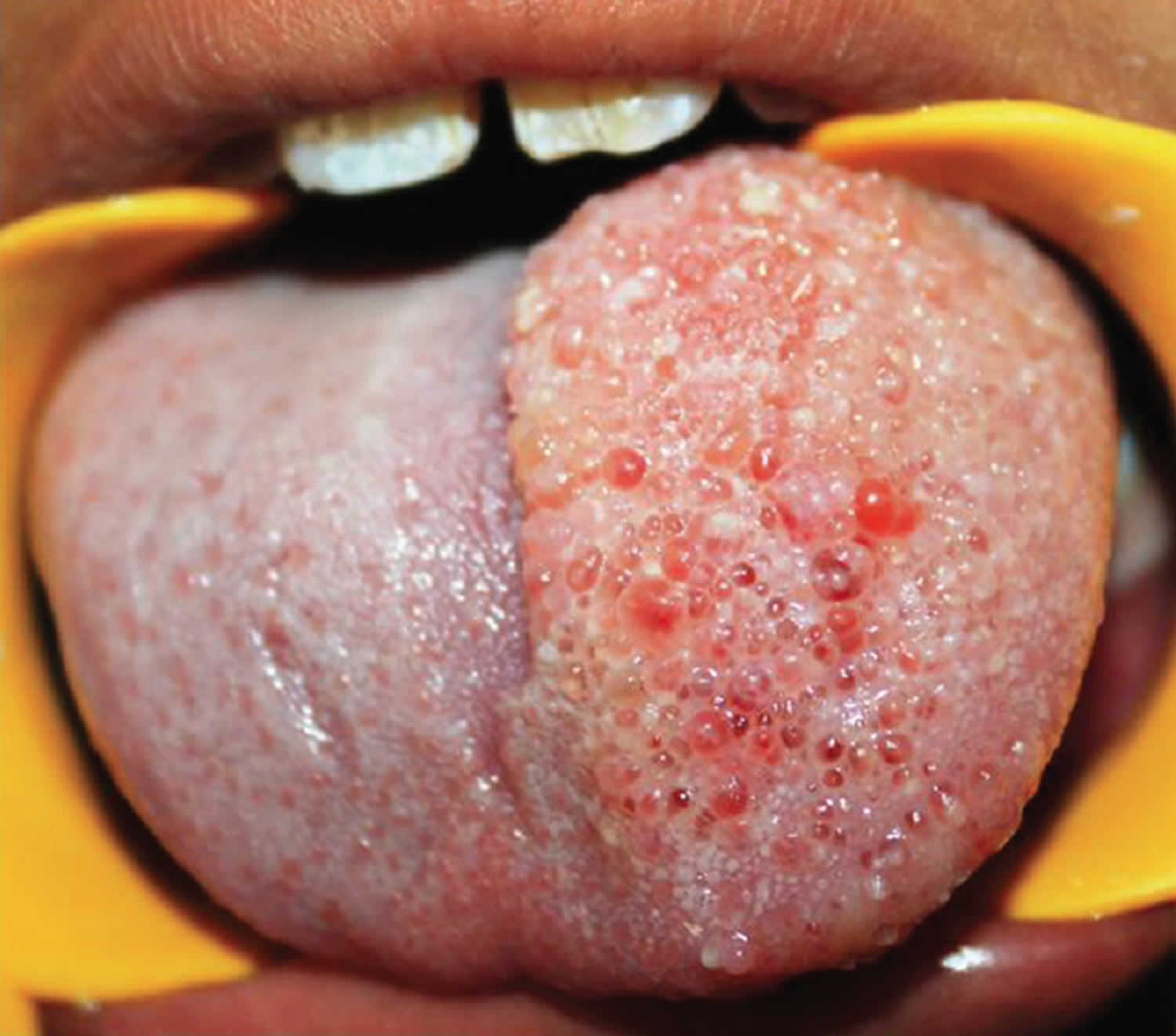
Footnote: An 8-year-old boy reported with a chief complaint of growth on tongue since 6 years. Patient’s mother noticed the growth when the patient was of 2-year-old, which was initially smaller in size and gradually increased to present size. The boy also complains of slurred speech and bleeding from the growth. The boy also reported difficulty in swallowing and chewing due to growth. On examination, multiple papular appearances was seen on the left dorsal half of the tongue of size approximately 4 cm × 3 cm, which was exactly within the midline and not crossing the midline, extending anteriorly from the tip of the tongue to posteriorly 0.5 cm from circumvallate papillae. The surface appears to be irregular and granular. On palpation, growth was soft, nontender, and pebbly. Reduction in the tongue movements without any loss of sensory or motor functions. Diascopy test was carried out which was negative, and there were no palpable pulsations felt. Based on the history and clinical feature a provisional diagnosis of lymphangioma of the tongue was made, and excisional biopsy under general anesthesia was carried out. The excised specimen was sent for histopathological examination and diagnosed as lymphangioma. Based on history, clinical and histological features a final diagnosis of lymphangioma was made.
[Source 2 ]Figure 2. Cavernous lymphangioma of the male breast
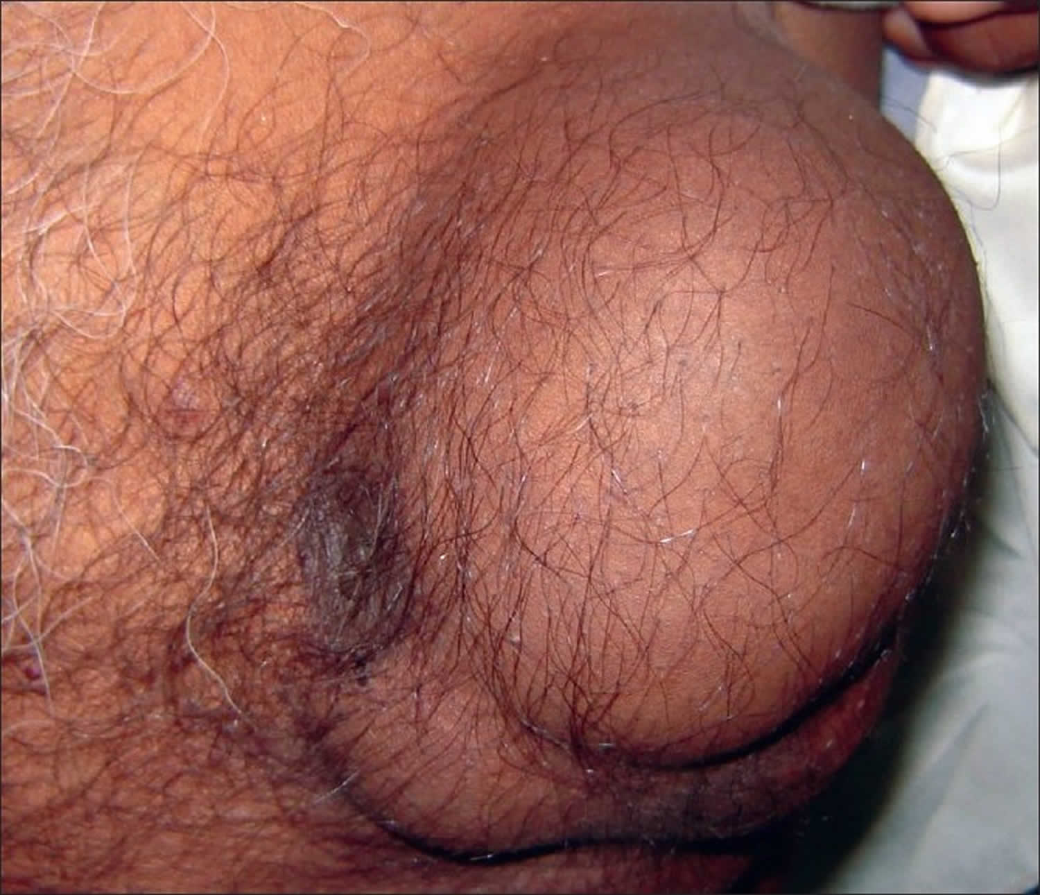
Footnote: 60-year-old male with lymphangioma of the left breast, which was present since childhood and had gradually increased to its present size of 7×7×5 cm. There was no history of nipple discharge. Physical examination revealed a well-circumscribed, firm, and nontender lump located largely in the upper outer quadrant. The nipple was located at the medial end of the lump and was not fixed or retracted. There was no axillary lymphadenopathy. Fine needle aspiration cytology (FNAC) from the breast lesion yielded blood-stained fluid from multiple sites. Smears showed lymphocytes and a few macrophages against a hemorrhagic background. A diagnosis of vascular malformation was suggested. The patient subsequently had excision biopsy of the lump.
[Source 3 ]Lymphangioma circumscriptum
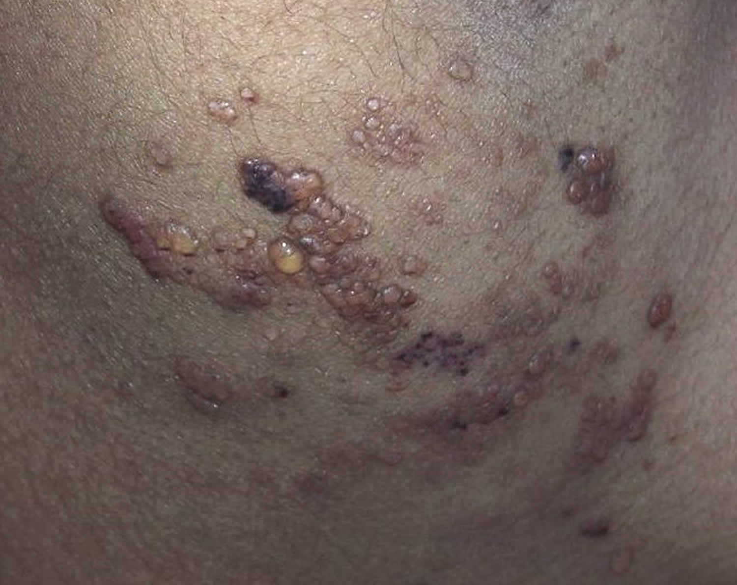
Lymphangioma circumscriptum is a ‘microcytic’ lymphatic malformation. Lymphangioma circumscriptum appears as a cluster of small firm blisters filled with lymph fluid, resembling frog-spawn. Because lymphangioma circumscriptum consist of a combination of blood and lymph elements, purple areas can be seen scattered within the vesicle-like papules. Lymphangioma circumscriptum range in color from clear to pink, dark red, brown or black and may become warty, especially when affecting a palm or sole. Lymphangioma circumscriptum is most commonly found on the shoulders, neck, underarm area, limbs, and in the mouth, especially the tongue. In the genital area, the surface can be verrucous, and the lesions may be mistaken for warts. The acquired form is most often found in the axilla, inguinal, and genital areas and there is often coexisting lymphedema. Associated symptoms may include pruritus, pain, burning, lymphatic drainage, infection, and aesthetic concerns.
Lymphangioma circumscriptum patches may become more prominent at puberty and may ooze or bleed. Occasionally they become infected.
Figure 3. Lymphangioma circumscriptum
Cystic lymphangioma
Cystic lymphangioma is also called cystic hygroma or lymphangioma cysticum, is a ‘macrocytic’ lymphatic malformation, and is composed of large fluid-filled spaces. Cystic lymphangioma appears as a skin colored, red or bluish, somewhat transparent, swelling under the skin. Cystic lymphangioma most often affects the neck, armpits or groin. Very large lesions may involve the chin and face, and occasionally interfere with eating or breathing. Cystic lymphangiomas can become infected, which may be life-threatening if the neck is involved.
Cystic lymphangioma is more common in some congenital disorders including Turner syndrome, hydrops fetalis, Down syndrome and fetal alcohol syndrome. So it may be advisable for babies with this type of lymphatic malformation to have chromosomal analysis performed.
An ultrasound scan is often performed to find out the extent of the abnormality. Characteristically, firm rounded spaces are detected. A cystic lymphangioma is distinguished from venous malformation and hemangioma by the appearance and lack of flow of the fluid.
In more complicated cases it may be necessary to perform Magnetic Resonance Imaging (MRI) or angiography to help plan treatment.
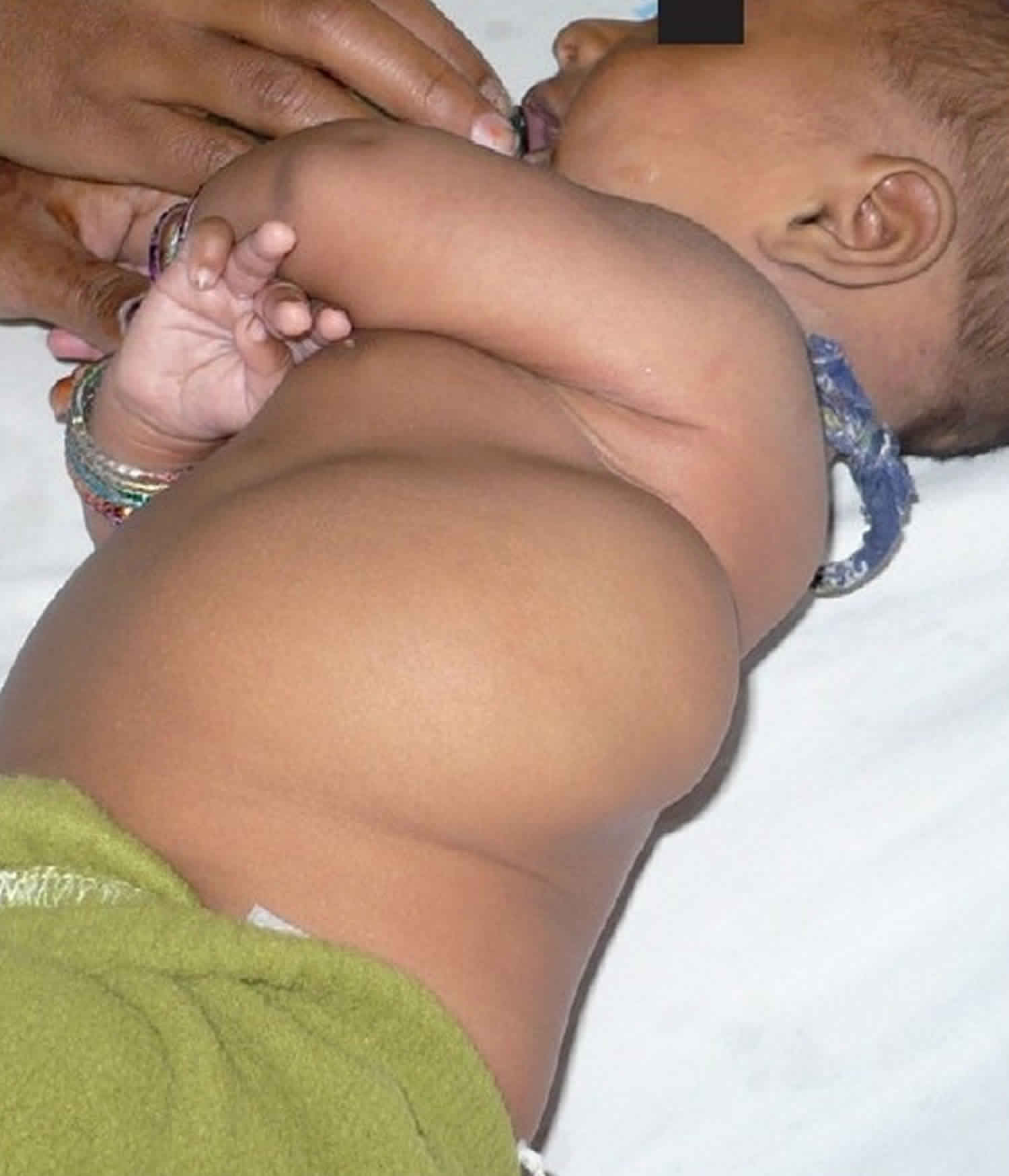
Figure 4. Cystic lymphangioma of lumbar region
Lymphangioma causes
Lymphangiomas result from congenital or acquired abnormalities of the lymphatic system. The congenital form typically occurs before the age of 5 years and is due to improper connection of lymphatic channels to the main lymphatic drainage duct. Acquired lymphangiomas occur as a consequence of any interruption of previously normal lymphatic drainage such as surgery, trauma, malignancy, and radiation therapy. Acquired lymphangiomas are not believed to have malignant potential; however, associated chronic lymphedema places the patient at risk for lymphangiosarcoma, which is an aggressive tumor with a dismal prognosis 1. Cutaneous angiosarcoma has also been reported in massive localized lymphedema in morbidly obese patients. Acquired lymphangiectasia can be painful, and poor lymphatic drainage may lead to bacterial infection.
Congenital (present at birth) lymphangiomas form due to blockage of the lymphatic system during fetal development, though the cause remains unknown 1. Cystic lymphangiomas are associated with genetic disorders, including trisomies 13, 18, and 21, Noonan syndrome, Turner syndrome, and Down syndrome.
Lymphangioma histopathology
On histopathologic examination, lesions of superficial lymphangioma consist of a collection of large lymphatic cisterns lying deep in the subcutaneous plane that communicate via dilated dermal lymphatic channels lined with endothelial cells. The overlying epidermis is usually acanthotic or hyperkeratotic and has an irregular elongation of rete pegs. No atypical vascular features, nuclear atypia, mitotic activity, or koilocytic changes typically exist. A mild to the moderate inflammatory infiltrate may be present. Cystic or cavernous lymphangiomas demonstrate large, irregular vascular spaces lined with a single layer of flattened endothelial cells within a fibroblastic or collagenous stroma, which may contain lymphocytes. Penetration of muscle may be seen.
Lymphangioma diagnosis
In most cases, a clinical diagnosis can be made based on the history and examination findings. As needed, dermoscopy and biopsy can be used to confirm the diagnosis and imaging may be warranted to assess the depth and extent of a lesion.
The dermoscopic examination can be helpful in distinguishing superficial lymphangioma from other cutaneous lesions. Two distinct dermoscopic patterns have been described: yellow lacunae surrounded by pale septa without the inclusion of blood and yellow to pink lacunae alternating with dark-red or bluish lacunae, representing the inclusion of blood. A more recently described dermoscopic finding is a “hypopyon-like” feature which consists of a color transition from dark to light in some lacunae. This occurs due to sedimentation of blood that separates cellular components to the bottom and serum to the upper part of the lacunae.
Lymphangioma treatment
Both superficial and deep lymphangiomas can be difficult to treat. Cystic lymphangiomas and cavernous lymphangiomas are generally removed surgically. Unfortunately, the deeper lesions are quite difficult to remove completely and sometimes regrow. Oral sirolimus (which inhibits angiogenesis) and sildenafil (which causes dilation of blood vessels) are undergoing investigation as a potential medical treatments for severe lymphatic malformations but may have significant adverse effects 5.
Destructive treatments with carbon dioxide (CO2) laser, long-pulsed Nd-YAG laser, and electrosurgery have been reported to improve symptoms 1. Cryotherapy, superficial radiotherapy, and sclerotherapy with 23.4% hypertonic saline are less commonly used modalities. Direct injection of a sclerosing agent, including 1% or 3% sodium tetradecyl sulfate, doxycycline or ethanol, can be made into lymphatic malformations. Compression may reduce swelling caused by lymphedema. Infection prevention is crucial.
Lymphangioma circumscriptum is benign (non-cancerous) and usually requires no treatment. Antibiotics may be given if secondary infection occurs (rare). If necessary it may be removed surgically, by dermabrasion or by laser. Lymphangioma circumscriptum have a tendency to recur after treatment. Treatment of the more deep seated components is difficult as radical surgery is necessary and recurrence is not uncommon.
Lymphangioma surgery
When feasible the treatment of choice for any type of lymphangioma, however, remains surgical excision. Wide local excision of the affected lymphatic channels is necessary as recurrence is common. Recurrence rates for surgical excision of lymphangioma circumscriptum have been reported to be as high 23% in follow-up periods up to 81 months. Surgical success rates are higher for small, superficial lymphangiomas.
References- Miceli A, Stewart KM. Lymphangioma. [Updated 2018 Oct 27]. In: StatPearls [Internet]. Treasure Island (FL): StatPearls Publishing; 2018 Jan-. Available from: https://www.ncbi.nlm.nih.gov/books/NBK470333
- Bhayya H, Pavani D, Avinash Tejasvi M L, Geetha P. Oral lymphangioma: A rare case report. Contemp Clin Dent 2015;6:584-7 http://www.contempclindent.org/text.asp?2015/6/4/584/169851
- Malhotra P, Bansal A, Chintamani, Saxena S. Cavernous lymphangioma of the male breast. Indian J Pathol Microbiol 2010;53:853-4 http://www.ijpmonline.org/text.asp?2010/53/4/853/71997
- Mirza B, Ijaz L, Saleem M, Sharif M, Sheikh A. Cystic hygroma: an overview. J Cutan Aesthet Surg. 2010;3(3):139-44. https://www.ncbi.nlm.nih.gov/pmc/articles/PMC3047730/
- Swetman GL, Berk DR, Vasanawala SS, Feinstein JA, Lane AT, Bruckner AL. Sildenafil for severe lymphatic malformations. N Engl J Med. 2012 Jan 26;366(4):384-6.


