What is mycetoma
Mycetoma is a chronic, granulomatous, subcutaneous, inflammatory disease caused by true fungi (eumycetoma) or filamentous bacteria (actinomycetoma, bacteria that produce filaments, like fungi) 1.
Mycetoma is classified as:
- Eumycetoma also called mycotic mycetoma — when it is caused by a fungus;
- Actinomycetoma (actinomycotic mycetoma) — when it is caused by filamentous bacteria from the order, actinomycetes.
Mycetoma is rare in the United States, but is commonly diagnosed in Africa, Mexico and India. Mycetoma occurs in the mycetoma belt stretching between the latitudes of 15 degrees South and 30 degrees North and is endemic in relatively arid areas 1. The organisms are present in the soil and may enter the subcutaneous tissue by traumatic inoculation. In these countries, it occurs most frequently in farmers, shepherds, and people living in rural areas. Frequent exposure to penetrating wounds by thorns or splinters is a risk factor 2. Mycetoma commonly affects adults aged 20 to 40 years, predominantly males. The foot is most commonly affected. The first symptom of the condition is generally painless swelling beneath the skin, which progresses to a nodule (lump) over several years 3. Eventually, affected people experience massive swelling and hardening of the affected area; skin rupture; and formation of sinus tracts (holes) that discharge pus and grains filled with organisms. Some affected people have no discomfort while others report itching and/or pain 2. Both forms of mycetoma present as a progressive, subcutaneous swelling, although actinomycetoma has a more rapid course. Multiple nodules develop which may suppurate and drain through sinuses, discharging grains during the active phase of the disease.
Eumycetoma is mainly caused by Pseudallescheria boydii (Scedosporium apiospermum), Madurella mycetomatis or Magnaporthe grisea 4. Madurella has been identified in cattle dung in rural East Africa 5. The color of the grains is sometimes helpful in pinpointing the exact etiologic agent. For example, the grains of Madurella mycetomatis or Magnaporthe grisea are typically black, while those of Pseudallescheria boydii and several actinomycetes are usually faint yellowish or white.
Actinomycetoma can be caused by the following:
- Nocardia species
- Actinomadura madurae
- Actinomadura pelletieri
- Streptomyces somaliensis
Consultation with specialists in infectious disease or tropical medicine is advised in areas of the world where mycetoma is unfamiliar. Diagnosis may involve radiology, ultrasonic imaging, cytology, culture, histology or immunodiagnosis. Actinomycetoma is amenable to treatment by antibiotics, preferably by combined drug therapy for long periods. Treatment varies based on the cause of mycetoma and may include antibiotics or antifungal medications 6. Eumycetoma is usually treated by aggressive surgical excision combined with medical treatment 1.
Who gets mycetoma?
The bacteria and fungi causing mycetoma have a worldwide distribution in the soil and plant material, but mycetoma is mostly found in tropical areas in south and south-east Asia, Latin America and Africa. Most cases are reported in the ‘Trans-African mycetoma belt” from western Africa, including Senegal and Sierra Leone, to eastern Africa, including Somalia and Sudan 7.
Mycetoma occurs most often in farmers, shepherds, Bedouins, nomads, and people living in rural areas. Frequent exposure to penetrating wounds by thorns or splinters is a risk factor, especially in combination with contaminated soil material.
Infection usually occurs after local trauma.
- Eumycetoma mainly affects middle-aged men that work as herdsmen or farmers, especially when walking barefoot.
- Mycetoma can also affect younger men and females.
- Actinomycetoma can also be related to oral infections, teeth or gum disease.
Mycetoma has no known vector or animal reservoir, and there are no reports of infections transmitted from person to person 8.
What causes mycetoma?
The causative agent can be suspected from the color of the grains.
Eumycetoma
- Dark or black grains
- Madurella mycetomatis
- Trematosphaeria grisea (formerly Madurella grisea)
- Exophiala jeanselmei
- Medicopsis romeroi (formerly Pyrenochaeta romeroi)
- Falciformispora senegalensis (formerly Leptosphaeria senegalensis)
- Falciformispora thompkinsii
- Curvularia lunata
- White, pail or unstained grains
- Acremonium spp.
- Fusarium spp
- Neotestudina rosatii
- Aspergillus nidulans
- Aspergillus flavus
- Microsporum ferrugineum
- Microsporum audouinii
- Microsporum langeronii
- Scedosporium apiospermum
- Scedosporium boydii (formerly Pseudallescheria boydii)
Actinomycetoma
- White to yellow grains
- Nocardia brasiliensis
- Nocardia otitidiscaviarum (formerly Nocardia caviae)
- Actinomadura madurae
- Pleurostomophora ochracea
- Brown grains
- Streptomcyes somaliensis
- Red to pink grains
- Actinomadura pelletieri
Mycetoma symptoms
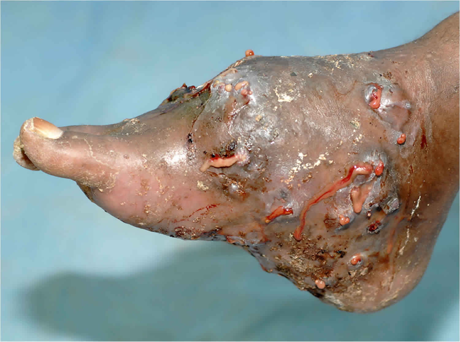
The classic clinical triad of mycetoma is tumor or soft tissue swelling, sinus tracts, and characteristic macroscopic grains 9. The grains typically represent aggregates of the infecting organisms. The earliest sign is often painless subcutaneous swelling. Some patients may give a history of a penetrating injury at the site of involvement.
Several years later, a painless subcutaneous nodule is observed. After some years, massive swelling of the area occurs, with induration, skin rupture, and sinus tract formation.
As the infection spreads to contiguous body parts, old sinuses close and new ones open.
Nearly 20% of patients with mycetoma experience associated pain, usually due to secondary bacterial infection or, less commonly, bone invasion.
Constitutional symptoms and signs of mycetoma are rare.
Patients may report a deep itching sensation.
Irrespective of the causal agent, the appearance of the mycetoma lesion is consistent, as follows:
- Initially, subcutaneous swelling is present.
- In a later phase, a subcutaneous nodule develops.
- Eventually, massive swelling with induration, rupture of the skin, and formation of sinus tracts occur.
In general, eumycetoma is more circumscribed and progresses slower than actinomycetoma.
Regional lymphadenopathy is unusual; when it does occur, it is due to one of the following:
- Lymphatic spread of mycetoma to regional nodes occurs in only 1-3% of affected patients.
- Secondary bacterial infection or a local immunologic reaction may enlarge the regional lymph nodes.
Lymphatic obstruction and fibrosis can cause lymphedema and erythema.
Pulmonary mycetoma has been found to develop and progress more rapidly in individuals infected with HIV.
How is mycetoma diagnosed?
Mycetoma is suspected when there is a typical triad of symptoms and signs:
- A painless firm subcutaneous mass
- Multiple sinus tracts
- A purulent or sero-purulent discharge that contains grains.
Dermoscopy may be useful for early stages of the disease detecting grains within skin lesions 10. Grains are conglomerates of fungi or bacteria disposed radially (like sun rays).
Tissue for microscopy and culture is collected by:
- Making a small incision and expressing the secretions;
- Using a needle and syringe to withdraw fluid.
The color of the grains can narrow the options of the causative bacterium or fungus. The grains are treated with potassium hydroxide (KOH) and then stained with Gram and PAS stains. On microscopy:
- Actinomycotic grains have very fine filaments
- Eumycotic grains contain thicker, branched filaments.
As some are slow-growing organisms, cultures at 25–30°C and 37°C are set for up to 6 weeks for both bacteria and fungi 11. When it is difficult to identify the responsible organism, molecular techniques such as PCR (polymerase chain reaction) can be used 7.
Skin biopsy may show typical histopathological features of mycetoma.
Imaging can be used to assess the depth of infection 12.
- Plain X-ray
- Ultrasound scan
- Computed tomography (CT)
- Nuclear magnetic resonance imaging (MRI).
Mycetoma treatment
Mycetoma cannot be cured without active treatment. In the treatment of mycetoma, antibiotic or antifungal therapy should be attempted first and may need to be combined with surgery 13, especially for eumycetoma lesions in the extremities 14. Mycetoma requires antibiotics (for actinomycetomas) or oral antifungals (for eumycetomas) for weeks, months or years. Damage to subcutaneous tissues (muscle, bones, joints or tendons) often persists, so local surgery (including amputation) and physiotherapy may be required 8. External beam radiotherapy in doses ranging from 3.5-14 Gy has been considered successful treatment in a few selected cases 15.
Actinomycetoma (single or in combination)
- Trimethoprim + sulphamethoxazole
- Amoxicillin + clavulanic acid
- Amikacin or gentamicin
- Doxycycline or minocycline
- Penicillin
- Dapsone
- Streptomycin
- Rifampicin
Eumycetoma (only one usually used)
- Ketoconazole
- Itraconazole
- Terbinafine
Surgery
Surgery is recommended for localized mycetoma lesions that can be excised completely without residual disability. Surgical reduction of large lesions can improve the patient’s response to medical treatment 16. However, partial surgical resection without subsequent use of appropriate antimicrobial or antifungal agents is prone to failure.
Mycetoma prognosis
Despite the need for prolonged treatment, prognosis is usually good with proper therapy. The longer the infection has been present prior to treatment, the more likely complications and disability will occur 17.
References- A.H. Fahal, Mycetoma: a thorn in the flesh, Transactions of The Royal Society of Tropical Medicine and Hygiene, Volume 98, Issue 1, January 2004, Pages 3–11, https://doi.org/10.1016/S0035-9203(03)00009-9
- Mycetoma. https://emedicine.medscape.com/article/211459-overview
- Mycetoma. https://rarediseases.info.nih.gov/diseases/3862/mycetoma
- Ahmed AO, van Leeuwen W, Fahal A, et al. Mycetoma caused by Madurella mycetomatis: a neglected infectious burden. Lancet Infect Dis. 2004 Sep. 4(9):566-74.
- de Hoog GS, Ahmed SA, Najafzadeh MJ, Sutton DA, Keisari MS, Fahal AH, et al. Phylogenetic findings suggest possible new habitat and routes of infection of human eumyctoma. PLoS Negl Trop Dis. 2013 May. 7(5):e2229
- Mycetoma. https://www.dermnetnz.org/topics/mycetoma
- Nenoff P, van de Sande W, Fahal A, et al. Eumycetoma and actinomycetoma – an update on causative agents, epidemiology, pathogenesis, diagnostics and therapy. J Eur Acad Dermatol Venereol 2015; 29: 1873 – 83. DOI: 10.1111/jdv.13008
- Zijlstra E, van de Sande W, Welsh O, et al. Mycetoma: a unique neglected tropical disease. Lancet Infect Dis 2016; 16: 100–12.
- Mycetoma Clinical Presentation. https://emedicine.medscape.com/article/211459-clinical
- Reis L, Lima B, Zillo F, et al. Dermoscopy assisting the diagnose of a mycetoma: case report and literature review. An Bras Dermatol 2014; 89: 832 – 3. DOI: 10.1590/abd1806-4841.20143008
- Van de Sande W, Fahal A, Goodfellow M, et al. Merits and pitfalls of currently used diagnostic tools in mycetoma. PLoS Negl Trop Dis 2014; 8(7): e2918. DOI: 10.1371/journal.pntd.0002918
- Van de Sande W, Fahal A, Goodfellow M, et al. Merits and pitfalls of currently used diagnostic tools in mycetoma. PLoS Negl Trop Dis 2014; 8(7): e2918. DOI: 10.1371/journal.pntd.0002918.
- Venkatswami S, Sankarasubramanian A, Subramanyam S. The madura foot: looking deep. Int J Low Extrem Wounds. 2012 Mar. 11(1):31-42.
- Ahmed AA, van de Sande WW, Fahal A, et al. Management of mycetoma: major challenge in tropical mycoses with limited international recognition. Curr Opin Infect Dis. 2007 Apr. 20(2):146-51.
- Falkson C, Sur R, Pacella J. External beam radiotherapy: a treatment option for massive haemoptysis caused by mycetoma. Clin Oncol (R Coll Radiol). 2002 Jun. 14(3):233-5.
- Maina AM, Macharia JT. Alleviating a Nomad’s Anguish: Successful Treatment of a Case of Leg Mycetoma-A Case Report. Case Rep Orthop. 2012. 2012:753174.
- Wadal A, Elhassan T, Zein H, et al. Predictors of post-operative mycetoma recurrence using machine-learning algorithms: the mycetoma research center experience. PLoS Negl Trop Dis 2016; 10: e0005007. DOI: 10.1371/journal.pntd.0005007


