What is ochronosis
Ochronosis is the bluish-black or grey-blue discoloration of the skin, especially the ear cartilage, the ocular (eye) tissue, and other body locations 1. This blue-black pigmentation usually appears after age 30. Ochronosis can occurs in individuals with alkaptonuria, an inherited metabolic disorder or because of exposure to various substances such as phenol, trinitrophenol, resorcinol, mercury, picric acid, benzene, hydroquinone, and antimalarials. Ochronosis typically occurs in adults, but has been reported in children. Ochronosis associated with alkaptonuria is caused by a mutation in the HGD gene, which results in the accumulation and deposition of homogentisic acid (HGA) in cartilage 2. The affected tissue becomes weak and brittle with time, leading to chronic inflammation, joint pain, and osteoarthritis.
Alkaptonuria is a rare inherited metabolic disorder with a prevalence of 1 case per 1 million population. Alkaptonuria occurs worldwide, with the highest frequency seen in Slovakia and Dominican Republic, in which the prevalence approaches 1 case per 19,000 inhabitants 3. Alkaptonuria is caused by deficiency of homogentisic acid oxidase (HGO), the only enzyme capable of catabolizing homogentisic acid (HGA). Alkaptonuria features a defect in the biochemical pathway by which phenylalanine and tyrosine are normally degraded into fumaric and acetoacetic acid. Alkaptonuria genetic defect is inherited in an autosomal recessive manner and is mapped to the HGO gene on arm 3q1, and 18 genetic missense mutations are known to cause homogentisic acid oxidase aberrations 4. This deficiency results in accumulation and deposition of homogentisic acid (HGA) in cartilage, causing the characteristic diffuse bluish black pigmentation. Interleukin 6 (IL-6) has demonstrated involvement in the pigmentation process of chondrocytes 5. These affected connective tissues become weak and brittle with time, leading to chronic inflammation, degeneration, and osteoarthritis.
People with alkaptonuria typically develop arthritis, particularly in the spine and large joints, beginning in early adulthood. Other features of this condition can include heart problems, kidney stones, and prostate stones.
Alkaptonuria can be diagnosed based on the symptoms, laboratory testing, and genetic testing. People with alkaptonuria their urine turn black when exposed to air. Alkaptonuria is present at birth and is often diagnosed by discoloration of the diapers. Alkaptonuria is often recognized at birth when parents note discoloration of the urine. However, up to 25% of patients with alkaptonuria do not have the characteristic dark urine staining, and many patients remain undiagnosed until adulthood. Nevertheless, many patients with this metabolic disorder are symptomless until ochronotic changes occur with bluish black pigmented patches in the sclera developing in patients aged 30-39 years 6. These ocular discolorations are located between the corneal margin and the inner canthus.
The fourth decade often marks the onset of thickening and blue-black or gray-blue discoloration of the ear cartilage. Other body locations that frequently display the alteration in skin hue are the eyelids, the forehead, the cheeks, the axillae, the genital region, the nail beds, the buccal mucosa, the larynx, the tympanic eardrum, and the tendons (most easily demonstrated by the patient making a fist).
Ochronotic arthropathy develops later with arthritic symptoms 7.
There is no specific treatment for ochronosis associated with alkaptonuria and treatment is based on the symptoms 2.
Exogenous ochronosis is when ochronosis is due to exposure to substances and the condition is not inherited 8. Exogenous ochronosis is associated with malarial drugs, skin-lightening creams and over-exposure to the sun. Other than the skin findings, there are no other health affects. Exogenous ochronosis is difficult to diagnose. There is no specific treatment for exogenous ochronosis. Several different treatment options are available including prescription skin creams, vitamins, and laser treatments 9.
Ochronosis causes
Alkaptonuria is a genetic disorder related to a deficiency of homogentisic acid oxidase 10.
Ochronotic pigmentation can develop from medications. Similar skin and cartilage alterations can be induced by quinacrine administration and at sites of quinine injections 11. Quinines directly inhibit homogentisic acid oxidase. Carbolic acid topical applications to cutaneous ulcers have also induced ochronotic skin alterations.
Exogenous ochronosis has been reported with topical applications of phenol and hydroquinones to the skin 12. In the case of hydroquinone, it is reported that 35% of black Africans exhibit ochronotic skin changes when using a 6-8% hydroquinone preparation over a prolonged period. Indeed, the prevalence among users of these skin lighteners has been stated to be 69% in a South African study. In African Americans, this cutaneous adverse effect of hydroquinones has also been reported, even when using 2% hydroquinone products. With exogenous ochronosis, the arthropathy seen with alkaptonuria does not occur.
Ochronosis symptoms
Most patients don’t have any symptoms throughout childhood or early adult life and it is not until they reach their 40’s that other signs of the disease start appearing. One of the earliest signs is thickening of the ear cartilage (the pinna feels noticeably thickened and flexible). In addition the skin turns a blue-black color. Earwax is often reddish-brown or jet-black. Gradually patients will suffer sore joints, leading to arthropathy (joint disease characterized by swelling and enlarged bones). Many body parts become affected due to the build-up of pigment deposits in bones and cartilage.
- Bones and cartilage of the lower back, knees, shoulders and hips are most affected. Firstly patients suffer low back pain with stiffness, followed by knee, shoulder and hip pain over the next 10 years. Cartilage becomes brittle and can break apart easily. In some cases this leads to spinal injuries such as prolapsed intervertebral discs.
- Deposits around the trachea (windpipe), larynx (voice box) and bronchi (air passages to the lungs) may cause shortness of breath and difficulty breathing.
- Deposits around the heart and blood vessels can calcify (harden) and lead to atherosclerotic plaques (hard spots in arteries).
- Pigmentation of the sclera of the eye usually occurs early on. This does not affect vision but appears as brown or grey deposits on the surface of the eye.
- Skin color changes are most apparent on areas exposed to the sun and where sweat glands are found. Areas most affected include the cheeks, forehead, armpits and genital regions. The skin takes on a blue-black speckled discoloration. Sweat produced has been found to stain clothes. Sometimes nails can be affected and turn a distinct brown color.
Ochronosis diagnosis
Most laboratory testing for alkaptonuria detects the alterations in the urine. Increased urinary levels of homogentisic acid (HGA) are characteristic of this metabolic disorder.
Elevated levels of HGA in the urine, blood, and other tissues can be determined by specific enzymatic and colorimetric tests, direct spectrophotometric methods, high-performance liquid chromatographic testing, and molecular techniques.
Other simple urinary studies include darkening of urine with the addition of sodium hydroxide, black reaction with FeCl3, and blackening of photographic emulsion paper with alkali added to urine.
Imaging Studies
In patients with ochronotic arthropathy, radiography and MRI help identify characteristic and diagnostic features, including articular space narrowing up to osseous ankylosis, calcifications, osteophytosis, and reactive sclerosis of the articular surfaces 13. Bone scintigraphy can be useful in evaluation, correlation with the clinical course, and follow-up of such patients 14.
Skin biopsy
Skin biopsy samples with hematoxylin and eosin staining reveal yellowish brown – pigmented bodies in the dermis that represent altered widened elastic fibers, as well as in macrophages, endothelial cells, apocrine glands, and epidermal basement membranes. The deposits do not lose their pigmentation after 3 days in 10% hydrogen peroxide. Furthermore, the ochronotic pigment reacts with all routine stains for melanin. Such deposits can also be seen in cartilage and elastic tissue.
Exogenous ochronosis reveals ochronotic collagen fibers leading to the formation of ochronotic colloid milium 15. The dermal cell infiltrate is variable but often granulomatous. Transfollicular elimination of these ochronotic fibers has been reported.
Experimental evidence from 2016 has shown the presence of serum amyloid A (SAA) in several alkaptonuric chondrocytes, classifying alkaptonuria as a secondary amyloidosis. SAA in alkaptonuric chondrocytes has been shown to localize to actin, vimentin, and β-tubulin cytoskeletal proteins 16.
Other Tests
Synovial fluid examination of affected joints shows characteristic frequent pigmented fibrillar connective tissue, which are golden-brown with microscopy, while being black on gross examination 17.
Arthroscopy can be used in diagnosing cases of ochronotic arthropathy 18.
Ochronosis treatment
Alkaptonuria is a lifelong disease. There is no cure for the condition. If alkaptonuria is diagnosed early on in life it is reasonable for patients to have a low-protein diet. This reduces the intake of amino acids phenylalanine and tyrosine, which in turn reduces the amount of homogentisic acid produced. Although not proven, this could potentially avoid or minimize complications later in life.
Vitamin C has been found to slow down the conversion of homogentisic acid to the polymeric deposits in cartilage and bone. A dose of up to 1g/day is recommended for older children and adults. Nitisinone, an enzyme inhibitor that mediates the formation of homogentisic acid is being used in restricted experimental treatments 19.
Vitamin E and N-acetyl cysteine have been examined as novel potential therapies to prevent damage to articular cartilage 20.
Life expectancy is normal although patients may be at increased risk of heart conditions and may require surgical treatments for spine, hip, knee and shoulder joint problems.
Exogenous cutaneous ochronosis has been successfully treated by laser.
Ochronosis prognosis
Patients with alkaptonuria can expect a normal life span; nevertheless, the complications of debilitating arthritis, cardiovascular compromise, and ochronotic skin alterations will occur. Additionally, chronic kidney disease affects the natural history of the disease by accelerating the onset of major complications. [5]
Mortality/morbidity
With the absence of homogentisic acid (HGA) oxidase in liver and kidney cells, HGA accumulates. The black urine of patients with alkaptonuria results from renal excretion of homogentisic acid (HGA), while ochronotic pigment is a sequela of HGA accumulation in the connective tissues of individuals who are affected.
In alkaptonuria, the accumulation of HGA inhibits collagen cross-linking by affecting a crucial enzyme in collagen synthesis, leading to a diminution of structural collagen integrity. This results in ochronotic arthropathy, which occurs in men aged in their fourth and fifth decades; women develop similar complications in their sixth decade. The larger joints are most affected with early calcification, narrowing, and collapse of the intervertebral discs. In addition to joint disease, reports suggest an increased incidence of cardiovascular disease due to cartilaginous changes of vessel walls.
Homogentisic acid oxidase requires atmospheric oxygen, ferrous ion, and sulfhydryl groups for normal function, and the enzyme is inhibited by quinones. HGA is colorless in solution but darkens on exposure to air, especially in the presence of alkali. Individuals with acidic urine may not demonstrate the very dark-colored urine characteristic of this condition.
Exogenous ochronosis
Exogenous ochronosis is when ochronosis is due to exposure to substances and the condition is not inherited 8. Exogenous ochronosis is associated with malarial drugs, skin-lightening creams and over-exposure to the sun. Other than the skin findings, there are no other health affects. Exogenous ochronosis is more typically seen in African and Afro-Caribbean populations due to the use of skin-bleaching products containing hydroquinone in attempts to lighten the appearance of the skin 21. In fact, the largest series of cases was from South Africa 22. Exogenous ochronosis was described in 28–35% of black population. It was then thought that the condition is reported exclusively among dark-skinned African individuals. However, recently published reports show that exogenous ochronosis is also seen in many fair-skinned individuals such as Europeans and Hispanics 23. Exogenous ochronosis is reported correspondingly low in the USA 24. The exact epidemiological figures are not available except for the US, which mentions an incidence of 22 cases in more than 50 years 25.
The incidence of exogenous ochronosis is said to be low in Asians, but cases are increasingly being reported from India 26, China 27, Thailand 28, Singapore 29. In a recent case series from Singapore, Tan described 15 new cases in 5 year duration 27. This indicates that exogenous ochronosis may occur across a wide spectrum of skin types, from type II or III to type V, and thus, is not limited to darker skin types 30. Recent increasing reports might be due to increased awareness of the condition.
Exogenous ochronosis is difficult to diagnose. The clinical diagnosis may be missed in early stages where it may mimic melasma. The diagnosis may not be confirmed in a number of patients due to lack of skin biopsy findings. This might have led to under-reporting of the cases of exogenous ochronosis 8. Thus, the actual incidence of exogenous ochronosis might be much higher than that reflected in the global literature.
Exogenous ochronosis causes
Various factors are necessary for the development of exogenous ochronosis are unprotected prolonged sun-exposure, prolonged use of skin-lightening agents mainly containing hydroquinone and, presence of a number of viable melanocytes, as suggested by Hull and Procter 31. Outdoor occupation, application of hydroquinone over a larger area or whole body and/or applying hydroquinone in large quantity may also predispose to this condition.
exogenous ochronosis has been reported following long-term application of skin-lightening creams which contains hydroquinone, phenol or resorcinol. Of these, hydroquinone has the strongest association and was described by Findlay et al. 32 in 1975. The condition is due to continual use of hydroquinone, generally, in higher concentrations more than 2%. An alcoholic solution containing hydroquinone preparations are said to more readily predispose to exogenous ochronosis than creams 33.
Some of the recent reports mention the occurrence of exogenous ochronosis even with as low concentrations of hydroquinone as 2% 34.
The lesions may develop gradually over 6 months to 3 years or longer 32. Two recent case reports from India mention the occurrence of exogenous ochronosis with use of 2% hydroquinone preparations for 7–8 years 35. All these reports, unsupervised usage of skin-lightening agents containing hydroquinone was a common highlighting feature.
Exogenous ochronosis symptoms
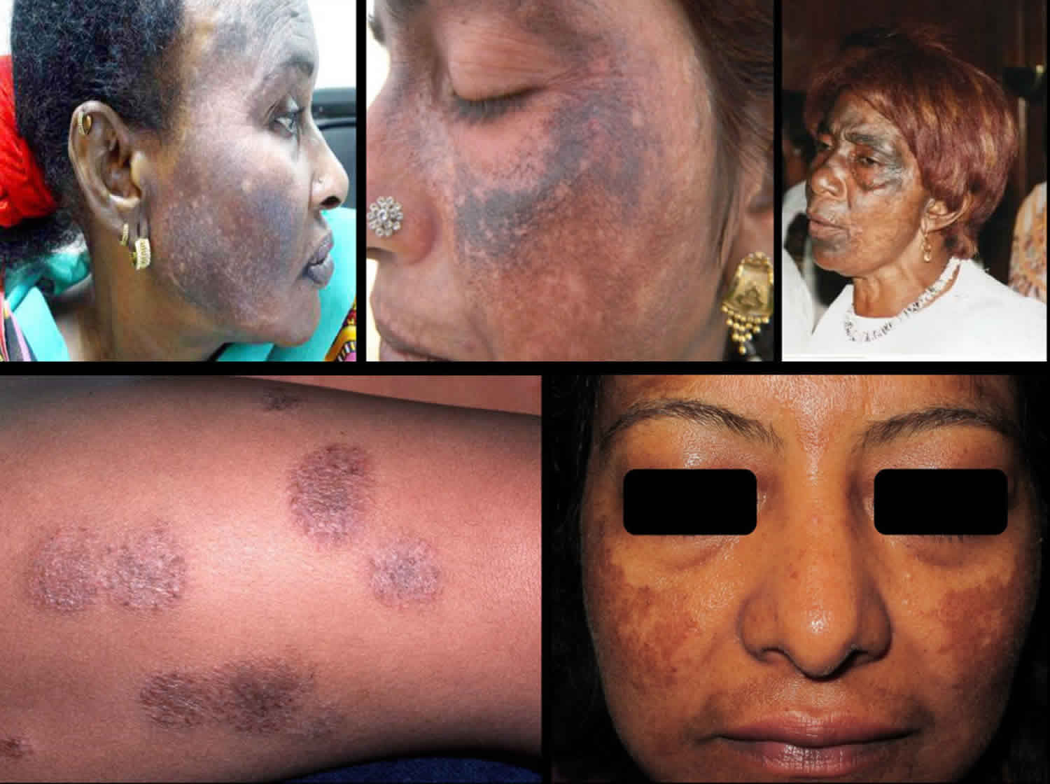
Exogenous ochronosis manifests as hyper-pigmentation in the photo-exposed regions 36. It occurs over osseous surfaces 37 often affecting the zygomatic 38 regions in a symmetrical pattern. The lesions are gray-brown or blue-black macules usually with hyperchromic, pinpoint, and caviar-like papules 32.
The most initial clinical classification was suggested in 1979 by Dogliotti and Leibowitz 39. They described exogenous ochronosis in three clinical stages as follows:
- Stage 1 – Erythema and mild hyper-pigmentation: This stage might be clinically indistinguishable from melasma
- Stage 2 – Progressive hyper-pigmentation pigmented colloid milium (caviar-like lesions) and scanty atrophy
- Stage 3 – Papulonodular (sarcoid-like) lesions. In 1986, Phillips et al. 40 graded and described the condition as:
- Mild – Coarsening and darkening of the skin
- Moderate – Large black papules; skin in between papules is of normal pigmentation
- Severe – Larger, coalescing, caviar-like papules, which are relatively dark in color.
In 1989, Hardwick et al. 41 in their epidemiological study based on clinical diagnosis graded exogenous ochronosis as:
- Grade 1 – Faint macular sooty pigmentation
- Grade 2 – Distinct macular stippling/small papules
- Grade 3 – Dark deposits and papules
- Grade 4 – Colloid milia (1 mm and greater)
- Grade 5 – Keloid-like nodules and cysts.
In 1991, Jordaan and Van Niekerk 37 described two extremes of exogenous ochronosis. The milder variant characterized by coarsening and darkening of skin and the severe one with caviar-like black papules and skin atrophy.
Exogenous ochronosis diagnosis
Histopathology, although invasive, remains a gold standard in the diagnosis of exogenous ochronosis, and confirms it beyond doubt. The pathognomic histopathological feature is the presence of the ochre-colored, banana-shaped fibers in the dermis. Homogenization and swelling of collagen bundles in the papillary and reticular dermis may also be seen 27. Speckled macular lesions or early caviar-like papules are preferred lesions for a skin biopsy to yield more accuracy.
Exogenous ochronosis treatment
Exogenous ochronosis has remained a very difficult condition to treat. Despite several modalities of treatments available, the results are often inconsistent, unpredictable and are often far from gratifying.
There are various modalities available for treatment as follows.
Nonpharmacological measures
Most important step is to stop the further use of the offending agent. Avid sun-protection is said to be a help in the prevention of further progress of the condition to some extent. Wide-brim hats, sun-goggles and appropriate sun-protective clothing are equally important.
Pharmacological treatments
Physical sunblocks and chemical sunscreens widely help in clinical improvement of the skin lesions of exogenous ochronosis, especially when combined with the other pharmacological and procedural approaches 42. Topical retinoid acid, glycolic acid, and a topical corticosteroid (low-potency creams) used judiciously often lead to considerable improvement in pigmentation 42. A solitary case report suggested the efficacy of tetracycline in the clearance of papular sarcoid-like ochronosis 43. Antioxidants, high doses of Vitamin E and C, may assist dilution of the pigment. Both Vitamin C and Vitamin E act as de-pigmentating agents. Both act synergistically along with antioxidants to provide photoprotection 44.
Procedural treatments
Chemical peeling with glycolic acid or tricarboxylic acid has been used for the treatment of exogenous ochronosis shows improvement in pigmentation 42. Few patients were treated with derma-abrasion 42. One case report mentions combination treatment of derma-abrasion and CO2 laser showing improvement 38.
In one study done, two patients with exogenous ochronosis were treated with a Q-switched 755-nm alexandrite laser at fluence of 6–8 J/cm² showed progressive lightening of pigmentation of the lesional skin and decreased dermal pigmentation on histological examination 45. Q-switched alexandrite laser is believed to enhance the clearance of ochronotic pigment fibers from dermis. The Q-switched ruby laser was also used in another study which showed improvement. Kramer et al. 46 treated exogenous ochronosis in a Hispanic woman with Q-switched 1064-nm neodymium-doped yttrium aluminium garnet (Nd: YAG) laser with satisfactory results. In another study, histologically proven cases of exogenous ochronosis were treated with Q-switched Nd: YAG laser which showed satisfactory improvement in dyschromia 47.
References- Ochronosis. https://emedicine.medscape.com/article/1104184-overview
- Alkaptonuria. https://rarediseases.org/rare-diseases/alkaptonuria
- Zatkova A. An update on molecular genetics of Alkaptonuria (AKU). J Inherit Metab Dis. 2011 Dec. 34 (6):1127-36.
- Felbor U, Mutsch Y, Grehn F, Müller CR, Kress W. Ocular ochronosis in alkaptonuria patients carrying mutations in the homogentisate 1,2-dioxygenase gene. Br J Ophthalmol. 1999 Jun. 83(6):680-3.
- Mistry JB, Jackson DJ, Bukhari M, Taylor AM. A role for interleukins in ochronosis in a chondrocyte in vitro model of alkaptonuria. Clin Rheumatol. 2016 Jul. 35 (7):1849-56.
- Ranganath LR, Cox TF. Natural history of alkaptonuria revisited: analyses based on scoring systems. J Inherit Metab Dis. 2011 Dec. 34 (6):1141-51.
- Zhao BH, Chen BC, Shao de C, Zhang Q. Osteoarthritis? Ochronotic arthritis! A case study and review of the literature. Knee Surg Sports Traumatol Arthrosc. 2009 Jul. 17(7):778-81.
- Bhattar PA, Zawar VP, Godse KV, Patil SP, Nadkarni NJ, Gautam MM. Exogenous Ochronosis. Indian J Dermatol. 2015;60(6):537–543. doi:10.4103/0019-5154.169122 https://www.ncbi.nlm.nih.gov/pmc/articles/PMC4681189
- Acquired hyperpigmentation disorders. https://www.uptodate.com/contents/acquired-hyperpigmentation-disorders
- Vilboux T, Kayser M, Introne W, et al. Mutation spectrum of homogentisic acid oxidase (HGO) in alkaptonuria. Hum Mutat. 2009 Dec. 30(12):1611-9
- Bruce S, Tschen JA, Chow D. Exogenous ochronosis resulting from quinine injections. J Am Acad Dermatol. 1986 Aug. 15(2 Pt 2):357-61.
- Bongiorno MR, Aricò M. Exogenous ochronosis and striae atrophicae following the use of bleaching creams. Int J Dermatol. 2005 Feb. 44(2):112-5.
- Perrone A, Impara L, Bruni A, Primicerio P, Marini M. Radiographic and MRI findings in ochronosis. Radiol Med. 2005 Oct. 110(4):349-58.
- Cortes Hernandez J, Ruiz-Oliva Ruiz F, Alonso Colmenares JI, Alvarez Ruiz S, Caton Santaren B, Alcorta Armentia MP. [Ochronotic arthropathy: the value of bone scintigraphy in alkaptonuria]. Rev Esp Med Nucl. 2004 May-Jun. 23(3):189-92.
- Gonul M, Cakmak SK, Kilic A, Gul U, Heper AO. Pigmented coalescing papules on the dorsa of the hands: pigmented colloid milium associated with exogenous ochronosis. J Dermatol. 2006 Apr. 33(4):287-90.
- Geminiani M, Gambassi S, Millucci L, Lupetti P, Collodel G, Mazzi L, et al. Cytoskeleton Aberrations in Alkaptonuric Chondrocytes. J Cell Physiol. 2016 Jul 25. 9999:1-11.
- Bhangle S, Panush RS, Berman E, Schumacher HR. Clinical images: Synovial fluid clues to ochronosis. Arthitis & Rheumatism. Feb 2012. 64:473-473.
- Gil JA, Wawrzynski J, Waryasz GR. Orthopedic Manifestations of Ochronosis: Pathophysiology, Presentation, Diagnosis, and Management. Am J Med. 2016 May. 129 (5):536.e1-6
- Suwannarat P, O’Brien K, Perry MB, Sebring N, Bernardini I, Kaiser-Kupfer MI, et al. Use of nitisinone in patients with alkaptonuria. Metabolism. 2005 Jun. 54(6):719-28
- Gil JA, Wawrzynski J, Waryasz GR. Orthopedic Manifestations of Ochronosis: Pathophysiology, Presentation, Diagnosis, and Management. Am J Med. 2016 May. 129 (5):536.e1-6.
- Benn EK, Alexis A, Mohamed N, Wang YH, Khan IA, Liu B. Skin Bleaching and Dermatologic Health of African and Afro-Caribbean Populations in the US: New Directions for Methodologically Rigorous, Multidisciplinary, and Culturally Sensitive Research. Dermatol Ther (Heidelb). 2016 Dec. 6 (4):453-459.
- DeCaprio AP. The toxicology of hydroquinone – Relevance to occupational and environmental exposure. Crit Rev Toxicol. 1999;29:283–330.
- Gil I, Segura S, Martínez-Escala E, Lloreta J, Puig S, Vélez M, et al. Dermoscopic and reflectance confocal microscopic features of exogenous ochronosis. Arch Dermatol. 2010;146:1021–5.
- Snider RL, Thiers BH. Exogenous ochronosis. J Am Acad Dermatol. 1993;28:662–4.
- Levitt J. The safety of hydroquinone: A dermatologist’s response to the 2006 Federal Register. J Am Acad Dermatol. 2007;57:854–72.
- Jain A, Pai SB, Shenoi SD. Exogenous ochronosis. Indian J Dermatol Venereol Leprol. 2013;79:522–3
- Tan SK. Exogenous ochronosis in ethnic Chinese Asians: A clinicopathological study, diagnosis and treatment. J Eur Acad Dermatol Venereol. 2011;25:842–50.
- Kullavanijaya P, Ophaswongse S, Silprachawong S. Exogenous ochronosis and pigmented colloid milium induced by bleaching skin cream. Environ Dermatol. 1998;5:20–5.
- Tan SK. Exogenous ochronosis – A diagnostic challenge. J Cosmet Dermatol. 2010;9:313–7
- Kadurina M, Dimitrov B, Tonev S. Hydroquinone-induced exogenous ochronosis. J Plast Dermatol. 2009;5:197–201.
- Hull PR, Procter PR. The melanocyte: An essential link in hydroquinone-induced ochronosis. J Am Acad Dermatol. 1990;22:529–31.
- Findlay GH, Morrison JG, Simson IW. Exogenous ochronosis and pigmented colloid milium from hydroquinone bleaching creams. Br J Dermatol. 1975;93:613–22.
- Levin CY, Maibach H. Exogenous ochronosis. An update on clinical features, causative agents and treatment options. Am J Clin Dermatol. 2001;2:213–7
- Martins VM, Sousa AR, Portela Nde C, Tigre CA, Gonçalves LM, Castro Filho RJ. Exogenous ochronosis: Case report and literature review. An Bras Dermatol. 2012;87:633–6.
- Gandhi V, Verma P, Naik G. Exogenous ochronosis after prolonged use of topical hydroquinone (2%) in a 50-year-old Indian female. Indian J Dermatol. 2012;57:394–5.
- O’Donoghue MN, Lynfield YL, Derbes V. Ochronosis due to hydroquinone. J Am Acad Dermatol. 1983;8:123.
- Jordaan HF, Van Niekerk DJ. Transepidermal elimination in exogenous ochronosis. A report of two cases. Am J Dermatopathol. 1991;13:418–24.
- Diven DG, Smith EB, Pupo RA, Lee M. Hydroquinone-induced localized exogenous ochronosis treated with dermabrasion and CO2 laser. J Dermatol Surg Oncol. 1990;16:1018–22.
- Dogliotti M, Leibowitz M. Granulomatous ochronosis – A cosmetic-induced skin disorder in blacks. S Afr Med J. 1979;56:757–60.
- Phillips JI, Isaacson C, Carman H. Ochronosis in black South Africans who used skin lighteners. Am J Dermatopathol. 1986;8:14–21.
- Hardwick N, Van Gelder LW, Van der Merwe CA, Van der Merwe MP. Exogenous ochronosis: An epidemiological study. Br J Dermatol. 1989;120:229–38.
- Levin CY, Maibach H. Exogenous ochronosis. An update on clinical features, causative agents and treatment options. Am J Clin Dermatol. 2001;2:213–7.
- Fisher AA. Tetracycline treatment for sarcoid-like ochronosis due to hydroquinone. Cutis. 1988;42:19–20.
- Burke KE. Interaction of Vitamins C and E as better cosmeceuticals. Dermatol Ther. 2007;20:314–21.
- Bellew SG, Alster TS. Treatment of exogenous ochronosis with a Q-switched alexandrite (755 nm) laser. Dermatol Surg. 2004;30(4 Pt 1):555–8.
- Kramer KE, Lopez A, Stefanato CM, Phillips TJ. Exogenous ochronosis. J Am Acad Dermatol. 2000;42(5 Pt 2):869–71.
- Tan SK. Exogenous ochronosis – Successful outcome after treatment with Q-switched Nd: YAG laser. J Cosmet Laser Ther. 2013;15:274–8.


