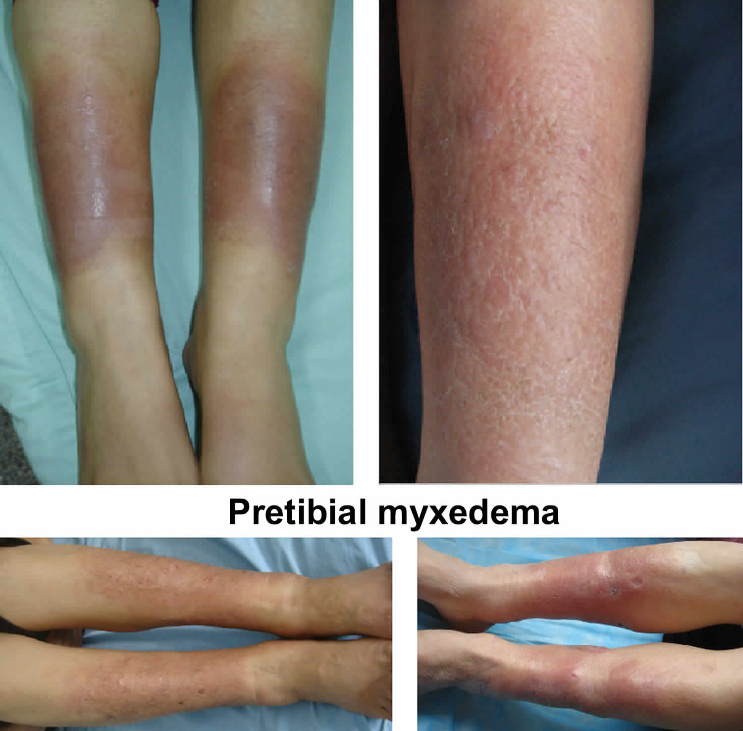
What is pretibial myxedema
Pretibial myxedema is also called thyroid dermopathy, localized myxedema and infiltrative dermopathy, is a form of diffuse mucinosis in which there is an accumulation of excess glycosaminoglycans in the dermis and subcutis of the skin, usually as a component of thyroid disease. Although pretibial myxedema is most commonly seen on the shins (pretibial areas) and is characterized by swelling and lumpiness of the lower legs. , it may occur anywhere on the skin, especially the ankle, dorsum of the foot, knees, shoulders, elbows, upper back, pinnae, nose, and neck 1.
Pretibial myxedema is nearly always associated with autoimmune thyroid disease, that is Graves disease. Pretibial myxedema occurs in 0.5-4.3% of patients with Graves disease. Graves disease is an autoimmune disorder that affects the thyroid gland in which there are antibodies to TSH-R activate the receptor, often causing an increase in circulating thyroid hormone. This is hyperthyroidism or thyrotoxicosis. Symptoms of thyrotoxicosis include weight loss, palpitations, sweating (hyperhidrosis) and tremors. Glycosaminoglycans also called mucopolysaccharides, are complex carbohydrates that are important for tissue hydration and lubrication. The main glycosaminoglycan in pretibial myxedema is hyaluronic acid, which is made by cells called the fibroblasts.
Pretibial myxedema has also been reported, but much less frequently, in patients with Hashimoto thyroiditis, primary hypothyroidism, and euthyroidism. Peak incidence occurs in the fifth to sixth decades of life.
Women are affected more frequently than men, with a female-to-male ratio of 3.5:1. Pretibial myxedema may occur in children and young adults, but most cases occur in older adults, with a peak age at onset in the fifth to sixth decades of life.
The classic triad of signs of Graves disease is:
- Pretibial myxedema
- Ophthalmopathy (prominent eyes due to deposition of myxedema behind the orbit)
- Acropachy (swelling of distal digits with overgrown nail plates that may lift off the nail bed; similar to clubbing)
Pretibial myxedema:
- Affects 0.5–4.3% of patients with Graves disease; it is seen in up to 13% in those with severe eye disease
- Has also been seen in patients with Hashimoto thyroiditis, primary hypothyroidism (underactive thyroid), and euthyroidism (normal thyroid function)
- Is associated with high serum concentrations of TSH-R antibodies
- Is most common in people between the ages of 40 and 60
- More commonly affects females, with a female:male ratio of 3.5:1
The lesions of pretibial myxedema are primarily of cosmetic concern, although severe elephantiasic forms may lead to significant limb enlargement and impair function.
Surgical treatment should be avoided because scarring may aggravate the dermopathy, and benefits are equivocal.
Local application of corticosteroids remains the mainstay of treatment.
Compression wraps or stockings that provide 20-40 mm Hg of pressure can be useful as an adjunctive therapy.
Types of pretibial myxedema
- Diffuse, non-pitting edema (swelling) – the most common form
- Plaque form – raised plaques on a background of non-pitting edema
- Nodular form – sharply circumscribed tubular or nodular lesions
- Elephantiasic form – nodular lesions with pronounced lymphedema (swelling due to accumulation of lymphatic tissue fluid). Lesions may coalesce to give the entire extremity an enlarged, warty appearance. This form is rare.
What causes pretibial myxedema?
Pretibial myxedema is generally considered a skin manifestation of thyroid disease. The exact mechanism for the deposition of glycosaminoglycans in the skin of the lower legs is uncertain.
Pretibial myxedema is likely to be due to a combination of the following:
- An immunological process in which thyroid stimulating hormone receptor (TSH-R) antibodies bind to and stimulate fibroblasts to increase glycosaminoglycan production.
- A cellular process in which fibroblasts are activated indirectly through sensitised T lymphocytes (immune cells). T cells sensitised to a common antigen (likely TSH-R antibodies) could infiltrate dermal tissue and release cytokines (cellular messenger proteins), which activate dermal fibroblasts to produce glycosaminoglycans.
- A mechanical process, where myxedema accumulates in areas of trauma, after prolonged standing, and in dependent sites. This theory may explain why it occurs in the pretibial area. Dependent swelling could result in pooling of immune cells and proteins, increasing the disease effects.
Pretibial myxedema pathophysiology
Pretibial myxedema occurs as a result of the deposition of hyaluronic acid in the dermis and subcutis. The precise cause of this phenomenon remains uncertain. A leading theory proposes that fibroblasts are stimulated to produce abnormally high amounts of glycosaminoglycan under the influence of cytokines due to exposure to thyrotropin receptor antibody (TRAB) and antigen-specific T cells. Thyrotropin receptor antibody-binding sites are found in the plasma membranes of fibroblasts derived from the skin of patients with pretibial myxedema. Thyrotropin receptor antibody is present in the serum of most patients with pretibial myxedema (80-100%), but it has also been found in the serum of patients without pretibial myxedema 2.
Research published in 2006 suggests that it may not be just the high level of glycosaminoglycans, but the change in percentage of the constituents of the glycosaminoglycans in the blood that leads to the development of pretibial myxedema. Thyroid hormones, by means of their influence on prostaglandin metabolism, alter the synthesis and degradation of glycosaminoglycans. Prostaglandin degradation may be what is changed in the course of Graves disease, based on findings that glycosaminoglycan synthesis is reduced, as is extracellular matrix assembly in vitro with exposure to T3 excess 3.
Cell-mediated immunity, using differentially expressed T-cell surface receptors in localized pretibial myxedema, has also been proposed as having a causative role 4. The fact that pretibial myxedema frequently develops in areas of injury suggests that trauma may contribute to local fibroblast activation. In addition, extrathyroid manifestations of Graves disease often occur in the skin and eyes—fibroblasts within the orbits and skin were found to have phenotypic differences from other fibroblasts throughout the body.
Pretibial myxedema symptoms
Pretibial myxedema can appear before, during, or after the thyrotoxic state. Pretibial myxedema is not related to thyroid function. The onset of pretibial myxedema is most commonly seen 12–24 months after the diagnosis of Graves disease, but it may occur before or after the onset of thyrotoxicosis. Pretibial myxedema in the absence of Graves disease is uncommon. Most patients who develop pretibial myxedema also have Graves ophthalmopathy, with the onset of dermopathy typically following the onset of ophthalmopathy by 6-12 months. The natural history of pretibial myxedema is not well defined. Available data indicate that about 10-26% of patients eventually experience complete remission, and about 24% have partial remission. Rare cases of pretibial myxedema without ophthalmology have been recorded 5.
Skin lesions or areas of non-pitting edema appear on the anterior or lateral aspects of the legs or in sites of old or recent trauma in patients with Graves disease.
Otherwise unexplained skin lesions or areas of non-pitting edema occur in patients with thyroid disease.
- It is most commonly found on the pretibial areas, the dorsum of the feet, or in sites of prior trauma.
- It is usually asymptomatic and more of a cosmetic concern, but can be itchy or sore.
- Early lesions are bilateral, firm, non-pitting, asymmetrical plaques or nodules; they may coalesce to form scaly, thickened and hardened skin areas.
- Hair follicles are often prominent giving a peau d’orange (orange peel) texture.
- The overlying skin may be discolored, in colors that range from a violet tinge to a slightly pigmented yellow-brown
- There may be localized hyperhidrosis (increased sweating) and/or hypertrichosis (increased hair growth) over the affected skin.
Pretibial myxedema diagnosis
Diagnosis of pretibial myxedema is made by taking a history and finding characteristic clinical appearance on examination of the patient.
Skin biopsy is rarely necessary for diagnosis, especially if there is a history of hyperthyroidism, or Graves ophthalmopathy.
If a biopsy is done, histopathology typically shows deposition of mucin (glycosaminoglycans) throughout the dermis and subcutis. Deposited mucin promotes dermal edema by promoting the retention of fluid in the skin. This results in compression/occlusion of small peripheral lymphatics and lymphedema.
The biopsy also shows attenuation of collagen fibers – the collagen fibers are frayed, fragmented and widely separated. Stellate (star-shaped) fibroblasts are often observed, but the number of fibroblasts remains normal. Often a mild, superficial lymphocytic infiltrate around blood vessels is seen, and the overlying epidermis may show hyperkeratosis (increased scale).
Thyroid-stimulating hormone (TSH) levels may be abnormally high, normal, or low, depending on whether the underlying thyroid disease has been recognized and treated.
Thyrotropin receptor antibody (TRAB) levels are elevated in about 80-100% of patients with pretibial myxedema 6.
Pretibial myxedema treatment
Pretibial myxedema is often asymptomatic and mild, and may require no treatment at all.
If symptomatic, treatment options include:
- Minimization of risk factors:
- Avoid tobacco
- Reduce weight
- Normalize thyroid function
- Compression stockings, which can be worn to improve lymphedema
- Mid to high potency topical corticosteroid, which are usually recommended under occlusion (eg plastic cling-film wrap) nightly or every other night to enhance effect
Other therapies reported to be successful include:
- Intralesional corticosteroids
- Systemic steroids
- Pentoxifylline
- Octreotide
- Rituximab
- Plasmapheresis
- Intravenous immunoglobulin
- Surgical excision has been successful in case reports, but it is generally not recommended due to the risk of pretibial myxedema developing in injured skin
Pretibial myxedema prognosis
Pretibial myxedema prognosis is generally quite good. Most patients with asymptomatic pretibial myxedema do not require treatment or follow-up.
The myxedema clears up completely in the majority of patients with mild disease.
Even with more severe disease, it resolves in more than half of patients after several years. The elephantiasic form is the most difficult to treat, and is the least likely to clear up.
The likelihood of remission depends on the severity of the initial disease rather than its treatment.
References- Doshi DN, Blyumin ML, Kimball AB. Cutaneous manifestations of thyroid disease. Clin Dermatol. 2008 May-Jun. 26 (3):283-7.
- Kamath C, Young S, Kabelis K, Sanders J, Adlan MA, Furmaniak J, et al. Thyrotrophin receptor antibody characteristics in a woman with long-standing Hashimoto’s who developed Graves’ disease and pretibial myxoedema. Clin Endocrinol (Oxf). 2012 Sep. 77(3):465-70.
- Komosinska-Vassev K, Winsz-Szczotka K, Olczyk K, Kozma EM. Alterations in serum glycosaminoglycan profiles in Graves’ patients. Clin Chem Lab Med. 2006. 44(5):582-8.
- Heufelder AE, Bahn RS, Scriba PC. Analysis of T-cell antigen receptor variable region gene usage in patients with thyroid-related pretibial dermopathy. J Invest Dermatol. 1995 Sep. 105(3):372-8.
- Sendhil Kumaran M, Dutta P, Sakia U, Dogra S. Long-term follow-up and epidemiological trends in patients with pretibial myxedema: an 11-year study from a tertiary care center in northern India. Int J Dermatol. 2015 Aug. 54 (8):e280-6.
- Ai J, Leonhardt JM, Heymann WR. Autoimmune thyroid diseases: etiology, pathogenesis, and dermatologic manifestations. J Am Acad Dermatol. 2003 May. 48 (5):641-59; quiz 660-2.


