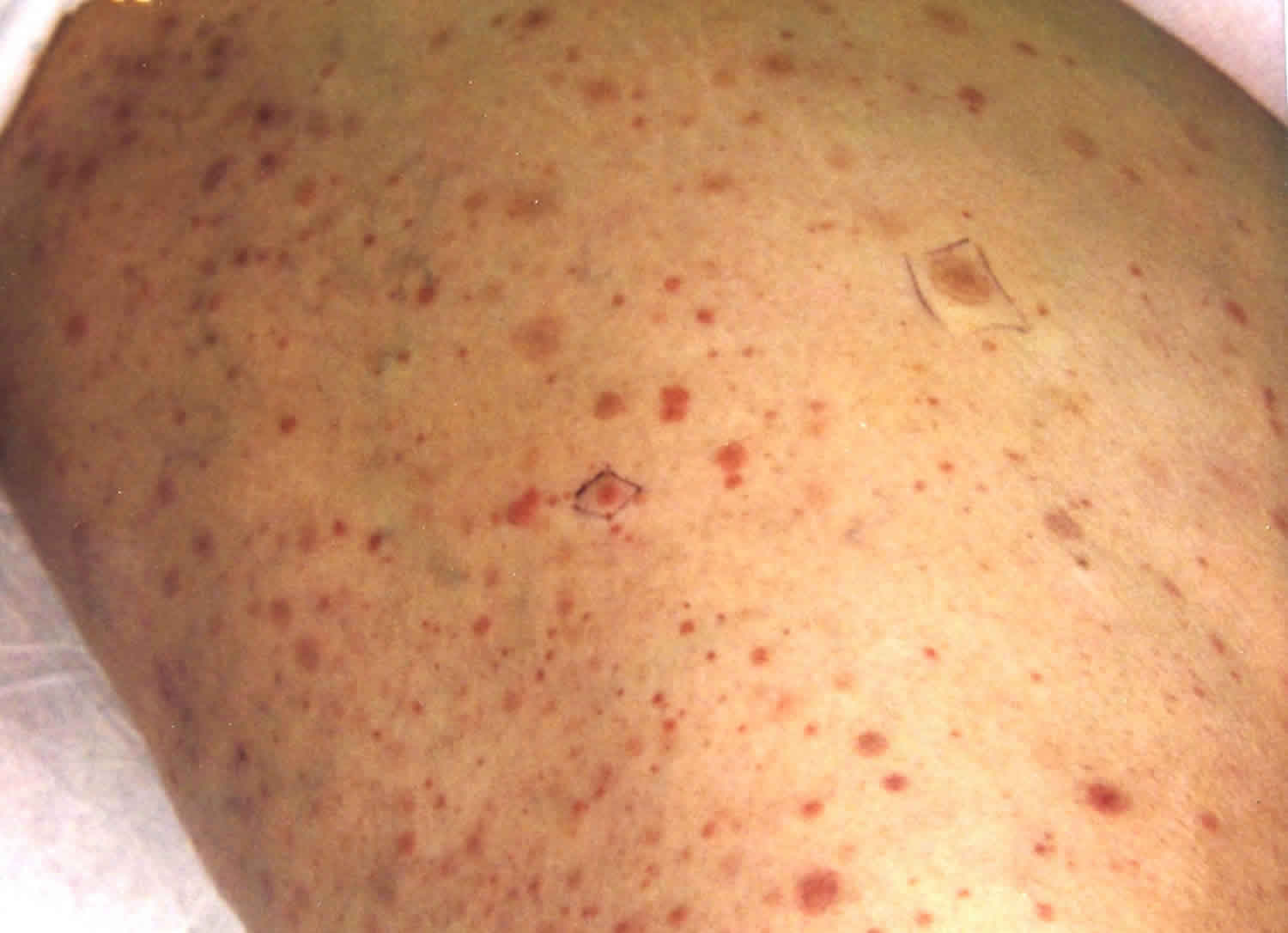
Petechiae
Petechiae are pinpoint non-blanching bleeding into the skin that measure less than 2 mm in size which affect the skin and mucous membranes 1. A non-blanching spot is one that does not disappear after brief pressure is applied to the area. Petechiae are small, purpuric lesions up to 2mm across. Purpura is the name given to non-blanching spot that measures greater than 2 mm of the skin or mucous membranes due to bleeding (hemorrhage) from small blood vessels.
Petechial rashes are a common presentation to the pediatric emergency department. Non-blanching rashes can be a great cause for concern for parents and physicians alike. Therefore, careful assessment and evaluation must be undertaken to formulate a sensible management plan 2, 3, 4.
Petechial rashes result from areas of hemorrhage into the dermis. Derangements in the normal hemostasis can result in petechiae along with a variety of other clinical findings. The primary pathophysiological causes of petechiae and purpura are thrombocytopenia, platelet dysfunction, disorders of coagulation, and loss of vascular integrity. Some clinical pictures result in petechial lesions from a combination of these mechanisms 5.
If your rash is accompanied with fever or chills, you should see a physician. It may be something as simple as a 24-hour virus, or it could indicate something more serious. There are a number of dangerous conditions that are urgent, and then there are some serious skin conditions that are not necessarily urgent. This distinction may not be easy for a patient to make, so it is necessary to see a physician.
Some signs and symptoms that may indicate that a rash is dangerous include:
- Feeling ill
- Fever
- Chills
- Diarrhea
- Muscle aches or pains
- Presence of blisters
Purple marks on the skin that do not blanch on pressure (i.e, when you put pressure on the area with your thumb and then release, the marks do not fade) may be a sign of inflammation of the blood vessels. Purple marks that do not blanch may fall into one of two categories, known as of purpura and petechiae.
If platelets and other clotting functions are not working properly, it could indicate a serious situation and manifest as skin with purple, pinpoint dots that do not blanch with pressure. Finally, if you have irregular spots that have changed over a relatively short period of time, a serious underlying systemic condition should be suspected.
If your rash is accompanied by any of the above-mentioned symptoms, please seek medical help as soon as possible.
Petechiae causes
There are many causes of a petechial rash in a child to be considered. Invasive meningococcal disease caused by Neisseria meningitidis, is the priority in the differential diagnosis to consider on initial presentation 1. Consequently, a child with fever and a petechial rash requires urgent and comprehensive assessment. Multiple studies have shown that the rate of IMD has reduced following the introduction of meningococcal vaccines into childhood immunization schedules and that the low prevalence of invasive meningococcal disease suggests that most children presenting with a petechial rash have less serious pathologies. However, given its associated morbidity and mortality, it should remain at the forefront of the clinician’s mind when assessing the child with fever and petechiae 6.
Causes can be classified into the following categories:
Infective
- Viral: Enterovirus, Parvovirus B19, Dengue
- Bacterial: Meningococcal, scarlet fever, infective endocarditis
- Rickettsial: Rocky Mountain Spotted fever
- Congenital: TORCH. TORCH infection includes Toxoplasmosis, Other (syphilis, varicella-zoster, parvovirus B19), Rubella, Cytomegalovirus (CMV), and Herpes infections 7
Trauma
- Accidental injury
- Non-accidental injury
- Increased pressure following bouts of coughing, vomiting or straining
Hematological and Malignancy
- Leukemia
- Idiopathic thrombocytopenic purpura (ITP)
- Thrombocytopenia with absent radius (TAR) syndrome
- Fanconi anemia
- Disseminated intravascular coagulation (DIC): Disseminated intravascular coagulation (DIC) also known as consumption coagulopathy is a rare but serious disorder that causes abnormal blood clotting throughout the body’s blood vessels. It is caused by another disease or condition, such as an infection or injury, that makes the body’s normal blood clotting process become overactive. Disseminated intravascular coagulation may develop quickly over hours or days, or more slowly. Signs and symptoms may include bleeding, bruising, low blood pressure, shortness of breath, or confusion. Complications can be life-threatening and include bleeding or multiple organ failure. Disseminated intravascular coagulationC that develops quickly usually requires emergency treatment in the hospital. In treating DIC, your doctor will treat the disease that is causing DIC. Your doctor may also give you medicines to prevent blood clots, or blood products such as platelets or clotting factors to stop bleeding.
- Hemolytic uremic syndrome (HUS): Hemolytic uremic syndrome is a condition involving red blood cells and your kidneys. Hemolytic uremic syndrome happens after you get an infection in your digestive system. Hemolytic-uremic syndrome often occurs after a gastrointestinal infection with E. coli bacteria (Escherichia coli O157:H7). However, hemolytic uremic syndrome has also been linked to other gastrointestinal infections, including shigella and salmonella. Hemolytic uremic syndrome has also been linked to nongastrointestinal infections. The infection makes toxins that destroy red blood cells. These damaged cells block the kidneys’ filtering system and can cause your kidneys to fail. Anyone can get hemolytic uremic syndrome, but it is more common in children and older adults. Many people who get an E. coli infection do not develop hemolytic uremic syndrome. For you to be diagnosed with hemolytic uremic syndrome, the infection must cause 3 disorders:
- Hemolytic anemia(not enough red blood cells).
- Thrombocytopenia (not enough platelets).
- Renal (kidney) failure.
- Splenomegaly
- Neonatal alloimmune thrombocytopenia
Vasculitis and inflammatory conditions
- Henoch Schonlein Purpura
- Systemic Lupus Erythematosus (SLE)
Connective Tissue Disorder
- Ehlers Danlos syndrome
Congenital
- Wiskott Aldrich syndrome
- Glanzmann Thrombasthenia
- Bernard-Soulier syndrome
Other
- Drug reaction
- Vitamin K deficiency
- Chronic liver disease
Petechiae diagnosis
A detailed history and physical examination are paramount for every child presenting with petechiae. Key features in the history include time of onset, anatomical pattern and a detailed chronological account of any other symptoms, e.g., fever, coughing, vomiting, any recent upper respiratory tract infection (URTI) or gastroenteritis, and any sick contacts. A rapidly spreading rash is more concerning for invasive meningococcal disease in an unwell child with a fever. A recent viral infection (upper respiratory tract infection or gastroenteritis) is common in idiopathic thrombocytopenic purpura, Henoch Schonlein Purpura, and hemolytic uremic syndrome. Petechiae confined to above the nipple line are associated with bouts of vomiting or coughing. It is also important to ask about any bleeding from mucosal surfaces such as gingival bleeding, epistaxis, melena, among others. As always, vaccination status should be confirmed.
On examination, a complete set of observations and neurological status should be regularly monitored. A full systemic examination should be completed, including cardiac, respiratory, abdominal, otorhinolaryngological, and neurological (if concerns of invasive meningococcal disease). The skin should be thoroughly examined from head to toe, and the pattern of rash should be clearly documented. Demarcating areas of petechiae with a skin marker can be helpful for monitoring progression of the rash in clinical practice.
The age of the child can be useful in reaching the most likely diagnosis, for example; a neonate with petechiae could have a neonatal alloimmune thrombocytopenia or a TORCH infection, and Henoch Schonlein Purpura is more common in the 2 to 5 year age range.
Patterns of concerning symptoms and signs presenting with petechiae include but are not limited to:
- Fever (pyrexia), tachycardia and rapidly spreading petechiae: invasive meningococcal disease
- Pallor, bruising, weight loss, lymphadenopathy: Malignancy
- Hypertension: Renal disease associated with hemolytic-uremic syndrome, Henoch Schonlein Purpura or SLE (Systemic Lupus Erythematosus)
- Unusual patterns of petechiae with bruising, a history that is inconsistent or signs or injury or neglect.
Petechiae tests
Investigations to diagnose the cause of a petechial rash depend on the clinical presentation and can differ from one pediatric emergency department to another. Adhering to the local protocol is advised. In general, investigations will depend on the location of petechiae, associated pyrexia or clinical suspicion for any of the concerning patterns of signs and symptoms. A healthy child with scattered petechiae of obvious causation, e.g., known trauma or petechiae confined to above the nipple line may not require any investigations. At the very least, the healthy child, as described, should be observed for 4 hours before discharge.
- Complete blood count (CBC) to check platelet number, a raised or decreased white cell count or decreased hemoglobin
- If concerns about invasive meningococcal disease or other infection: C-reactive protein, blood culture
- Coagulation profile, urea, electrolytes, and liver function tests may be considered in some cases (disseminated intravascular coagulation [DIC], invasive meningococcal disease, Henoch Schonlein Purpura, hemolytic uremic syndrome). A prolonged prothrombin time can indicate factor deficiencies, Vitamin K deficiency, disseminated intravascular coagulation [DIC], liver or renal disease
- Urine dipstick and microscopy are useful when renal causes are part of the differential (Henoch-Schönlein purpura, hemolytic uremic syndrome, SLE), to check for proteinuria in particular
- Further tests may be later requested when a more specific diagnosis is narrowed down.
- Additional studies could include a fecal occult blood test, serology for hepatitis B and C, serum protein electrophoresis, anti-streptolysin O titre, and measurement of antinuclear antibodies, antineutrophil cytoplasmic antibodies, rheumatoid factor, serum immunoglobulins, and cryoglobulins. Abdominal ultrasound and renal biopsy may also be indicated.
Petechiae treatment
Many patients attending the pediatric emergency department with petechial rashes will not require any specific treatment. In fact, if a child remains well after a period of observation, with no spreading of the rash, a normal platelet count and no physical signs or signs of infection on blood tests, they may be discharged home. If invasive meningococcal disease is likely, urgent intravenous antibiotics as per local guidelines should be administered, with close observations after admission to the ward. Some patients may be given a dose of antibiotics pre-hospital if high clinical suspicion of invasive meningococcal disease is present. If specific diagnoses are made, for example, Henoch Schonlein Purpura or idiopathic thrombocytopenic purpura (ITP), and there is no risk of going home, the child may be discharged with an appointment to return to the appropriate outpatient department and condition specific education. Other conditions will require admission and treatment, for example, urgent referral to oncology inpatient services for a patient with pancytopenia and a likely malignant diagnosis 8.
References- McGrath A, Barrett MJ. Petechiae. [Updated 2019 Jun 8]. In: StatPearls [Internet]. Treasure Island (FL): StatPearls Publishing; 2020 Jan-. Available from: https://www.ncbi.nlm.nih.gov/books/NBK482331
- Iba T, Watanabe E, Umemura Y, et al. Sepsis-associated disseminated intravascular coagulation and its differential diagnoses. J Intensive Care. 2019;7:32. Published 2019 May 20. doi:10.1186/s40560-019-0387-z https://www.ncbi.nlm.nih.gov/pmc/articles/PMC6528221
- Clark WF, Huang SS. Introduction to therapeutic plasma exchange. Transfus. Apher. Sci. 2019 Jun;58(3):228-229.
- Ranganathan D, John GT. Therapeutic Plasma Exchange in Renal Disorders. Indian J Nephrol. 2019 May-Jun;29(3):151-159.
- Galera P, Dulau-Florea A, Calvo KR. Inherited thrombocytopenia and platelet disorders with germline predisposition to myeloid neoplasia. Int J Lab Hematol. 2019 May;41 Suppl 1:131-141.
- Sargentini-Maier ML, De Decker P, Tersteeg C, Canvin J, Callewaert F, De Winter H. Clinical pharmacology of caplacizumab for the treatment of patients with acquired thrombotic thrombocytopenic purpura. Expert Rev Clin Pharmacol. 2019 Jun;12(6):537-545.
- TORCH Infections. Toxoplasmosis, Other (syphilis, varicella-zoster, parvovirus B19), Rubella, Cytomegalovirus (CMV), and Herpes infections. Curr Womens Health Rep. 2002 Aug;2(4):253-8. https://www.ncbi.nlm.nih.gov/pubmed/12150751
- Jelusic M, Sestan M, Cimaz R, Ozen S. Different histological classifications for Henoch-Schönlein purpura nephritis: which one should be used? Pediatr Rheumatol Online J. 2019 Feb 28;17(1):10.


