
What is pulmonary stenosis
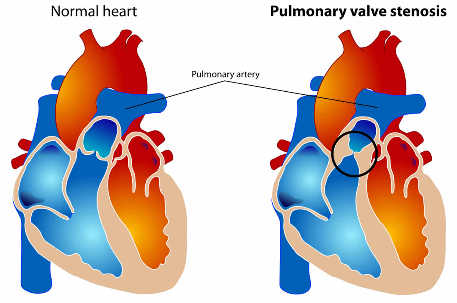
Pulmonary stenosis or pulmonary valve stenosis means that your pulmonary valve may be too thick, narrowed and stiff to open easily. The pulmonary valve separates the right ventricle (one of the chambers in your heart) and the pulmonary artery. The pulmonary artery carries oxygen-poor blood from your heart to your lungs. With pulmonary valve stenosis, your right ventricle can also become thicker and more muscular (hypertrophied) as it has to work harder to pump blood through the pulmonary valve to your lungs. Stenosis, or narrowing, occurs when the pulmonary valve cannot open wide enough. As a result, less blood flows to your lungs. If the pulmonary valve is severely narrowed, this can limit how much exercise your child can manage.
Pulmonary valve stenosis may occur alone or with other heart defects that are present at birth. The condition can be mild or severe.
Most children have only mild pulmonary stenosis and don’t need any treatment even when they are older, but the pulmonary valve can become more narrowed
as time goes by. But for others, your pulmonary valve can become more narrowed as time goes by and will need treating. You will need to take your child for
regular check-ups with your child’s cardiologist, even if they appear appears to be well and doesn’t have any symptoms.
Babies who are diagnosed before birth usually have a severe form of pulmonary stenosis and will usually need treatment shortly after birth.
If your pulmonary valve is severely narrowed, you will need treatment shortly after your diagnosis is made. Many young people have a procedure called balloon valvuloplasty, but if your pulmonary valve is not suitable for this procedure then surgery will be required.
Talk to your doctor if you or your child has:
- Shortness of breath
- Chest pain
- Fainting
If you have pulmonary stenosis or another heart problem, prompt evaluation and treatment can help reduce your risk of complications.
Normal pulmonary valve anatomy
The pulmonary valve is made up of three thin pieces of tissue called cusps that are arranged in a circle. With each heartbeat, the valve opens in the direction of blood flow — into the pulmonary artery and continuing to the lungs — then closes to prevent blood from flowing backward into the heart’s right ventricle.
In pulmonary valve stenosis one or more of the cusps may be defective or too thick, or the cusps may not separate from each other properly. If this happens, the valve doesn’t open correctly, restricting blood flow.
Figure 1. Pulmonary valve anatomy

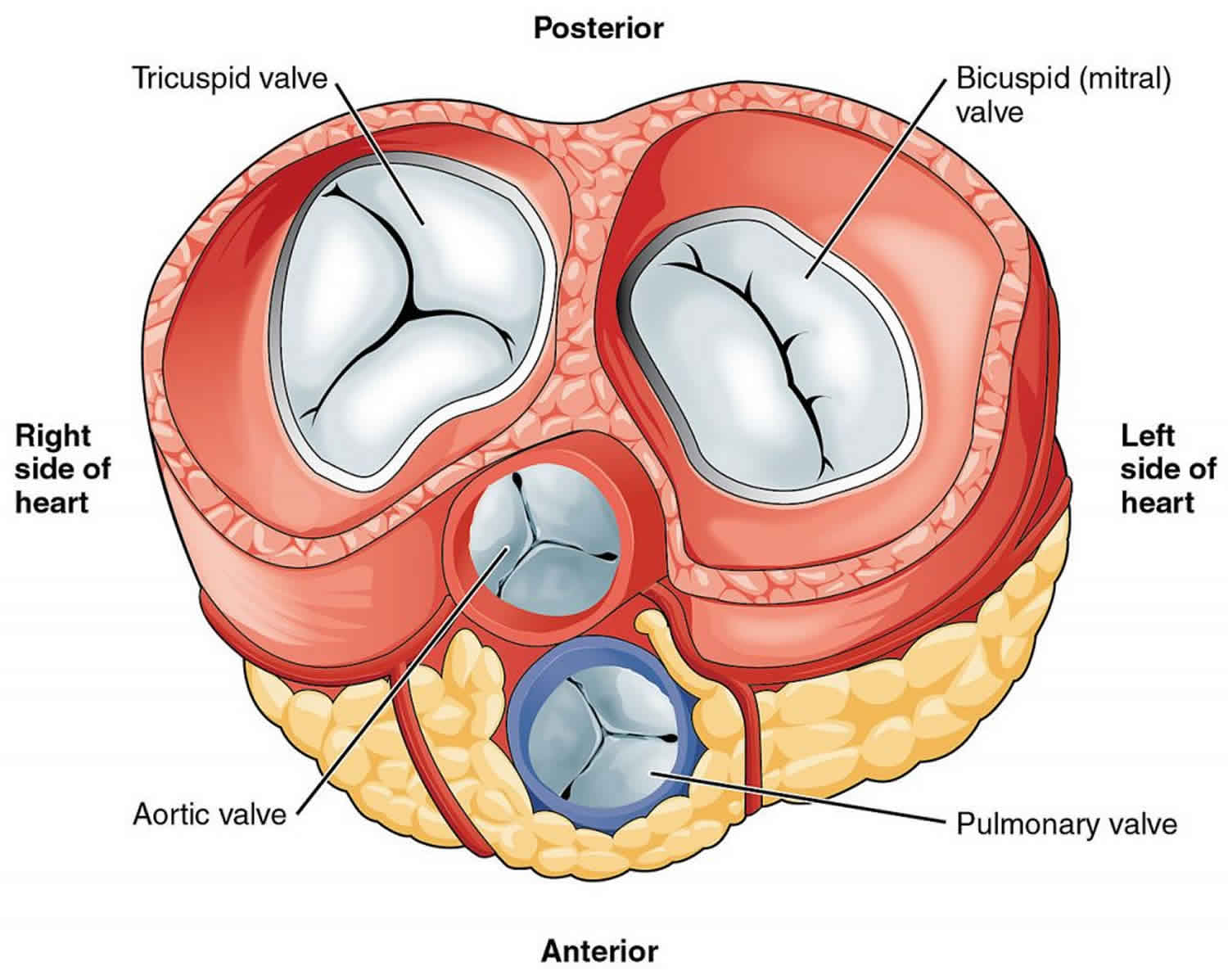
Figure 2. Pulmonary stenosis
What other conditions are associated with pulmonary stenosis?
Pulmonary stenosis mostly occurs on its own, without other abnormalities. However, sometimes children with pulmonary stenosis may also have a genetic abnormality such as Noonan syndrome.
Sometimes children with pulmonary stenosis also have other heart abnormalities. Your child’s cardiologist will tell you if this is the case for your child.
What happens as my child grows up?
Those who have not needed any treatment by the time they are fully grown usually don’t ever need treatment for their pulmonary stenosis. However, in some rare cases the pulmonary valve can become narrower in later adult life. Balloon valvuloplasty is usually effective in adult life just as it is in children.
After any kind of treatment for pulmonary stenosis, the pulmonary valve never works completely normally, and will leak to some extent. This leak is normally nothing to worry about, but there is a very small chance that some children might need surgery to replace or repair their valve in later life.
Pulmonary stenosis causes
Pulmonary valve stenosis is most often present at birth (congenital) when the pulmonary valve doesn’t grow properly during fetal development. It is caused by a problem that occurs as the baby develops in the womb before birth. It’s not known what causes the pulmonary valve to develop abnormally, but genes may play a role. Babies who have the condition may have other congenital heart abnormalities, as well. In some cases, the problem runs in families.
Sometimes other medical conditions or having an artificial valve can cause pulmonary stenosis.
- Carcinoid syndrome. This syndrome — a combination of signs and symptoms, including flushing of the skin and diarrhea — results from the release of a chemical, serotonin, from growths called carcinoid tumors in the digestive system.
- Rheumatic fever. This complication of an infection caused by streptococcus bacteria, such as strep throat, may injure the heart valves.
Risk factors for pulmonary stenosis
Because pulmonary valve stenosis usually develops before birth, there aren’t many known risk factors. However, certain conditions and procedures can increase your risk of developing pulmonary valve stenosis later in life, including:
- Carcinoid syndrome
- Rheumatic fever
- Noonan syndrome
- Pulmonary valve replacement
Pulmonary stenosis symptoms
Pulmonary valve stenosis signs and symptoms vary, depending on the extent of the obstruction. Most children with pulmonary stenosis won’t have any symptoms at all, and will appear perfectly well. Usually the only sign of pulmonary stenosis is a heart murmur (an extra sound from the heart), which may be picked up during a routine medical check.
If your child has significant pulmonary stenosis, they may feel tired when playing or doing physical activity. In some rare cases where it is very severe, the child may have fainting episodes. If this happens, you should let your child’s doctor know immediately.
Pulmonary valve stenosis signs and symptoms may include:
- Heart murmur — an abnormal whooshing sound heard using a stethoscope, caused by turbulent blood flow
- Fatigue
- Shortness of breath, especially during exertion
- Chest pain
- Loss of consciousness (fainting)
- Abdominal distention
- Bluish color to the skin (cyanosis) in some people
- Poor appetite
- Chest pain
- Poor weight gain or failure to thrive in infants with a severe blockage
- Sudden death
Pulmonary valve stenosis complications
Pulmonary stenosis may be associated with the following:
- Infection. People with heart valve problems, such as pulmonary stenosis, have a higher risk of developing bacterial infections in the inner lining of the heart (infective endocarditis) than people without heart valve problems.
- Heart-pumping problems. In severe pulmonary stenosis, the heart’s right ventricle must pump harder to force blood into the pulmonary artery. Pumping of the right ventricle against increased pressure causes the muscular wall of the ventricle to thicken (right ventricular hypertrophy). Eventually, the heart becomes stiff and may weaken.
- Heart failure. If the right ventricle is unable to pump efficiently, heart failure develops. This results in swelling of the legs and abdomen and can cause fatigue and shortness of breath.
- Irregular heartbeat (arrhythmia). People with pulmonary stenosis are more likely to have an irregular heartbeat. Unless the stenosis is severe, irregular heartbeats associated with pulmonary stenosis usually aren’t life-threatening.
To reduce your risk of getting infective endocarditis:
- Keep your teeth and mouth clean and have regular check-ups with a dentist
- Avoid body piercing and tattooing
- Never inject recreational drugs
Pulmonary stenosis diagnosis
In most cases, pulmonary stenosis is not diagnosed until after the baby is born, but some severe cases may be detected before birth using an echocardiogram. This is an ultrasound scan of the heart and it won’t hurt your baby at all.
Pulmonary valve stenosis is often diagnosed in childhood, but sometimes it isn’t detected until later in life. If your doctor hears a heart murmur during a routine checkup and suspects pulmonary stenosis, he or she may then use a variety of tests to confirm the diagnosis.
- Echocardiogram. Sound waves bounce off your heart and produce moving images that can be viewed on a video screen. This test is useful for checking the structure of the pulmonary valve, the location and severity of the narrowing (stenosis), and right ventricle size and function. Doctors may also perform a 3-D echocardiogram.
- Electrocardiogram (ECG or EKG). During this procedure, patches with wires (electrodes) are placed on your chest, wrists and ankles. The electrodes measure electrical activity in your heart, which is recorded on paper. This test helps determine if the muscular wall of your right ventricle is thickened (right ventricular hypertrophy).
- Other imaging tests. MRI and CT scans are sometimes used to confirm the diagnosis of pulmonary valve stenosis.
- Cardiac catheterization. During this procedure, your doctor inserts a thin, flexible tube (catheter) into an artery or vein in your groin and threads it up to your heart or blood vessels. Dye injected through the catheter makes your blood vessels visible on X-rays. Doctors also use cardiac catheterization to measure the blood pressure in the heart chambers and blood vessels. Doctors generally use this test only if they suspect that you or your child will need a balloon valvuloplasty, a procedure that can be done at the same time as cardiac catheterization.
Pulmonary stenosis treatment
Pulmonary valve stenosis is classified as mild, moderate or severe, depending on a measurement of the blood pressure difference between the right ventricle and pulmonary artery. Mild pulmonary stenosis that isn’t causing symptoms doesn’t usually require treatment, just routine checkups.
If the pulmonary valve is severely narrowed, your child will need treatment shortly after diagnosis. It is very rare for a child with pulmonary stenosis to need open-heart surgery, most cases can be treated using a procedure called balloon valvuloplasty.
Balloon valvuloplasty
Balloon valvuloplasty is a procedure that stretches open your narrowed pulmonary valve. Balloon valvuloplasty is usually done under a general anesthetic. A catheter (a thin, hollow tube) with a small collapsed balloon at its tip is inserted into a vein at the top of your child’s leg. This is guided up the vein into the right side of the heart and across the narrowed valve. Using X-ray, the balloon is positioned in the narrow valve, and is then inflated, stretching the valve open. The balloon is then deflated and removed.
Balloon valvuloplasty does not make your pulmonary valve normal and it does not always work. However in many cases it can widen your narrowed pulmonary, helping to delay the need for surgery.
Following the balloon valvuloplasty procedure, your pulmonary valve may start to leak to some extent – meaning that some blood flows back into your right ventricle instead of to your lungs. This leak is usually small and doesn’t need treating, but some people may go on to need surgery to repair or replace their pulmonary valve in later life.
What happens after a balloon valvuloplasty?
Your child may have to stay in hospital for a few days afterwards. If the narrowing has been only partly relieved by the balloon valvuloplasty, it’s advisable the procedure be repeated at a later date. Because the thickened heart muscle itself can cause some narrowing, it is not always possible to tell if the procedure has been successful straight away. It can take several weeks for the thickened muscle to return to normal.
What are the risks of balloon valvuloplasty?
Valvuloplasty is a very effective form of treatment and the fatality risks are very low. However, do be aware that in new-born babies with severe pulmonary stenosis, the risk will be greater. Your child’s cardiologist will be able to talk through the details with you.
In a small proportion of children, the thickened heart muscle doesn’t return to normal and the muscle itself can obstruct the normal flow of blood. If this causes significant narrowing inside the heart, your child will need to have surgery to remove some of the muscle.
Valve repair or valve replacement surgery
Sometimes the pulmonary valve cannot be stretched open using the balloon valvuloplasty procedure, and open-heart surgery is needed to carry out a valvotomy or pulmonary valve replacement surgery. This involves having a general anesthetic. During the operation, the heart is stopped and the function of the heart is taken over by a ‘heart-lung machine’, which makes sure that blood is still pumped around your child’s body. The surgeon will then open the heart and make a small cut in the narrowed valve, to allow more blood to flow through. The heart is then restarted. After the operation, your child will have a scar down the center of their chest, along the breast bone.
Sometimes because the pulmonary valve is very small, a human donor valve (called a homograft valve) is used to replace your pulmonary valve. Sometimes the replacement of your pulmonary valve can be done by keyhole surgery rather than open-heart surgery.
What happens after pulmonary valve stenosis surgery?
Your child will need to stay in hospital for a few days after the surgery. You will have to bring your child back to see your child’s pediatric cardiologist.
What are the risks of pulmonary valve stenosis surgery?
The good news is, surgery for pulmonary stenosis carries a very low fatality risk. The risk of major complications such as brain damage is also very small. Other uncommon complications – such as a small amount of fluid collecting around the heart or lungs – can also occur after the operation, but these are rarely serious.
Lifestyle and home remedies
While there’s little you can do to prevent pulmonary valve stenosis, you can take steps to ensure you won’t develop complications of your condition.
Your doctor will also likely recommend regular follow-up appointments to evaluate your condition.
Preventive antibiotics
In most cases, you won’t need to take antibiotics to prevent an infection of the inner lining of the heart (infective endocarditis). Your doctor will recommend antibiotics if you’ve had endocarditis before or if you’ve had a pulmonary valve replacement.
Heart-healthy lifestyle
Adopting a heart-healthy lifestyle decreases your risk of developing other types of heart disease, such as a heart attack. Lifestyle changes to talk to your doctor about include:
- Quitting smoking
- Eating a heart-healthy diet, such as a variety of fruits and vegetables, low-fat dairy products, whole grains, and lean meat
- Maintaining a healthy weight
- Regular physical activity
Pregnancy
Pregnancy generally isn’t a problem for women who have mild and moderate pulmonary valve stenosis. If you have severe pulmonary valve stenosis, the risks of complications during labor and delivery are higher than those for women without the condition. If necessary, it’s possible to undergo a balloon valvuloplasty during pregnancy.
Pulmonary stenosis prognosis
People with mild disease rarely get worse. However, those with moderate to severe disease will get worse. The outcome is often very good when surgery or balloon dilation is successful. Other congenital heart defects may be a factor in the outlook.
Most often, the new valves can last for decades. However, some will wear out and need to be replaced.


