Radial tunnel syndrome
Radial tunnel syndrome is a painful condition caused by pressure on the radial nerve — one of the three main nerves in your arm. The most common place for compression of the radial nerve is at the elbow where the nerve enters a tight tunnel made by muscle, bone, and tendon. Radial tunnel syndrome occurs by intermittent compression on the radial nerve from the radial head to the inferior border of the supinator muscle, without obvious extensor muscle weakness 1. Compression could happen in five different sites but the arcade of Frose is the most common area that radial nerve is compressed 1. Radial tunnel syndrome is more prevalent in women with the age of 30 to 50 years old.
Radial tunnel syndrome is a condition that should considerered as presenting in elbow and forearm pains 2. Radial tunnel syndrome is diagnosed with lateral elbow and dorsal forearm pain that may radiate to the wrist and dorsum of the fingers 1. A painful point exists one inch distal to the lateral epicondyle and over the radial tunnel which exacerbates with resisted active extension of the wrist and third finger could confirm the diagnosis. To diagnosis radial tunnel syndrome, clinical examination is more important than paraclinic tests such as electrodiagnsic test and imaging studies. The exact site of the pain which can more specified by the Rule of Nine test (see Figure 6 below) and weakness of the third finger and wrist extension are valuable physical exams to diagnosis. MRI studies my show muscle edema or atrophy along the distribution of the posterior interosseous nerve. Although non-surgical treatments such as rest, NSAIDs, injections and physiotherapy do not believe to have permanent relief, but it is justified before surgery is considered. Surgery could diminish pain and symptoms in 67 to 93 percents of patients completely 1.
Radial nerve anatomy
The radial nerve arises from ventral rami of C5 to C8 (+/- T1) and is a continuation of the posterior cord of the brachial plexus and is the largest branch of the brachial plexus, innervating almost the entire posterior side of the upper limb and provides a motor function to the extensor muscles of the forearm, wrist, fingers, and thumb.
Within the axilla, the radial nerve has three branches: sensory posterior cutaneous nerve of the arm and motor branch to the long and medial head of the triceps 3. The radial nerve passes anterior to the subscapularis, latissimus dorsi and teres major. This is the first possible site of compression. An anomalous muscle, the accessory subscapularis-teres-latissimus is reported in literature to be another cause of compression of the radial nerve at this level 4. Another potential cause of compression, as described by Spinner 5 is penetration of the nerve directly by the subscapular artery more distally in the axilla, forming a neural loop.
The radial nerve enters the posterior compartment of the arm by traveling through the triangular interval bounded by the teres major superiorly, the long head of the triceps medially and the lateral head of the triceps laterally. The radial nerve then exits the axilla, courses through the lateral head of triceps brachii and winds around the posterior humerus in the radial groove accompanied by the profunda brachii artery, sending branches to the triceps brachii. Lorem et al. 6 described this as another possible site of entrapment. Familial radial nerve entrapment syndrome 7 also occurs secondary to compression at the lateral head of the triceps. Intermittent compression of the nerve may also occur secondary to genetic defect in Schwann cell myelin metabolism 8. The radial nerve gives off the following branches in the upper arm – the inferior lateral cutaneous nerve of the arm, posterior cutaneous nerve of the forearm and motor branches to the lateral head of triceps and anconeus. The radial nerve is in close proximity to the humerus in the spiral groove and is easily identified on ultrasound in its short axis overlying the humerus between the lateral and medial heads of the triceps. The radial nerve then courses from the posterior to anterior compartment of the arm by piercing the lateral intermuscular septum which is another possible site of entrapment.
The radial nerve then curves anteriorly around the lateral epicondyle at the elbow, where it divides into a superficial and a deep branches (posterior interosseous nerve) under the margin of the brachioradialis muscle in the lateral border of the cubital fossa.
The deep branch (posterior interosseous nerve) is predominantly motor and passes between the two heads of the supinator muscle to access and supply muscles in the extensor muscles on the forearm. The deep branch (posterior interosseous nerve) winds around the radial head, lying between two heads of the supinator muscle and then travels on the dorsal surface of the interosseous membrane. The superficial layer of the supinator muscle forms the arcade of Frohse which is the most common site of entrapment neuropathy causing the radial tunnel syndrome 9. Two anomalous courses of the posterior interosseous nerve have been reported in the literature by Woltman and Learmonth. First is passage of the nerve within the substance of the supinator, and the other involves a branch travelling superficial to the supinator brevis 10. The origin of the radial tunnel is where the deep branch (lateral) courses over the radio-humeral joint. The tunnel ends at the distal edge of the superficial supinator where the posterior interosseous nerve is formed. Radial tunnel syndrome can be caused by the arcade of Frohse, fibrous fascial bands coursing superficial to the nerve or the vascular leash of Henry 11 which is formed by the recurrent radial vessels. The posterior interosseous nerve supplies the abductor pollicis longus, the extensor pollicis brevis, the extensor indicis proprius and the extensor pollicis longus.
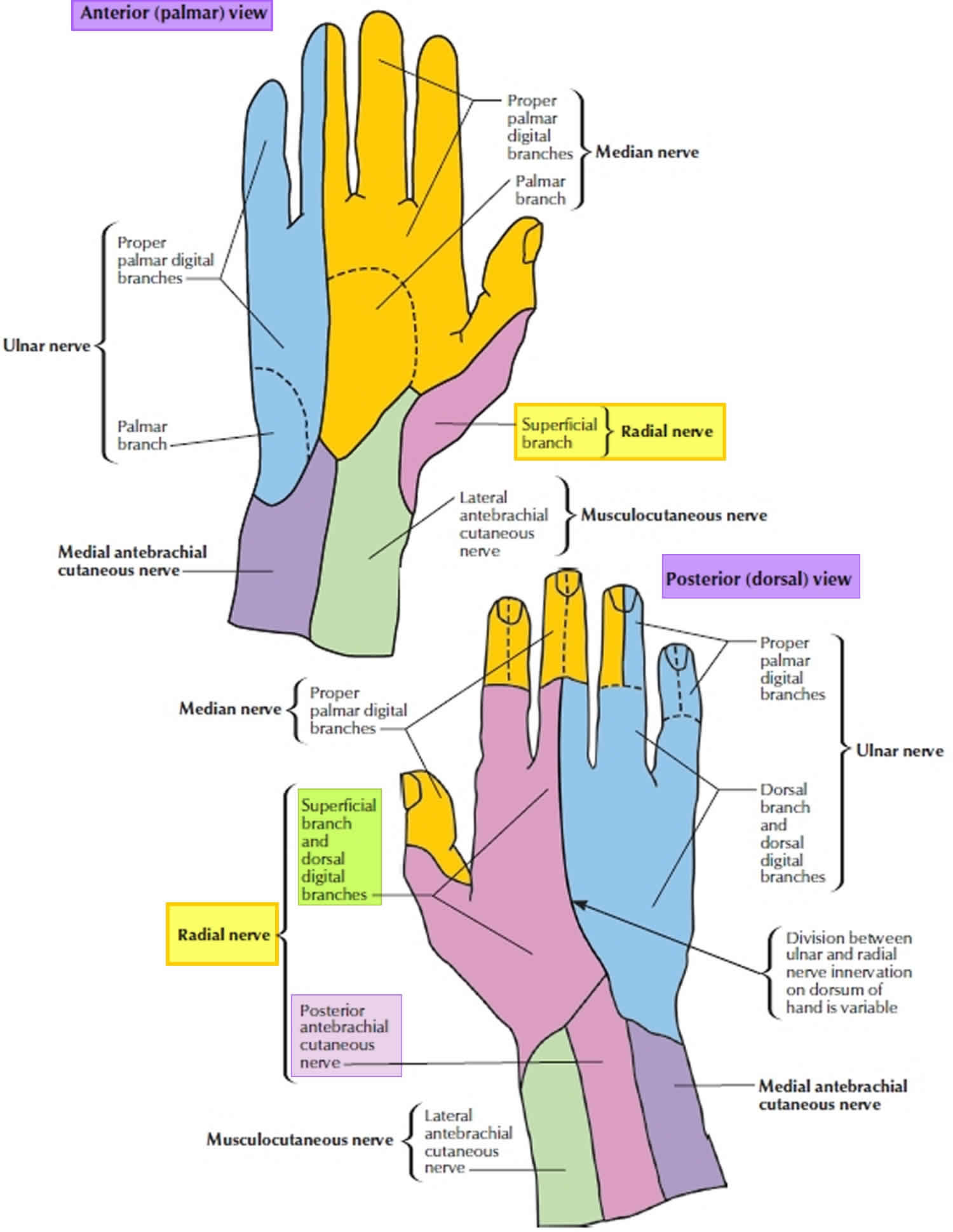
The superficial branch of the radial nerve is sensory. The superficial radial nerve descends medially along the lateral edge of the radius under cover of the brachioradialis muscle and in association with the radial artery. Approximately two-thirds of the way down the forearm, the superficial branch of the radial nerve passes laterally and posteriorly around the radial side of the forearm deep to the tendon of the brachioradialis . The superficial radial nerve continues into the hand where it pierces the lateral fascia to enter the anatomical snuffbox and gives sensory innervation to the dorsal surface of three and a half digits on the radial aspect. It also innervates the extensor digitorum, the extensor digiti minimi and the extensor carpi ulnaris muscles.
Figure 1. Radial nerve
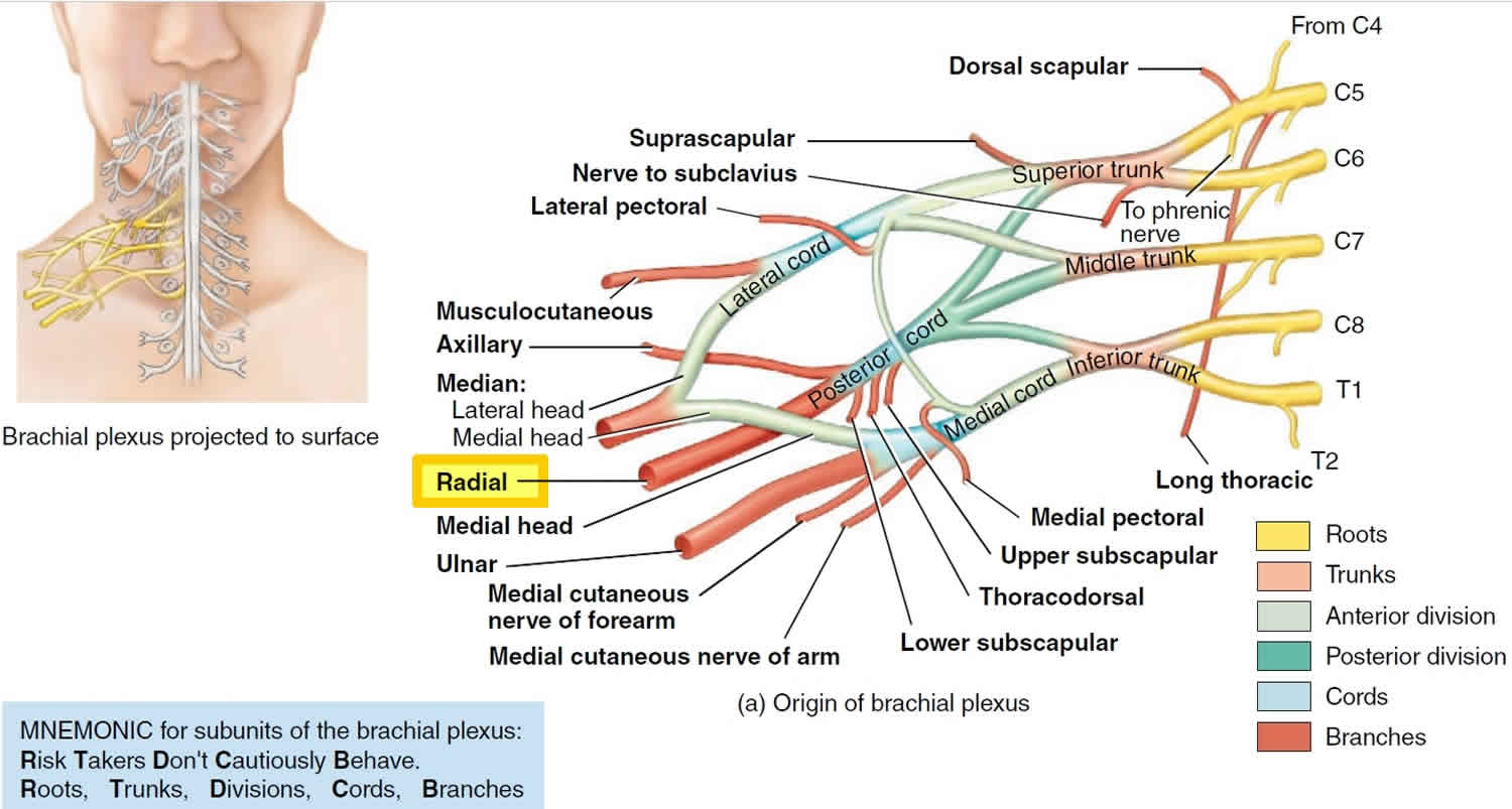
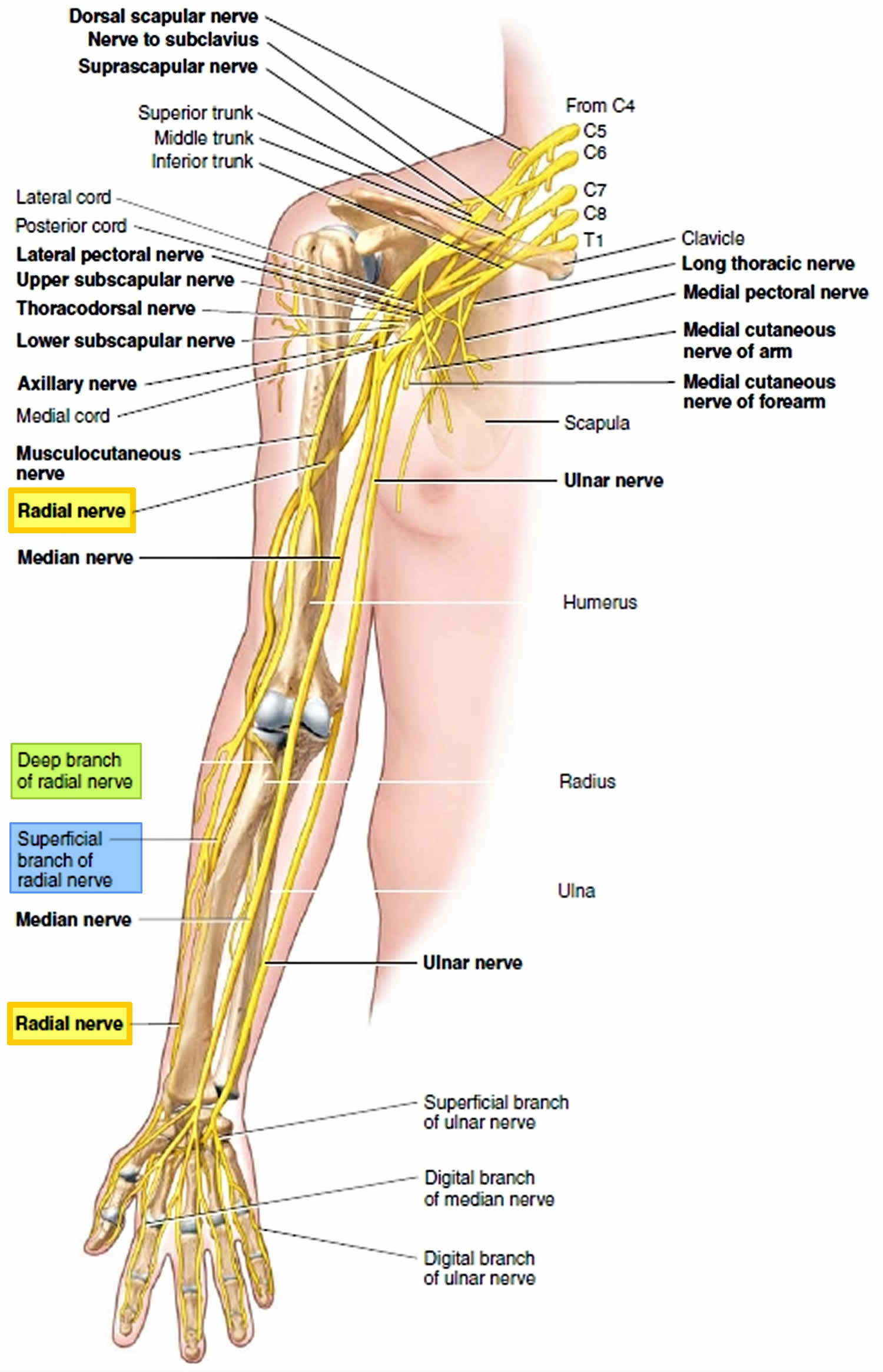
Figure 2. Radial nerve in the arm and posterior shoulder
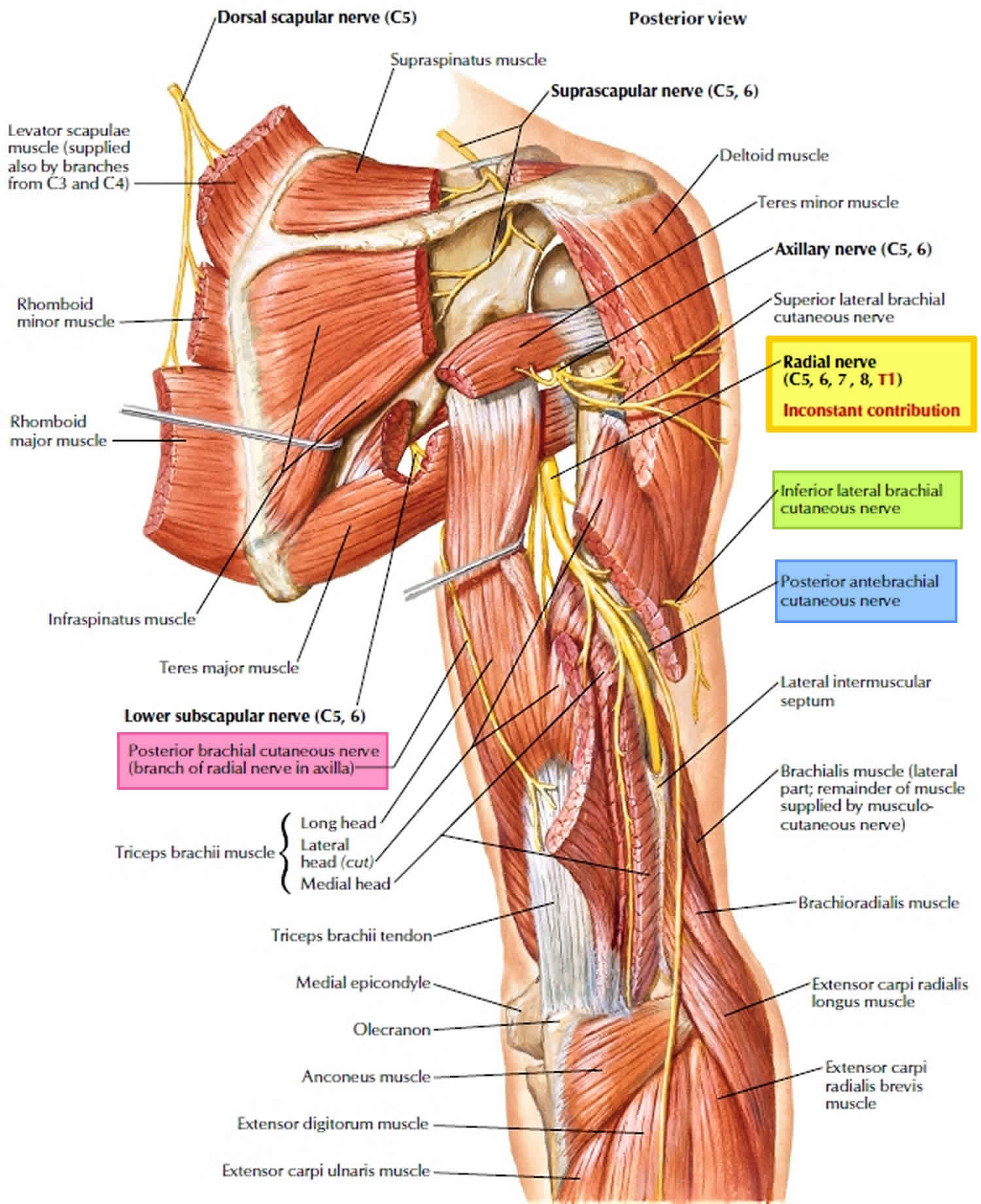
Figure 3. Radial nerve in the forearm
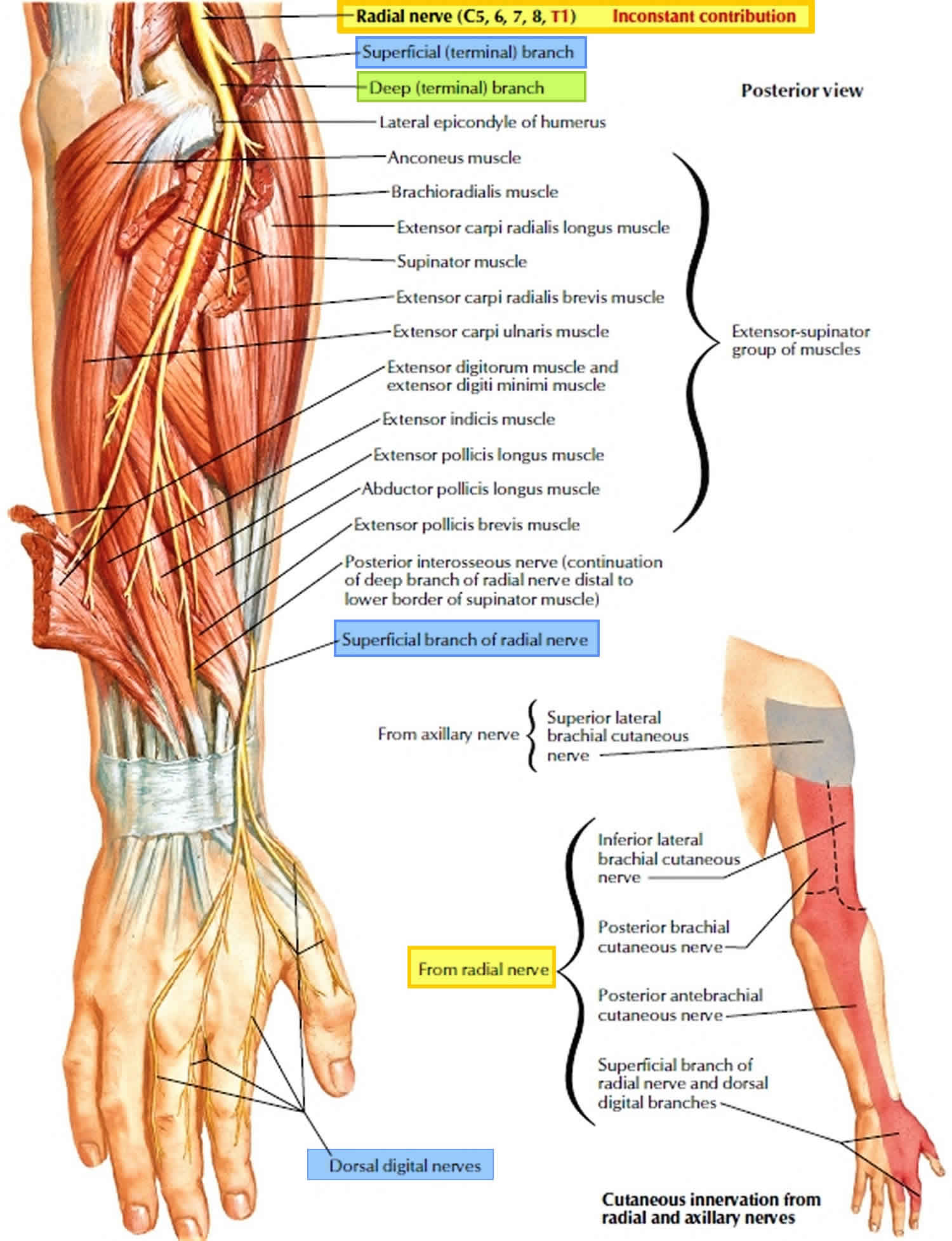
Figure 4. Radial nerve distribution
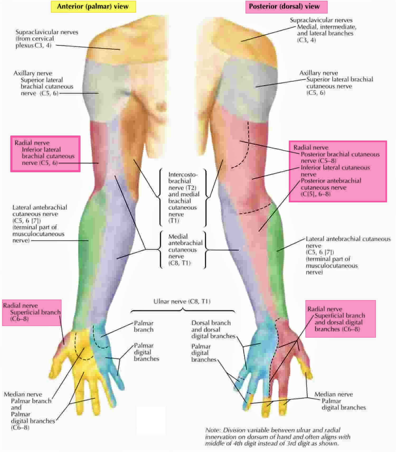
Figure 5. Radial nerve innervation
Radial tunnel syndrome causes
The anatomic radial tunnel extends from the radial head to the inferior border of the supinator muscle 12, the boundaries is formed by the supinator, extensor carpi radialis longus, extensor carpi radialis brevis, and brachioradialis muscles 13. Table 1 demonstrate the potential compression sites of the radial nerve.
Table 1. Potential radial nerve compression sites that may lead to radial tunnel syndrome:
| Order | Location | Basis for compression | Sensory involvement |
|---|---|---|---|
| 1 | Lateral elbow joint in radial head | Osteoarthritis or fibrous bands anterior to the radiocapitellar joint or synovitis of the radiocapitellar joint. | May engage sensory area |
| 2 | Leash of Henry | An arcade of anastomosing branches of the recurrent radial artery at the radial neck | May engage sensory area |
| 3 | Fibrous edge of the extensor carpi radialis brevis (ECRB) | The leading (medial proximal) edge of the extensor carpi radialis brevis | May engage sensory area |
| 4 | The arcade of Frohse | The proximal edge of the superficial layer of supinator muscle | Does not engage sensory area |
| 5 | Distal edge of radial tunnel | The distal edge of supinator muscle | Does not engage sensory area |
The entrapment of the radial nerve and its deep branch can occur at five different sites within the radial tunnel (Table 1) and most have identified it to be due to the arcade of Frohse 14. The reported female to male occurrence ratios vary from 1:1 to 6:1 15. The patients are typically between 30 to 50 years old at the time of diagnoses of radial tunnel syndrome 15. Radial tunnel syndrome usually involving the dominant side. In the study by Roles et al 16, 35 patients out of the 36 with the diagnosis of radial tunnel syndrome were right hand dominant. Bilateral involvement in a radial tunnel syndrome patient is rare. Sarhadi et al 17 only report one bilateral radial tunnel syndrome out of 26 patients. History of previous surgical procedure is a common finding in radial tunnel syndrome. Bolster et al. 15 reported 5 out of 12 patients with the diagnosis of radial tunnel syndrome had previous surgical intervention on the ipsilateral upper extremity for various pathologies such as shoulder instability, trigger finger, osteoarthritic finger joint, and carpal tunnel syndrome. In the study performed by Sotereanos et al 18, 7 out of 35 patients with the diagnosis of radial tunnel syndrome had previous surgical treatments for carpal tunnel syndrome, carpal tunnel syndrome with cuncurrent cubital tunnel syndrome, or DeQuervain’s tenosynovitis on the same side. Other contributing factors of radial tunnel syndrome occurrence may include trauma and heavy manual labor (13, 16). 13 out of 35 patients in Sotereanos’ study had history of elbow or forearm trauma 18. Roquelaure et al. 19, proposed an increased risk for radial tunnel syndrome in factory workers in their study with patients who perform effort intensive forearm extensions in pronation or supination.
Radial tunnel syndrome symptoms
Determining the exact location of the pain in the forearm, is the primary step in evaluating for radial tunnel syndrome 1. The main clinical feature of radial tunnel syndrome is a localized tenderness over the radial nerve 5 cm distal to the lateral epicondyle 1. Patients typically report aggravated pain at nights that may interfere with sleeping 1. The pain can also become more severe when increased traction is applied to the nerve by extending the elbow, pronating the forearm, or flexing the wrist. Two accepted clinical tests to confirm the diagnosis include exacerbation of the pain with resisted supination with the other being increased pain in the proximal radial forearm and over the radial tunnel when the wrist is hyperextended against resistance 15. Studies have concluded the pain elicited with resisted middle finger extension as a pathognomonic sign in diagnosing of radial tunnel syndrome 15. However, other studies noted the pain is rarely induced during resisted extension of the middle finger 20.
Radial tunnel syndrome diagnosis
Electromyography (EMG) and nerve conduction velocity (NCV) results are typically normal in patients diagnosed with radial tunnel syndrome. Electromyography (EMG) and nerve conduction velocity (NCV) studies in patients with radial tunnel syndrome are typically negative 21. Previously, studies have been performed to demonstrate nerve conduction abnormality after different active maneuvers. Rosén et al 22 compared the motor latency at rest and with active resisted supination but they could not find difference in these two different conditions. Kupfer et al. 23 measured differences in motor latency in forearm neutral, supination, and pronation positions. In the study 24, patients who previously had a confirmed diagnose of radial tunnel syndrome had a greater average differential latency compared with controls which formed the basis for the recommended differential latency of 0.30 ms or greater as diagnostic criteria for radial tunnel syndrome.
The Rule of Nine (Figure 6) is another valuable test in diagnosis of radial tunnel syndrome. Developed by Loh et al 25, the Rule of Nine is utilized when evaluating patients with non-specific elbow and proximal forearm pain. The Rule of Nine test is administered by subdividing the anterior, proximal forearm just distal to the elbow crease in to nine circular regions arranged in a 3×3 grid. The grid is about the size of a half dollar (Figure 6). Patients are asked to determine each place as painful, uncomfortable, or nothing when pressure is applied to the individual areas. Three medial pressure circles are the control areas and expected to be free of pain and discomfort. Tenderness on the two proximal circles at the lateral column indicates radial nerve irritation. The posterior interosseous branch of radial nerve lies more distally and between the two heads of supinator muscle. Thus, pressure on the third distal circle would not irritate the radial nerve and it could be used as a control site for radial tunnel syndrome in addition to the medial column 26. In the middle column, the two distal circles overlie the route of median nerve, and pain and tenderness in this area indicates a high level of median nerve irritation 26.
Figure 6. Rule of nine test
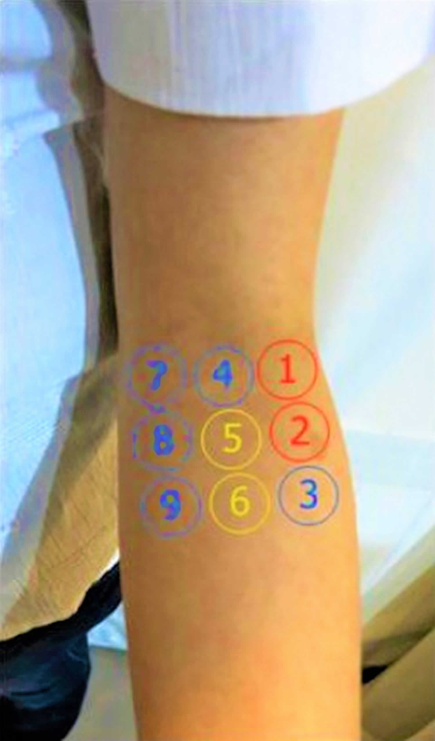
Footnote: The rule of nine test: Volar side of left proximal forearm, distal to elbow crease is divided to nine pressure points in three columns. Tenderness over two proximal lateral circles (red circles) indicates radial nerve irritation while tenderness over pressure points of 5 and 6 (yellow circles) indicates proximal median nerve irritation. Three medial points are control area.
[Source 25 ]Figure 7. Physical tests for Radial Tunnel Syndrome
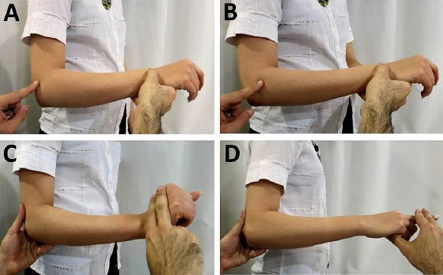
Footnote: Physical tests for Radial Tunnel Syndrome. A and B: Different sites of pain in radial tunnel syndrome and lateral epicondylitis. Tenderness in lateral epicondylitis is directly on the lateral epicondyle while in radial tunnel syndrome the tenderness is 5 cm distal to the epicondyle. C: Increased pain in the proximal radial forearm and over the radial tunnel, on hyperextension of the wrist against resistance. D: Pain during resisted extension of the middle finger is a valuable test in diagnosis of radial tunnel syndrome.
[Source 1 ]Imaging studies
MRI studies of patients with radial tunnel syndrome usually show no pathology but in some cases they may show muscle edema or atrophy along the distribution of the radial and posterior interosseous nerves (finger extensors, supinator and less, pronator muscles) but the validity of the MRI findings is controversial 27. Ferdinand et al. reported abnormal MRI findings in 21 out of 25 patients with radial tunnel syndrome. The most common finding was muscle denervation along the posterior interosseous nerve distribution within the supinator muscle 28. However some studies stated that MRI imaging does not have a key role in the radial tunnel syndrome diagnosis and workup 27.
Radial tunnel syndrome differential diagnosis
Pain on the dorsal forearm that worsens at night and arm fatigue are typical presentation of radial tunnel syndrome. However these symptoms are not specific to radial tunnel syndrome and diagnosing of radial tunnel syndrome based on the presentation is difficult 15. Another confounding factor in diagnosis of radial tunnel syndrome is the lack of confirmatory clinical tests. Therefore, radial tunnel syndrome is a diagnosis of exclusion and is dependent on clinical signs and symptoms.
The preliminary step in evaluation of radial tunnel syndrome requires the exclusion of proximal nerve pathology such as inflammation or trauma of the brachial plexus. Brachial plexus injuries can present as pain in the shoulder and upper limb. In addition, biceps muscle and its tendon may induce elbow pain that can be mistaken for radial tunnel syndrome. Tendinopathy is a commonly associated with a rotator cuff tear or shoulder disorders 29.
Ultrasound, magnetic resonance imaging (MRI), and electromyography can be utilized to rule out other pathologies that can underlie lateral elbow pain. Lateral epicondylitis, posterior interosseous nerve syndrome, osteoarthritis of the radioapitellar joint, impingement of the articular branch of the radial nerve, synovitis of the radiocapitellar joint, a muscle tear of the extensor carpi radialis brevis, and posterior plica impingement are diseases that can present with lateral elbow pain 26.
Lateral epicondylitis, which is differentiated from radial tunnel syndrome by the tenderness directly on the lateral epicondyle, has been identified concurrently in several studies to 21 – 41 % 18. Conversely, radial tunnel syndrome has been estimated to be present in 5% of patients diagnosed with lateral epicondylitis 30. Experts believe that in some patients the pain associated with lateral epicondylitis is because the extensor carpi radialis brevis (ECRB) lies next to the posterior interosseous nerve and the fact that the posterior interosseous nerve carries afferent impulses may explain the pain from the degenerated extensor carpi radialis brevis (ECRB) tendon.
Motor signs such as digital and thumb extensor motor weakness distinguish posterior interosseous nerve syndrome from radial tunnel syndrome. Even in complete posterior interosseous nerve palsy, the function of the extensor carpi radialis longus muscle and the ability to extend and radially deviate the wrist are preserved 31. posterior interosseous nerve syndrome also does not present with pain, which is the predominant complaint in patients with radial tunnel syndrome 32.
The pain due to the irritation of the superficial branch of the radial nerve can be mimicked by De Quervain’s tenosynovitis syndrome 26.
Radial tunnel syndrome treatment
The goals of treatment in radial tunnel syndrome management are pain resolution and encouraging the patient to return to work and previous activities 33. Both surgical and non-surgical treatments are available for management of radial tunnel syndrome 29.
Non-surgical treatment
Common non-surgical methods include immobilization of the wrist with splinting or casting, anti-inflammatory medication, ultrasound massage, and physical therapy 15. Radial nerve block using a local anesthetic injection through the radial tunnel in may provide a partial or complete resolution of the symptoms. Activity modifications such as avoiding prolonged elbow extension, forearm pronation, and wrist flexion are recommended as a part of the non-operative management. However, the success rate of conservative treatments is in doubt. Steven et al report only 4 out of 15 patients with the diagnosis of radial tunnel syndrome had improvement with non-surgical treatments 34. Jui-Tien Lee 30 reports similarly poor results as well. One study reported a single injection of 2 mL 1% lidocaine and 40 mg of triamcinolone in 1 mL of carrier achieved 72% pain relief after 6 weeks and 64% for a period of more than two years 17. Surgical intervention is recommended if the symptoms do not improve with three months of conservative treatments 31.
Radial tunnel syndrome exercise
Exercises to help the radial nerve slide through the tunnel at the elbow can improve symptoms. Stretching and strengthening the muscles of the forearm can also help to relieve pain and tenderness.Following a well-structured conditioning program will help you return to daily activities, as well as sports and other recreational pastimes. This exercise program for radial tunnel syndrome should be continued for 6 to 12 weeks, unless otherwise specified by your doctor or physical therapist. After your recovery, these exercises can be continued as a maintenance program.
- Do not ignore pain: You should not feel significant pain during an exercise. If your elbow pain steadily worsens, if the exercises increase the pain, or if the pain does not improve after you have performed the exercises for 6 to 12 weeks, talk to your doctor or physical therapist.
- Ask questions: If you are not sure how to do an exercise, or how often to do it, contact your doctor or physical therapist.
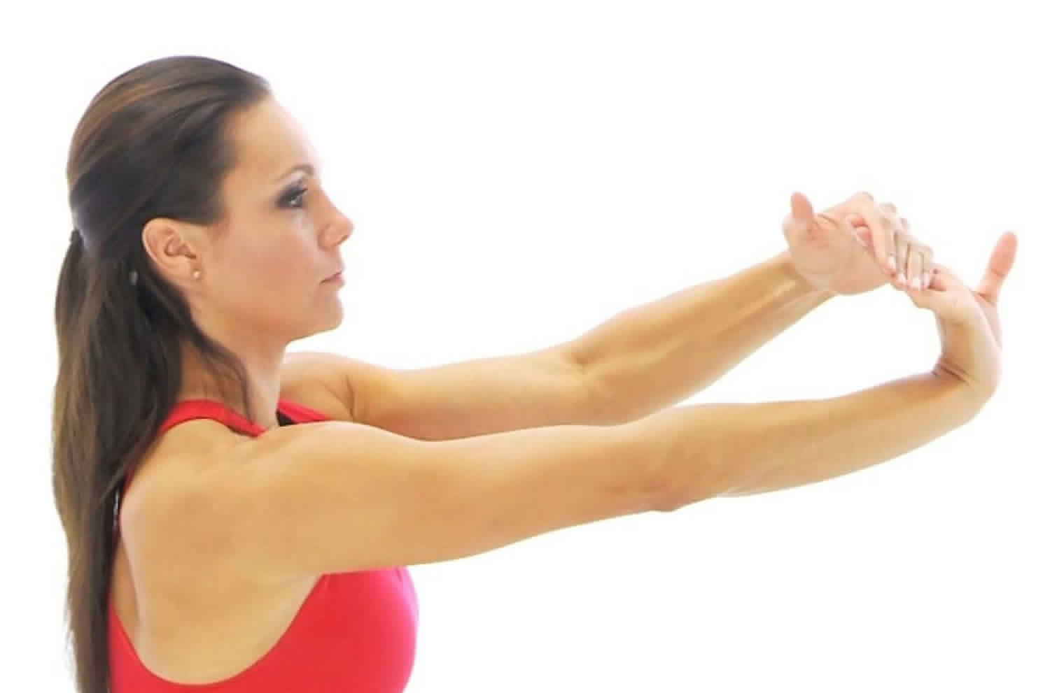
Wrist Extension Stretch
Wrist Extension Stretch should be done throughout the day, especially before activity. After recovery, Wrist Extension Stretch should be included as part of a warm-up to activities that involve
gripping, such as gardening, tennis, and golf.
- Tip: Do not lock your elbow.
Step-by-step directions
- Straighten your arm and bend your wrist back as if signaling someone to “stop.”
- Use your opposite hand to apply gentle pressure across the palm and pull it toward you until you feel a stretch on the inside of your forearm.
- Hold the stretch for 15 seconds.
- Repeat 5 times, then perform this stretch on the other arm.
Figure 8. Wrist Extension Stretch
Wrist Flexion Stretch
Wrist Flexion Stretch should be done throughout the day, especially before activity. After recovery, Wrist Flexion Stretch should be included as part of a warm-up to activities that involve
gripping, such as gardening, tennis, and golf.
- Tip: Do not lock your elbow.
Step-by-step directions
- Straighten your arm with your palm facing down and bend your wrist so that your fingers point down.
- Gently pull your hand toward your body until you feel a stretch on the outside of your forearm.
- Hold the stretch for 15 seconds.
- Repeat 5 times, then perform this stretch on the other arm.
Figure 9. Wrist Flexion Stretch

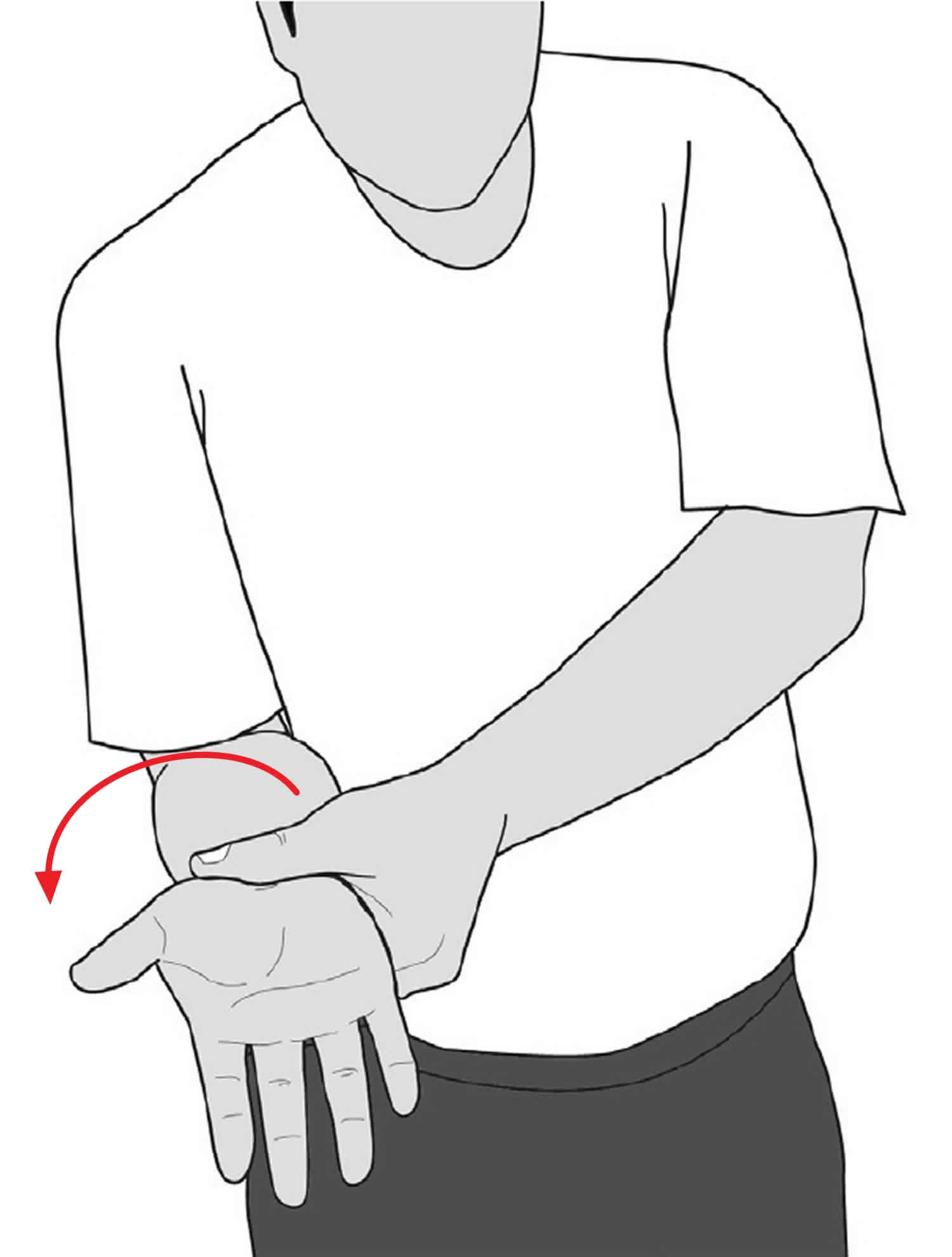
Wrist Supination
Wrist Supination stretch will help with activities that require a “palm up” position, gripping an object, and/or twisting (such as when using a screwdriver).
- Tip: Be sure to hold at your wrist – not your hand – to turn your forearm.
Step-by-step directions
- Bend your elbow at the side of your body with your palm facing the ceiling.
- Use your opposite hand to hold at your wrist and gently turn your forearm further into the palm-up position until you feel a stretch.
- Hold the stretch for 15 seconds.
- Repeat 5 times, then perform this stretch on the other arm.
Figure 10. Wrist Supination
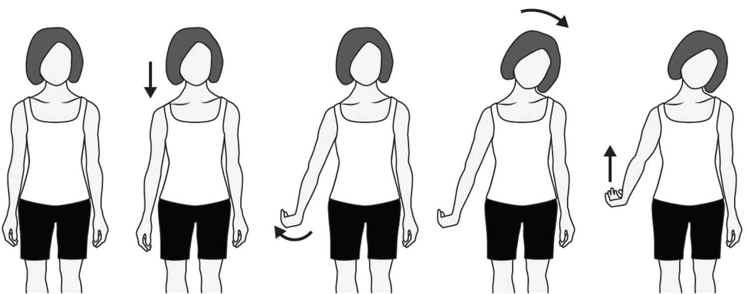
Radial Nerve Glides
Radial Nerve Glides help to restore nerve motion. This exercise will help the radial nerve glide normally through structures that are putting pressure on the nerve.
- Tip: Stretch only to the point where you feel tension.
Step-by-step directions
- Stand comfortably with your arms loose at your sides.
- Drop your shoulder down and reach your fingers toward the floor.
- Internally rotate your arm (thumb toward your body) and flex your wrist with the palm up.
- Gently tilt your head away from the side you are stretching.
- Raise your arm up and away from your body as you continue to flex your wrist and tilt your head.
- Hold each position of the glide for 3 to 5 seconds.
Figure 11. Radial Nerve Glides
Radial tunnel syndrome surgery
In 1983, Steven H 34 reported the first series of patients with radial tunnel syndrome treated surgically with the success rate of 14 out of 15 patients (93.4%). A patient whose symptoms do not respond to the non-surgical treatment is a good candidate for the surgical decompression 15. The decompression is performed for the radial nerve and both of its branches: the posterior interosseous nerve and the superficial branch of the radial nerve. The success rate for decompressing of patients suffering from radial tunnel syndrome was estimated between 10 to 95% 15. Regardless of the surgical approaches, release of the nerve from the arcade of Frohse and ligating the radial recurrent blood vessels is essential 14.
Transbrachioradialis and anterior approaches are the more frequently used approaches used today among the several known approaches to surgical release of the radial nerve. Bolster et al. 15 demonstrated in a study involving 12 patients that 11 patients (91.6%) were satisfied with the transbrachioradial approach.
Dorsal approach between mobile wad and finger extensors (Thompson)
The forearm is positioned in pronation and the incision is made one inch distal to the lateral epicondyle and extended along the midpoint of the wrist. The posterior cutaneous nerve of the forearm, usually located anterior to the incision, should be identified and protected. Then, the mobile wad, formed by the muscle bellies of the brachioradialis, extensor carpi radialis longus and brevis, is identified and dissected from the rest of the extensor muscles. A well-defined yellowish fascial which is more prominent at distal is the landmark which separates extensor carpi radialis berevis and extensor digitorom commonis mucles from each other. After separating the two muscles bluntly, the underlying supinator muscle, with its characteristic shiny oblique fibers, is identified. The arcade of Frohse is identified as a tendinous band at proximal side of supinator muscle. Initially the radial recurrent blood vessels (leash of Henry) should be ligated just proximal to the arcade of Frohse. The arcade of Frohse is released, and the superficial head of the supinator muscle is divided totally to ensure that the inferior margin of the supinator is completely released. If surgical treatment of associated lateral epicondylitis is desired, the incision can be extended proximally.
Dorsal approach between brachioradialis and wrist extensors
The skin incision is similar to dorsal (Thompson) approach. Unlike the previous approach, the interval between the brachioradialis and extensor carpi radialis longus is selected. The posterior cutaneous nerve of the forearm should be identified and protected. After identification of fascial interval between the two muscles, there belies divided from each other and the supinator mascle is exposed. The rest of the dissection can be continued bluntly to expose the arcade of Frohse. Some investigators advocate the release of the extensor carpi radialis brevis tendon at the lateral epicondyle from its origin to treat any associated lateral epicondylitis as well.
Anterior approach
While the forearm is in supination, a curvilinear incision is chosen; beginning before the lateral epicondyle and extending distally along the groove between the brachioradialis muscle and the biceps. The incision is extended between the mobile wad and brachioradialis where the radial nerve exists. After identification of the radial nerve, the nerve is followed distally as it bifurcates into the superficial radial nerve and the posterior interosseous nerve. The arcade of Frohse is released and the radial recurrent blood vessels are ligated. The entire length of the supinator muscle is visualized and completely released.
Since an anterior approach has the capability of exposure of the radial nerve both proximal and distal to the elbow, it is more useful in cases with probable compression of the radial nerve proximal to the elbow.
Transmuscular brachioradialis-splitting approach
In this approach, the incision is slightly anterior to the previous incision described with Thompson. After skin incision, the Brachioradialis fascia is released longitudinally and the muscle fibers are split along the muscle fibers bluntly until the radial nerve is exposed. The arcade of Frohse and the superficial head of the supinator muscle are divided completely.
Radial tunnel syndrome prognosis
Although non-surgical treatment shows a reliable outcome, surgical treatment is more promising. Posterior interosseous nerve release alone showed a 39% to 95% success rate while this amount in release of both posterior interosseous nerve and superficial branch of the radial nerve was equal to 67% to 92% 35. In the Stanley et al. 26 study, they concluded that if the patient could not recover completely over the period of 9 months, the surgery should be considered unsuccessful. Some studies emphasized at decompression of both posterior interosseous nerve and superficial branch of the radial nerve together for better outcome.
References- Moradi A, Ebrahimzadeh MH, Jupiter JB. Radial Tunnel Syndrome, Diagnostic and Treatment Dilemma. Arch Bone Jt Surg. 2015;3(3):156–162. https://www.ncbi.nlm.nih.gov/pmc/articles/PMC4507067
- Moradi A, Ebrahimzadeh MH, Ring D. Nonspecific Arm Pain. Arch Bone Jt Surg. 2013;1(2):53–8.
- Agarwal A, Chandra A, Jaipal U, Saini N. A panorama of radial nerve pathologies- an imaging diagnosis: a step ahead. Insights Imaging. 2018;9(6):1021-1034. https://www.ncbi.nlm.nih.gov/pmc/articles/PMC6269333/
- Kameda Y. An anomalous muscle (accessory subscapularis-teres-latissimus muscle) in the axilla penetrating the brachial plexus in man. Acta Anat (Basel) 1976;96:513–533. doi: 10.1159/000144700.
- Spinner M. Management of nerve compression lesions of the upper extremity. In: Omer GE Jr, Spinner M, editors. Management of Peripheral Nerve Problems. Philadelphia: WB Saunders; 1980. pp. 569–587.
- Lotem M, Fried A, Levy M, Solzi P, Najenson T, Nathan H. Radial palsy following muscular effort: a nerve compression syndrome possibly related to a fibrous arch of the lateral head of the triceps. J Bone Joint Surg Br. 1971;53:500–506. doi: 10.1302/0301-620X.53B3.500.
- Lubahn JD, Lister GD. Familial radial nerve entrapment syndrome: a case report and literature review. J Hand Surg Am. 1983;8:297–299. doi: 10.1016/S0363-5023(83)80163-4.
- Mayer RF, Garcia-Mullin R. Hereditary neuropathy manifested by pressure palsies: Schwann cell disorder? Trans Am Neurol Assoc. 1968;93:238–240.
- Moradi A, Ebrahimzadeh MH, Jupiter JB. Radial tunnel syndrome, diagnostic and treatment dilemma. Arch Bone Jt Surg. 2015;3(3):156–162.
- Woltman HW, Learmonth JR (1934) Progressive paralysis of the nervous interosseous dorsalis. Brain 57:25–31
- Hazani R, Engineer NJ, Mowlavi A, Neumeister M, Lee WPA, Wilhelmi BJ. Anatomic landmarks for the radial tunnel. Eplasty. 2008;8:e37.
- Lister GD, Belsole RB, Kleinert HE. The radial tunnel syndrome. J Hand Surg Am. 1979;4(1):52–9.
- Portilla Molina AE, Bour C, Oberlin C, Nzeusseu A, Vanwijck R. The posterior interosseous nerve and the radial tunnel syndrome: an anatomical study. Int Orthop. 1998;22(2):102–6.
- Clavert P, Lutz JC, Adam P, Wolfram-Gabel R, Liverneaux P, Kahn JL. Frohse’s arcade is not the exclusive compression site of the radial nerve in its tunnel. Orthop Traumatol Surg Res. 2009;95(2):114–8.
- Bolster MA, Bakker XR. Radial tunnel syndrome: emphasis on the superficial branch of the radial nerve. J Hand Surg Eur. 2009;34(3):343–7.
- Roles NC, Maudsley RH. Radial tunnel syndrome: resistant tennis elbow as a nerve entrapment. J Bone Joint Surg Br. 1972;54(3):499–508.
- Sarhadi NS, Korday SN, Bainbridge LC. Radial tunnel syndrome: diagnosis and management. J Hand Surg Br. 1998;23(5):617–9.
- Sotereanos DG, Varitimidis SE, Giannakopoulos PN, Westkaemper JG. Results of surgical treatment for radial tunnel syndrome. J Hand Surg Am. 1999;24(3):566–70.
- Roquelaure Y, Raimbeau G, Saint-Cast Y, Martin YH, Pelier-Cady MC. Occupational risk factors for radial tunnel syndrome in factory workers. Chir Main. 2003;22(6):293–8.
- Hagert CG, Lundborg G, Hansen T. Entrapment of the posterior interosseous nerve. Scand J Plast Reconstr Surg. 1977;11(3):205–12.
- Verhaar J, Spaans F. Radial tunnel syndrome. An investigation of compression neuropathy as a possible cause. J Bone Joint Surg Am. 1991;73(4):539–44.
- Rosén I, Werner CO. Neurophysiological investigation of posterior interosseous nerve entrapment causing lateral elbow pain. Electroencephalogr Clin Neurophysiol. 1980;50(1-2):125–33.
- Kupfer DM, Bronson J, Lee GW, Beck J, Gillet J. Differential latency testing: a more sensitive test for radial tunnel syndrome. J Hand Surg Am. 1998;23(5):859–64.
- van den Ende KI, Steinmann SP. Radial Tunnel Syndrome. J Hand Surg Am. 2010;35(6):1004–6.
- Loh YC, Lam WL, Stanley JK, Soames RW. A new clinical test for radial tunnel syndrome-the Rule-of-Nine test: a cadaveric study. J Orthop Surg (Hong Kong) 2004;12:83–6.
- Stanley J. Radial Tunnel Syndrome: A Surgeon’s Perspective. J Hand Ther. 2006;19(2):180–4.
- Ferdinand BD, Rosenberg ZS, Schweitzer ME, Stuchin SA, Jazrawi LM, Lenzo SR, et al. MR Imaging Features of Radial Tunnel Syndrome: Initial Experience. Radiology. 2006;240(1):161–8.
- Thomas SJ, Yakin DE, Parry BR, Lubahn JD. The anatomical relationship between the posterior interosseous nerve and the supinator muscle. J Hand Surg Am. 2000;25(5):936–41.
- Smith RS. The upper limb in primary care Part 1: Upper and lower arm, elbow. Reports Rheumatic Dis. 2012;6(11):1–10.
- Lee JT, Azari K, Jones NF. Long term results of radial tunnel releasee the effect of co-existing tenn is elbow, multipl e compressio n syndromes and workers ‘ compensation. J Plast Reconstr Aesthet Surg. 2008;61(9):1095–9.
- Dang AC, Rodner CM. Unusual Compression Neuropathies of the Forearm, Part I: Radial Nerve. J Hand Surg Am. 2009;34(10):1906–14.
- Naam NH, Nemani S. Radial tunnel syndrome. Orthop Clin North Am. 2012;43(4):529–36.
- Cleary CK. Management of radial tunnel syndrome: a therapist’s clinical perspective. J Hand Ther. 2006;19(2):186–91.
- Moss SH, Switzer HE. Radial tunnel syndrome: A spectrum of clinical presentations. J Hand Surg Am. 1983;8(4):414–20.
- De Smet L, Van Raebroeckx T, Van Ransbeeck H. Radial tunnel release and tennis elbow: disappointing results? Acta Orthop Belg. 1999;65(4):510–3.


