Rhinosporidiosis
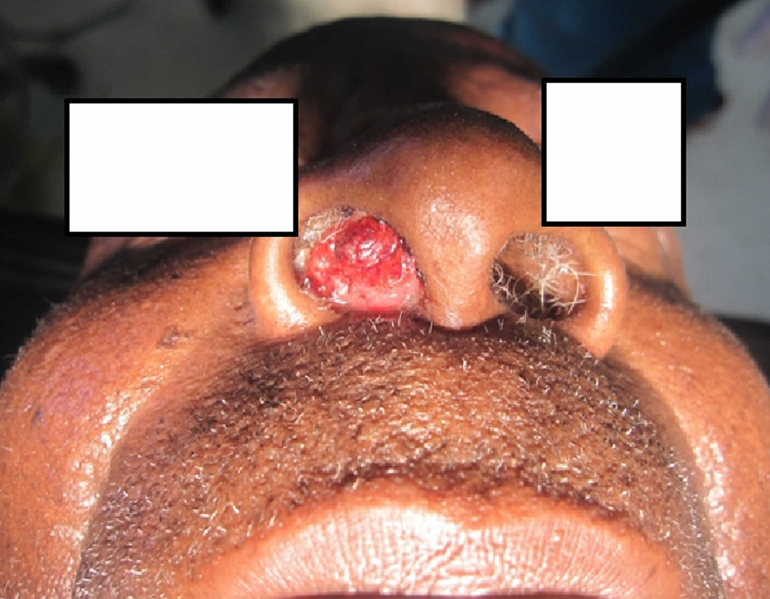
Rhinosporidiosis is a chronic granulomatous tropical disease of the mucous membranes and in rare cases, the skin or internal organs caused by Rhinosporidium seeberi, an enigmatic microorganism of disputed taxonomy 1. Rhinosporidiosis usually manifests as vascular friable polyps that arise from the nasal mucosa or external structures of the eye 2. Rhinosporidiosis usually affects mainly the nasal mucosa (70%), nasopharynx (6%) and conjunctiva 3. Rhinosporidium seeberi has not been isolated from the environment and has no known natural host or reservoir; consequently, it has been difficult to classify. Originally considered to be a protozoan and subsequently a fungus, Rhinosporidium seeberi is currently classified as an aquatic mesomycetazoan (“between fungi and animals”), on the basis of phylogenetic analysis of 18S ribosomal ribonucleic acid (rRNA) gene 4. This classification is not without controversy, as other researchers have presented data that Rhinosporidium seeberi is a cyanobacterium, further demonstrating the difficulties that arise when working with pathogens that cannot be maintained in the laboratory setting 5. Other molecular work has demonstrated evidence that Rhinosporidium seeberi may have host-specific strains (eg, human vs dog vs swan) 6.
Originally described in Argentina in 1900 7, rhinosporidiosis is most prevalent in India and Sri Lanka, followed by countries in South America and Africa 8. Rhinosporidiosis cases from the United States and Southeast Asia, as well as scattered occurrences throughout the world, have also been reported. Most cases of rhinosporidiosis occur in persons from or residing in the Indian subcontinent or Sri Lanka. In addition to humans, rhinosporidiosis has been noted in cats, cattle, dogs, ducks, goats, horses, mules, parrots, and swan 9. In Africa, rhinosporidiosis has been documented in Cameroon, Democratic Republic of the Congo, Côte d’Ivoire, Kenya, Malawi, South Africa, Tanzania, Uganda, and Zambia, predominantly as conjunctival disease 10. One previous report documented 3 cases of rhinosporidiosis in Rwanda: 2 in the nose (in a 60-year-old man in 1975 and in a 16-year-old young woman in 1977) and 1 in the conjunctiva (in a 3-year-old boy in 1986) 11.
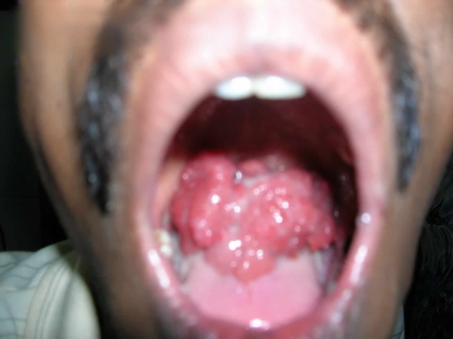
Figure 1. Nasal rhinosporidiosis
Footnote: Polypoidal nasal mass which had grown up gradually and stuffed the right nasal cavity.
[Source 12 ]Figure 2. Conjunctival rhinosporidiosis
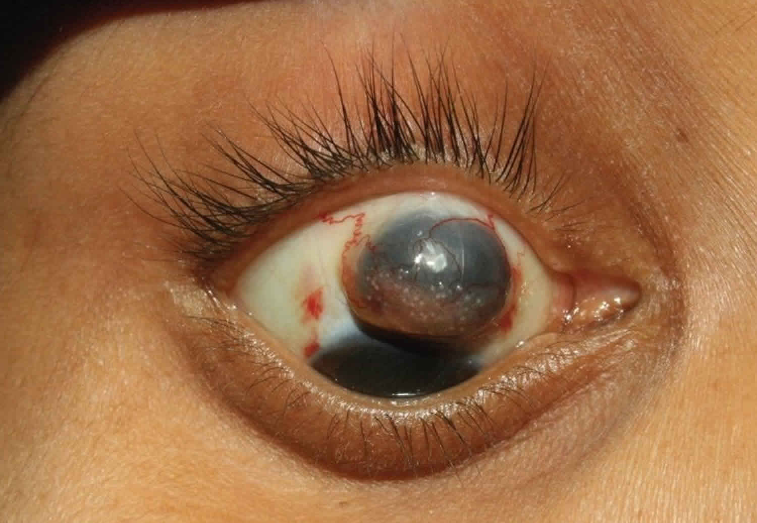
Footnote: Preoperative photograph showing spherules of rhinosporidiosis in the superior bulbar conjunctiva with staphyloma.
[Source 13 ]Rhinosporidiosis causes
The etiologic agent of rhinosporidiosis, Rhinosporidium seeberi, has traditionally been considered a fungus. Recent 18S ribosomal ribonucleic acid (rRNA) gene analysis has placed Rhinosporidium seeberi into a novel group of aquatic parasites of the class Mesomycetozoea, some of which cause similar diseases in amphibians and fish 14.
Most persons with rhinosporidiosis have had bathing or working exposure to stagnant water 15.
No immune deficiency has been associated with Rhinosporidium seeberi infection.
Rhinosporidiosis symptoms
Rhinosporidiosis is an infection that is typically limited to the mucosal epithelium. Infection usually results from a local traumatic inoculation with the organism. The disease progresses with the local replication of Rhinosporidium seeberi and associated hyperplastic growth of host tissue and a localized immune response.
Soft polyps may develop on the nose or eye. These polyps are pink to deep red, are sessile or pedunculated, and are often described as strawberrylike in appearance. Because the polyps of rhinosporidiosis are vascular and friable, they bleed easily upon manipulation. This appearance results from sporangia, which are visible as gray or yellow spots in the vascular polypoid masses.
Infection of the nose and nasopharynx is observed in 70% of persons with rhinosporidiosis; infection of the palpebral conjunctivae or associated structures (including the lacrimal apparatus) is observed in 15% 16.
Other structures of the mouth, upper airway, and eye may be sites of disease. Disease of the skin, ear, larynx, trachea, bronchi, genitals, and rectum has also been described 17. Genital rhinosporidiosis has been described in the vagina, penile urethra or meatus, and scrotum 18. Dissemination with cutaneous and multisite disease is also reported, but this is much less common. Isolated cases of dissemination involving deep organs have been rarely reported 19.
Nasal rhinosporidiosis may present with unilateral nasal obstruction or epistaxis. Other symptoms may include local pruritus, coryza with sneezing, rhinorrhea, and postnasal discharge (drip) with cough. Patients often report a sensation that a foreign body is present in their nasal canal.
Eye involvement is initially asymptomatic. Increased tearing may be reported as the disease progresses. Photophobia, redness, and secondary infection may occur.
Skin lesions begin as papillomas that gradually become verrucous.
Rhinosporidiosis complications
Complications of rhinosporidiosis include extremely rare, life-threatening dissemination, local secondary bacterial infection, and recurrence.
Rhinosporidiosis diagnosis
Apart from history and clinical findings, histopathology of the biopsied tissues from the lesion is mandatory for definitive diagnosis of rhinosporidiosis. Rhinosporidiosis is diagnosed by identifying the typical structures of Rhinosporidium seeberi directly on microscopic examination. This includes examination of smears of macerated tissue or histology of prepared biopsy sample sections 20.
The organism can be observed with typical fungal stains (eg, Gomori methenamine silver [GMS], periodic acid-Schiff [PAS]), as well as with standard hematoxylin and eosin (H&E) staining.
Smears can also be observed with potassium hydroxide (KOH) preparation.
Serologic testing
Serologic testing immunoblot or enzyme-linked immunosorbent assay [ELISA]) identification of antirhinosporidial antibody has been developed and used for epidemiologic studies in endemic areas, but this testing is not available for or routinely used in patient diagnosis 21.
Rhinosporidiosis treatment
Surgical excision remains the mainstay of treatment for rhinosporidiosis lesions. Wide, complete and meticulous excision of the polyp followed by thorough electro-cautery of the lesion’s base is recommended 22. Recurrence has been reported with simple excision. It is hypothesized that cauterization of the lesion’s base may abate recurrence resulting from spillage of endospores on the adjacent mucosa 23.
Besides surgery, a variety of adjuvant medical therapies have been tried in treatment of rhinosporidiosis. These include drugs like griseofluvin, amphoterecin B and dapsone (4, 4-diaminodiphenyl sulphone) 24. This drug may be useful in individuals with multisite rhinosporidiosis. However, by far there has been no tangible success with medical therapy 25.
Rhinosporidiosis prognosis
The prognosis of rhinosporidiosis is excellent, except with dissemination. Patients with rhinosporidiosis should be advised that recurrence is possible. They should be instructed to return for further evaluation if symptoms recur or new symptoms arise.
Cases of dissemination or more extensive disease have been associated with a prior history of both treated and untreated nasal disease. Accordingly, ensure that patients are instructed to (1) be vigilant for a recurrence of symptoms or new lesions and (2) promptly consult with their physician if these are noted.
References- Izimukwiye, A. I., Mbarushimana, D., Ndayisaba, M. C., Bigirimana, V., Rugwizangoga, B., & Laga, A. C. (2019). Cluster of Nasal Rhinosporidiosis, Eastern Province, Rwanda. Emerging Infectious Diseases, 25(9), 1727-1729. https://dx.doi.org/10.3201/eid2509.190021
- Rhinosporidiosis. https://emedicine.medscape.com/article/227734-overview
- Pal DK, Moulik D, Chowdhury MK. Genitourinary rhinosporidiosis. Indian J Urol. 2008;24(3):419–421. doi:10.4103/0970-1591.42632 https://www.ncbi.nlm.nih.gov/pmc/articles/PMC2684352
- Vilela R, Mendoza L. The taxonomy and phylogenetics of the human and animal pathogen Rhinosporidium seeberi: a critical review. Rev Iberoam Micol. 2012;29:185–99.
- Vilela R, Mendoza L. The taxonomy and phylogenetics of the human and animal pathogen Rhinosporidium seeberi: A critical review. Rev Iberoam Micol. 2012 Apr 12.
- Silva V, Pereira CN, Ajello L, Mendoza L. Molecular evidence for multiple host-specific strains in the genus Rhinosporidium. J Clin Microbiol. 2005 Apr. 43(4):1865-8.
- Seeber GR. A new sporozoan parasite of man. Two cases found in nasal polyps. Thesis presented to qualify for the degree of doctor of medicine [in Spanish]. Buenos Aires: Universidad de Buenos Aires, Facultad de Ciencias Médicas, Imprenta y Libreria Boullosa; 1900.
- Almeida FA, Feitoza LM, Pinho JD, Mello GC, Lages JS, Silva FF, et al. Rhinosporidiosis: the largest case series in Brazil. Rev Soc Bras Med Trop. 2016;49:473–6.
- Kennedy FA, Buggage RR, Ajello L. Rhinosporidiosis: a description of an unprecedented outbreak in captive swans (Cygnus spp.) and a proposal for revision of the ontogenic nomenclature of Rhinosporidium seeberi. J Med Vet Mycol. 1995 May-Jun. 33(3):157-65.
- Kaimbo KW, Parys-Van Ginderdeuren R. Conjunctival rhinosporidiosis: a case report from a Congolese patient. Bull Soc Belge Ophtalmol. 2008;309–310:19–22.
- Gigase P, Kestelyn P. Further African cases of rhinosporidiosis. Ann Soc Belg Med Trop. 1993;73:149–52.
- Uledi S, Fauzia A. Human nasal rhinosporidiosis: a case report from Malawi. Pan Afr Med J. 2011;9:27. https://www.ncbi.nlm.nih.gov/pmc/articles/PMC3215549
- Jacob P, Rose JS, Hoshing A, Chacko G. Tectonic corneal graft for conjunctival rhinosporidiosis with scleral melt. Indian J Ophthalmol. 2011;59(3):251–253. doi:10.4103/0301-4738.81046 https://www.ncbi.nlm.nih.gov/pmc/articles/PMC3120253
- Mendoza L, Taylor JW, Ajello L. The class mesomycetozoea: a heterogeneous group of microorganisms at the animal-fungal boundary. Annu Rev Microbiol. 2002. 56:315-44.
- Arseculeratne SN, Sumathipala S, Eriyagama NB. Patterns of rhinosporidiosis in Sri Lanka: comparison with international data. Southeast Asian J Trop Med Public Health. 2010 Jan. 41(1):175-91.
- Pushker N, Kashyap S, Bajaj MS, Meel R, Sood A, Sharma S, et al. Primary lacrimal sac rhinosporidiosis with grossly dilated sac and nasolacrimal duct. Ophthal Plast Reconstr Surg. 2009 May-Jun. 25(3):234-5.
- Deshpande AH, Agarwal S, Kelkar AA. Primary cutaneous rhinosporidiosis diagnosed on FNAC: a case report with review of literature. Diagn Cytopathol. 2009 Feb. 37(2):125-7.
- Sasidharan K, Subramonian P, Moni VN, Aravindan KP, Chally R. Urethral rhinosporidiosis. Analysis of 27 cases. Br J Urol. 1987 Jan. 59(1):66-9.
- Adiga BK, Singh N, Arora VK, Bhatia A, Jain AK. Rhinosporidiosis. Report of a case with an unusual presentation with bony involvement. Acta Cytol. 1997 May-Jun. 41(3):889-91.
- Arseculeratne SN, Sumathipala S, Eriyagama NB. Patterns of rhinosporidiosis in Sri Lanka Comparison with international data. Southeast Asian J Trop med Public health. 2010;41(1):175–191.
- Sudasinghe T, Rajapakse RP, Perera NA, Kumarasiri PV, Eriyagama NB, Arseculeratne SN. The regional sero-epidemiology of rhinosporidiosis in Sri Lankan humans and animals. Acta Trop. 2011 Oct-Nov. 120(1-2):72-81.
- Kaimbo wa Kaimbo D, Parys-Van Ginderdeuren R. A case report from a Congolese patient. Bull Soc Belge Ophtalmol. 2008;309-310:19–22.
- Godwins O Echejoh, A gabus N Manasseh, Matthew N Tanko, et al. Nasal rhinosporidiosis. J Natl Med Assoc. 2008;100(6):713–5.
- Madke B, Mahajan S, Kharkar V, Chikhalkar S, Khopkar U. Disseminated cutaneous with nasopharyngeal rhinosporidiosis: Light microscopy changes following dapsone therapy. Australas J Dermatol. 2011 May. 52(2):e4-6.
- Nayak S, Acharjya B, Devi B, Sahoo A, Singh N. Disseminated cutenous rhinosporidiosis. Indian J Dermatol venereol leprol. 2007;73:185–7.


