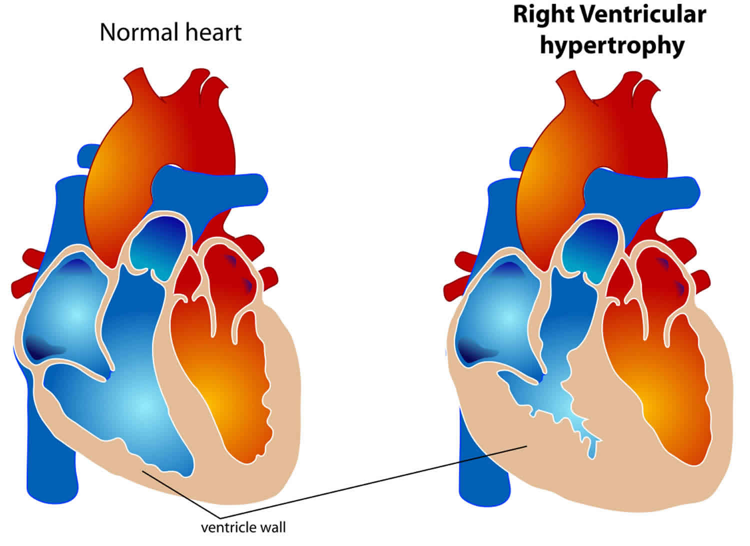
Right ventricular hypertrophy
Right ventricular hypertrophy is an abnormal enlargement or pathologic increase in muscle mass of the right ventricle in response to pressure overload, most commonly due to severe lung disease 1. The right ventricle is considerably smaller than the left ventricle and produces electrical forces that are largely obscured by those generated by the larger left ventricle 2.
Size and function of the right ventricle are adversely affected by the following:
- Pulmonary hypertension with or without left ventricular dysfunction
- Conditions that affect the tricuspid valve leading to significant tricuspid regurgitation
The right ventricle is composed of inflow (sinus) and outflow (conus) regions, separated by a muscular ridge, the crista supraventricularis. The inflow region includes the tricuspid valve, the chordae/papillary muscles as well as the body of the right ventricle. The right ventricle body boundaries are formed by the right ventricle free wall, extending from the interventricular septum’s anterior and posterior aspects. The standard septal curvature convexes toward the right ventricle cavity and imparts a crescent shape to the right ventricle when cross-sectioned. The right ventricle’s interior surface is heavily trabeculated; this feature along with the moderator band and more apical insertion of the tricuspid valve-annulus impart key morphologic differences that distinguish the right ventricle from the left ventricle by echocardiography. In contrast, the infundibulum is a smooth, funnel-shaped outflow portion of the right ventricle that ends at the pulmonic valve. Thus, the right ventricle has a complex geometry, with traditional right ventricle free-wall thickness of 0.3-0.5 cm, imparting greater distensibility and larger cavity volumes in the right ventricle versus the left ventricle, despite lower end-diastolic filling pressures. This translates to an right ventricle ejection fraction (RVEF) that is typically 35% to 45% (versus 55% to 65% in the left ventricle) yet generates the identical stroke volume as the left ventricle.
Changes in preload, afterload, and intrinsic contractility of the ventricle influence the systolic function of the right ventricle, like the left ventricle. Differences in right ventricle muscle fiber orientation dictate that the body of the right ventricle shortens symmetrically in the longitudinal and radial planes; thus, longitudinal shortening accounts for a much larger proportion of right ventricle ejection than in the left ventricle. The relatively conspicuous right ventricle shortening along the longitudinal axis can measure right ventricle systolic function using uncomplicated techniques that do not require geometric assumptions or meticulous endocardial definition, both being known limitations to the noninvasive assessment of right ventricle systolic function.
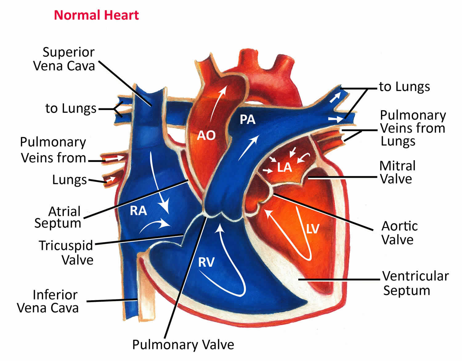
How the Heart Works
Your heart is a muscular double pump about the size of the palm of your hand with two functions (see Figure 1):
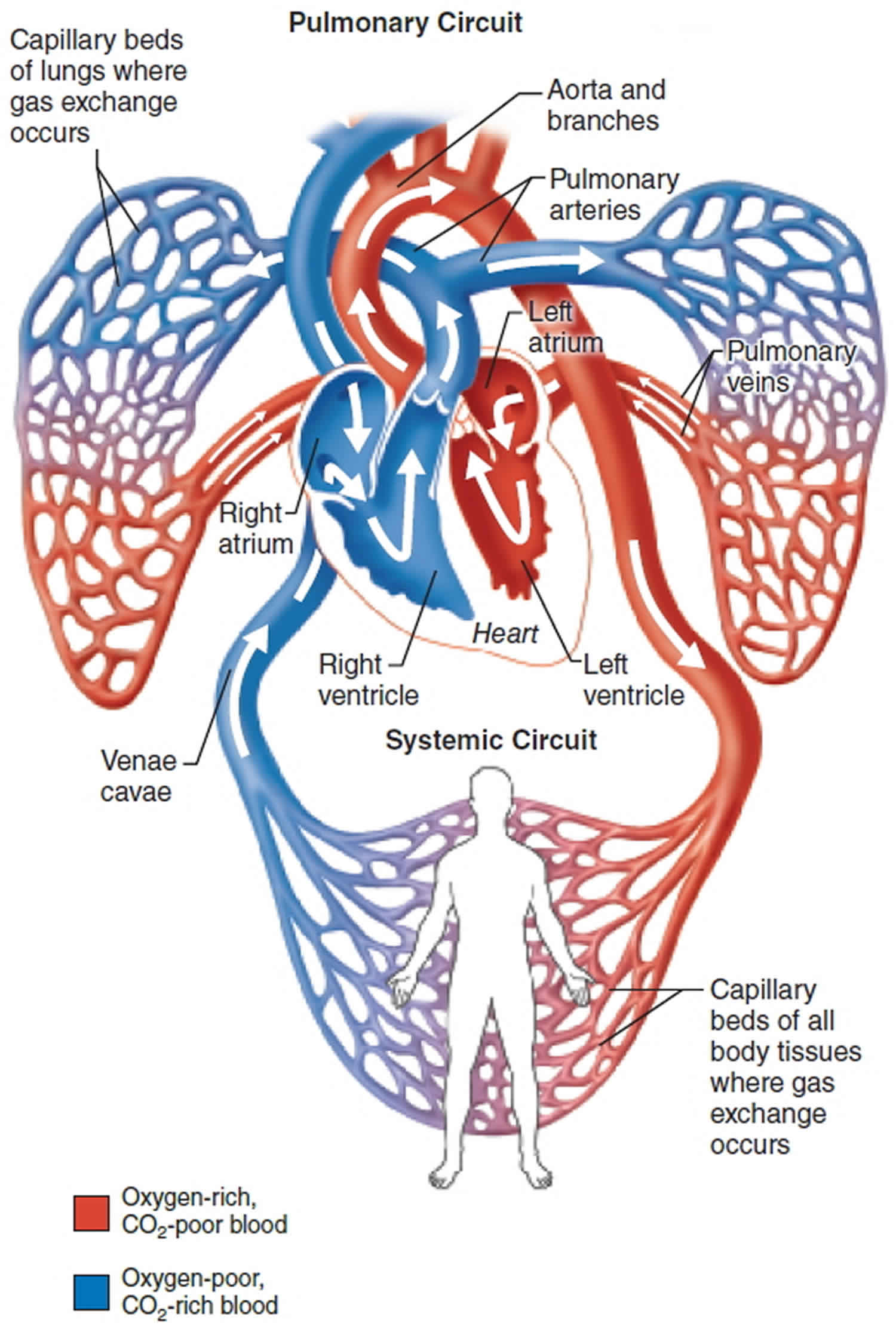
- Right side of the heart receives oxygen-poor blood from the body tissues and then pumps this blood to the lungs to pick up oxygen and dispel carbon dioxide. The blood vessels that carry blood to and from the lungs form the pulmonary circuit (pulmonos = lung). The right side of the heart pumps blood through the lungs, enabling blood to pick up oxygen and unload carbon dioxide. Even while you are sleeping, your heart pumps 30 times its own weight each minute, which amounts to about 5 liters (5.3 qt) to the lungs and the same volume to the rest of the body. At this rate, your heart pumps more than about 14,000 liters (3600 gal) of blood in a day, or 5 million liters (1.3 million gal) in a year. You don’t spend all of your time sleeping, however, and your heart pumps more vigorously when you are active. Thus, the actual blood volume your heart pumps in a single day is much larger.
- Left side of the heart receives the oxygenated blood returning from the lungs and pumps this blood throughout the body to supply oxygen and nutrients to the body tissues. The blood vessels that transport blood to and from all body tissues and back to the heart form the systemic circuit. The left side of the heart pumps blood through an estimated 100,000 km (60,000 mi) of blood vessels, which is equivalent to traveling around the earth’s equator about three times.
The heart has four chambers 3, two on the right and two on the left:
- Two upper chambers are called atrium (two is called an atria). The atria collect blood as it flows into the heart.
- Two lower chambers are called ventricles. The ventricles pump blood out of the heart to the lungs or other parts of the body.
The heart also has four valves that open and close to let blood flow from the atria to the ventricles and from the ventricles into the two large arteries connected to the heart in only one direction when the heart contracts (beats). The four heart valves are:
- Tricuspid valve, located between the right atrium and right ventricle
- Pulmonary or pulmonic valve, between the right ventricle and the pulmonary artery. This artery carries blood from the heart to the lungs.
- Mitral valve, between the left atrium and left ventricle
- Aortic valve, between the left ventricle and the aorta. This aorta carries blood from the heart to the body.
Each valve has a set of flaps (also called leaflets or cusps). The mitral valve has two flaps; the others have three. Valves are like doors that open and close. They open to allow blood to flow through to the next chamber or to one of the arteries. Then they shut to keep blood from flowing backward. Blood flow occurs only when there’s a difference in pressure across the valves, which causes them to open. Under normal conditions, the valves permit blood to flow in only one direction.
The heart four chambers and four valves and is connected to various blood vessels. Veins are blood vessels that carry blood from the body to the heart. Arteries are blood vessels that carry blood away from the heart to the body.
The heart pumps blood to the lungs and to all the body’s tissues by a sequence of highly organized contractions of the four chambers. For the heart to function properly, the four chambers must beat in an organized way.
When the heart’s valves open and close, they make a “lub-DUB” sound that a doctor can hear using a stethoscope 4.
- The first sound—the “lub”—is made by the mitral and tricuspid valves closing at the beginning of systole. Systole is when the ventricles contract, or squeeze, and pump blood out of the heart.
- The second sound—the “DUB”—is made by the aortic and pulmonary valves closing at the beginning of diastole. Diastole is when the ventricles relax and fill with blood pumped into them by the atria.
Figure 1. Anatomy of human heart

Figure 2. Anatomy of the heart chambers
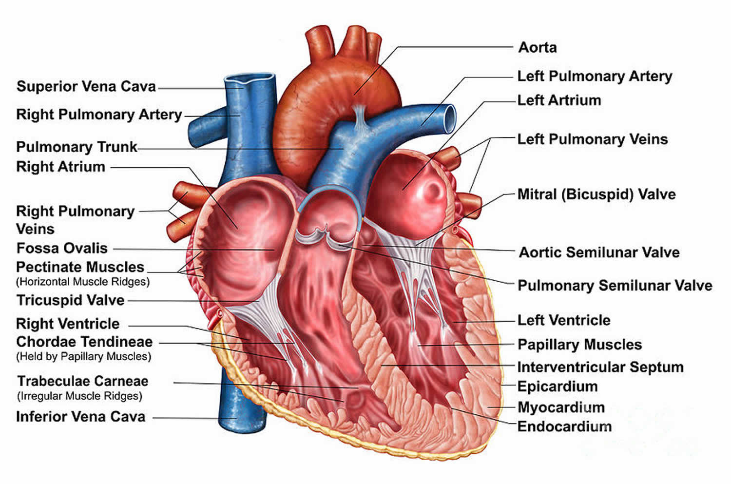
Figure 3. Heart function – double pump
Causes of right ventricular hypertrophy
The most common cause of right ventricular hypertrophy is severe lung disease. The disorders that induce pulmonary hypertension and secondary right ventricular hypertrophy include the following:
- Pulmonary arterial hypertension. Pulmonary arterial hypertension (PAH) is abnormally high blood pressure in the arteries carrying blood to the lungs, causing damage to the right side of the heart and heart failure. It is caused by an overgrowth of cells in the blood vessels of the lungs.
- Pulmonary hypertension owing to left heart disease
- Pulmonary hypertension from lung disease and/or hypoxia
- Chronic thromboembolic pulmonary hypertension
- Pulmonary hypertension with unclear multifactorial mechanisms
- Cardiac fibrosis
- Chronic obstructive pulmonary disease (COPD)
- Cystic fibrosis
- Mitral stenosis 5
- Pulmonic regurgitation
- Pulmonic stenosis
- Tetralogy of Fallot
- Ventricular septal defect
Causes of primary tricuspid regurgitation include:
- Direct valve injury secondary to cardiac instrumentation, chest trauma, Ebstein anomaly, rheumatic fever, infective and marantic endocarditis, right ventricular ischemia/infarction, tricuspid valve prolapse, connective tissue disorder, carcinoid syndrome and drug-induced tricuspid regurgitation.
Life threatening causes of right ventricular hypertrophy:
Life-threatening causes include conditions which may result in death or permanent disability within 24 hours if left untreated.
- Pulmonary embolism
Other less common causes of right ventricular hypertrophy are high altitude, left to right shunt, an atrial septal defect, anomalous pulmonary venous return, Eisenmenger syndrome, stenosis of the pulmonic valve or pulmonary artery, hyperthyroidism, cardiac fibrosis and cardiomyopathy affecting right ventricle, and athlete heart syndrome.
Right ventricular hypertrophy pathophysiology
The primary cause of significant pulmonary hypertension is almost always increased pulmonary vascular resistance. Increased flow alone (i.e., right ventricle output) does not usually cause significant pulmonary hypertension because the pulmonary vascular bed vasodilates and recruits vessels in response to increased flow. Similarly, increased pulmonary venous pressure (represented by the alveolar occlusion pressure) alone does not usually cause significant pulmonary hypertension. However, a chronic increase of either flow and/or pulmonary venous pressure can increase pulmonary vascular resistance. Numerous medical conditions can alter all variables:
- Increased pulmonary vascular resistance may be due to conditions associated with occlusive vasculopathy (remodeling and altered vascular tone) of the small pulmonary arteries and arterioles (conditions associated with pulmonary arterial hypertension), conditions that induce hypoxic vasoconstriction (hypoventilation syndromes and parenchymal lung disease), or conditions that decrease the cross-sectional area of the pulmonary vascular bed (pulmonary emboli, interstitial lung disease).
- Increased flow through the pulmonary vasculature may be due to congenital heart defects with left-to-right shunt (atrial septal defects, ventricular septal defects, patent ductus arteriosus), anemia, or liver cirrhosis.
- Increased pulmonary venous pressure may be due to left ventricular systolic or diastolic dysfunction, mitral valve disease, constrictive or restrictive cardiomyopathy or pulmonary venous obstruction (eg, pulmonary veno-occlusive disease).
Pulmonary arterial hypertension is a proliferative vasculopathy of the small pulmonary muscular arterioles (less than 50 microns) that is often characterized by vasoconstriction, hyperplasia, hypertrophy, fibrosis, and thrombosis involving all three layers of the vascular wall (intima, media, and adventitia). Patients with idiopathic pulmonary arterial hypertension and heritable variants may have a genetic predisposition to pulmonary arterial hypertension (bone morphogenetic protein receptor 2 mutations) with additional contributing mechanisms including vasoactive mediators, potassium channel dysfunction, and abnormal response to estrogen. The pathogenesis of other forms of pulmonary arterial hypertension such as drugs and toxins, connective tissue disease, congenital heart disease, human immunodeficiency virus, portopulmonary hypertension, schistosomiasis, pulmonary veno-occlusive disease, or persistent pulmonary hypertension of the newborn is poorly understood.
Tricuspid regurgitation is characterized by the backflow of blood into the right atrium during systole. Since the right atrium is relatively compliant, there are often no major hemodynamic consequences with mild or moderately severe tricuspid regurgitation. However, when tricuspid regurgitation is severe, right atrial and venous pressure rise and can result in the signs and symptoms of right-sided heart failure. In such patients, right ventricular pressure and/or volume overload frequently lead to right ventricular systolic dysfunction and a low forward cardiac output.
Right ventricular hypertrophy symptoms
Symptoms due to pulmonary hypertension (either primary or secondary) and/or severe tricuspid regurgitation disease include the following:
- Exertional chest pain (angina), usually due to subendocardial hypoperfusion caused by increased right ventricular wall stress and myocardial oxygen demand but occasionally caused by dynamic compression of the left main coronary artery by an enlarged pulmonary artery. This risk is greatest for patients with a pulmonary artery trunk at least 40 millimeters in diameter
- Peripheral edema due to right ventricular failure, increased right-sided filling cardiac pressures, and extracellular volume expansion.
- Exertional syncope due to the inability to increase cardiac output during activity or reflex bradycardia that is secondary to mechanoreceptor activation in the right ventricle.
- Anorexia and/or right upper quadrant abdominal pain due to passive hepatic congestion.
Uncommon symptoms include a cough, hemoptysis, and hoarseness (Ortner syndrome). Compression of the left recurrent laryngeal nerve by a dilated main pulmonary artery causes the hoarseness.
Physical examination
The most prominent features of the physical examination in patients with tricuspid regurgitation are those related to the regurgitant murmur and the development of right-sided heart failure. With severe right heart failure, the patient often looks cachectic, chronically ill, cyanotic, and occasionally jaundiced (reflecting hepatic dysfunction). If tricuspid regurgitation is due to left ventricular dysfunction, signs of left-sided heart failure may dominate. Patients with pulmonary hypertension may develop physical signs as they progress from pulmonary hypertension alone to pulmonary hypertension associated with right ventricular failure/hypertrophy. The systemic venous hypertension caused by right ventricular failure can lead to a variety of findings, such as elevated jugular venous pressure, a right ventricular third heart sound, and a prominent V wave in the jugular venous pulse. Hepatomegaly, a pulsatile liver, peripheral edema, ascites, and pleural effusion also may exist. Some patients with pulmonary hypertension due to severe chronic obstructive pulmonary disease (COPD) develop edema even in the absence of right heart failure.
Jugular veins
Distended and prominent jugular veins are apparent and reflect right atrial pressure elevation.
There is a presence of distinct “c-v” wave due to systolic regurgitation through the tricuspid valve into the right atrium. With the development of ventricular hypertrophy, a prominent “a” wave may emerge within the jugular venous pulse and may be accompanied by a right-sided fourth heart sound and either a left parasternal heave or a downward subxiphoid thrust.
Jugular venous distension may be more prominent with inspiration (Kussmaul sign), a result of the increase in venous return. This finding, however, may not be very obvious with marked venous distension.
Palpation
Palpation of the chest may reveal a dynamic right ventricular heave due to the dilated right ventricle. A sustained systolic left parasternal lift is most frequently appreciated in the presence of significant right ventricular hypertrophy. Long-standing, severe pulmonary arterial hypertension, whether precapillary (idiopathic pulmonary arterial hypertension or pulmonary valve stenosis) or postcapillary (mitral stenosis, cardiomyopathy), produces right ventricular hypertrophy and a sustained lower left parasternal lift. It may be associated with a palpable presystolic a wave preceding the right ventricular lift (heave); suggesting decreased right ventricular compliance. Epigastric and subxiphoid pulsations are usually abnormal and are related to right ventricular hypertrophy and dilation or the abdominal aortic aneurysm. However, in patients with emphysema, subxiphoid pulsations may not always indicate right ventricular hypertrophy.
A hyperdynamic but not sustained left parasternal systolic impulse may be palpable when right ventricular volume is increased, as with an atrial septal defect or tricuspid regurgitation. The left parasternal impulse becomes sustained during systole when pulmonary arterial hypertension is also present. When the tricuspid regurgitation is very severe, a pulsation may be detected along the right sternal border due to flow into the right atrium.
A left parasternal and mid-precordial systolic outward impulse, similar to that associated with right ventricular hypertrophy, can be palpable in the absence of right ventricular hypertrophy; for example, in patients with significant mitral regurgitation. The systolic pulsation appears to result from left atrial expansion due to mitral regurgitation pressing the anterior structures forward toward the anterior chest wall. In some patients with Ebstein’s anomaly, a right parasternal systolic outward movement, presumably due to a large, ventricularized right atrium, is appreciated.
Cardiac auscultation
Heart sounds
An S3, which may vary in intensity and with inspiration, is often associated with an extremely dilated right ventricle. An S4 may also be heard if there is significant right ventricular hypertrophy. The initial physical finding of pulmonary hypertension is ordinarily the increased intensity of the second heart sound’s pulmonic component which may become palpable. Patients with preserved right ventricular function, have a closely split or single second heart sound. Right ventricle failure (or a right bundle branch block) widens the splitting of the second heart sound. When there are other associated cardiac abnormalities, auscultatory findings of these conditions also may be appreciated.
Murmur
tricuspid regurgitation is classically associated with a holosystolic murmur that is best heard at the right or left mid sternal border or at the subxiphoid area. When the right ventricle is very enlarged, the murmur even may be appreciated at the apex. There is usually little radiation of the murmur, and a thrill is not palpable. However, the murmur of tricuspid regurgitation is often soft or absent, even when regurgitation is severe. Diastolic murmurs are usually absent in tricuspid regurgitation, although a diastolic rumble may be heard, particularly if there is associated tricuspid stenosis or when there is substantial blood flow across the tricuspid valve during diastole, which may occur with an atrial septal defect.
Maneuvers
Interventions that result in an increase in venous return, such as leg raising, exercise, or hepatic compression, will augment the murmur of tricuspid regurgitation. The murmur also may become louder after a premature beat and prolonged diastole. On the other hand, reducing venous return with standing or amyl nitrate will diminish the intensity of the murmur. In patients with pulmonary hypertension, the intensity of the murmur may change with changes in pulmonary artery pressure.
Respiratory variation in the intensity and duration of the murmur (Rivero-Carvallo sign) may be observed with mild to moderate tricuspid regurgitation. With inspiration, there is an increase in venous return to the right ventricle; as a result, the murmur of tricuspid regurgitation becomes louder and may become longer unless it is already holosystolic.
The respiratory variation may occasionally be augmented when the patient is standing and venous return is reduced. On the other hand, respiratory variation may not be appreciated in patients with severe tricuspid regurgitation or marked right ventricular enlargement and dysfunction.
Edema
Ascites and peripheral edema of variable severity may be present, and anasarca can occur in severe disease. There is frequent evidence of unilateral or bilateral pleural effusions, which are more common when the tricuspid regurgitation results from pulmonary hypertension secondary to a left-sided cardiac problem (valvular or myocardial).
Hepatomegaly
The liver is often enlarged and tender and may be pulsatile in severe tricuspid regurgitation. Occasionally, the systolic murmur may be transmitted to and heard over the liver (with a thrill) 6.
Right ventricular hypertrophy staging
Updated Classification of Pulmonary Hypertension (Fifth World Symposium held in 2013 in Nice, France)
1. Pulmonary arterial hypertension
- 1.a Idiopathic pulmonary arterial hypertension
- 1.b Heritable pulmonary arterial hypertension
- 1.b.1 BMPR-2 (bone morphogenic protein receptor type 2)
- 1.b.2 ALK-1, ENG (endoglin), SMAD9, CAV1 (caveolin-1), KCNK3
- 1.b.3 Unknown
- 1.c Drug and toxin-induced
- 1.d Associated with:
- 1.d.1 Connective tissue disease
- 1.d.2 Schistosomiasis
- 1.d.3 Congenital heart diseases
- 1.d.4 Portal hypertension
- 1.d.5 HIV (human immunodeficiency virus) infection
- Pulmonary veno-occlusive disease and/or pulmonary capillary hemangiomatosis
- Persistent pulmonary hypertension of the newborn (PPHN)
2. Pulmonary hypertension due to left heart disease
- 2.a Left ventricular systolic dysfunction
- 2.b Valvular disease
- 2.c Left ventricular diastolic dysfunction
- 2.d Congenital/acquired left heart inflow/outflow tract obstruction and congenital cardiomyopathies
3. Pulmonary hypertension due to lung diseases and/or hypoxia
- 3.a Interstitial lung disease
- 3.b Chronic obstructive pulmonary disease
- 3.c Other pulmonary diseases with mixed restrictive and obstructive pattern
- 3.d Alveolar hypoventilation disorders
- 3.e Sleep-disordered breathing
- 3.f Developmental lung diseases
- 3.g Chronic exposure to high altitude
4. Chronic thromboembolic pulmonary hypertension (CTEPH)
5. Pulmonary hypertension with unclear multifactorial mechanisms
- 5.a Systemic disorders: sarcoidosis, pulmonary histiocytosis, lymphangioleiomyomatosis
- 5.b Hematologic disorders: chronic hemolytic anemia, myeloproliferative disorders, splenectomy
- 5.c Metabolic disorders: glycogen storage disease, Gaucher disease, thyroid disorders
- 5.d Others: tumoral obstruction, fibrosing mediastinitis, chronic renal failure, segmental
Updated Clinical Classification of Pulmonary Arterial Hypertension Associated with Congenital Heart Disease (CHD)
Left-to-right shunts
- Correctable
- Non-correctable
Includes moderate to large defects; pulmonary vascular resistance is increased, systemic-to-pulmonary shunting is prevalent, cyanosis is not a feature.
Eisenmenger syndrome
- Includes extra- and intra-cardiac defects usually begins as systemic-to-pulmonary shunts and progress with time to a severe elevation of pulmonary vascular resistance and reversal (pulmonary-to-systemic) or bidirectional shunting; cyanosis, secondary erythrocytosis (due to hypoxia) and multi-organ involvement are present.
Post-operative pulmonary arterial hypertension
- Congenital Heart Disease is repaired, but pulmonary arterial hypertension can persist immediately after surgery or recur after surgery in the absence of significant postoperative hemodynamic lesions
Pulmonary arterial hypertension with coincidental congenital heart disease
- Marked increase in pulmonary vascular resistance in the presence of cardiac defects 7.
Right ventricular hypertrophy diagnosis
The focus in diagnosis is on underlying disease processes that progressed to right ventricular hypertrophy 8.
Right ventricular hypertrophy ECG criteria
Right axis deviation (axis greater than 90 to 100 degrees) is often present with right ventricular hypertrophy. There also may be associated right atrial overload and ST-segment and T-wave abnormalities in the right precordial leads (formerly called “right ventricular strain”), reflecting subendocardial ischemia or repolarization abnormalities of the right ventricular myocardium.
The right ventricular forces become predominant in patients with right ventricular hypertrophy (especially due to a pressure load as with pulmonic outflow obstruction or severe pulmonary hypertension), producing tall R waves in the right precordial leads (V1 and V2), and deep S waves in the left precordial leads (V5 and V6). Because of the increase in the amplitude of the R wave and decrease in the depth of the S wave, the R:S ratio in V1 greater than 1 is suggestive of right ventricular hypertrophy. Differential diagnosis of increased R:S ratio in adults includes right bundle branch block, posterior wall myocardial infarction, Wolff-Parkinson-White pattern (especially due to lateral or postero-lateral left ventricular pre-excitation), hypertrophic cardiomyopathy (septal hypertrophy), early precordial transition (counterclockwise rotation), normal or positional variant.
Obtaining an R:S ratio greater than 1 from a right-sided precordial lead (V3R or V4R) may be a more reliable indicator of right ventricular hypertrophy.
Thus, reported clues to the diagnosis of right ventricular hypertrophy include the following 1:
- Right axis deviation (greater than 90)
- R in V1 greater than 6 mm
- R in V1 + S in V5 or V6 greater than 10.5 mm
- R/S ratio in V1 greater than 1
- S/R ratio in V6 greater than1
- Late intrinsicoid deflection in V1 (greater than 0.035 seconds)
- Incomplete right bundle branch block
- ST-T wave abnormalities (“strain”) in inferior leads
- Right atrial hypertrophy/overload (“P pulmonale”)
- S greater than R in leads I, II, III, particularly in children (S1S2S3 pattern)
Patients with ventricular septal defect and EKG evidence of right ventricular hypertrophy require evaluation to determine the cause (pulmonary hypertension, pulmonary stenosis, or double-chambered right ventricle).
Figure 1. Right ventricular hypertrophy ECG criteria

Figure 2. Right ventricular hypertrophy ECG

Chest radiograph
Chest radiographs of patients with severe tricuspid regurgitation reveal cardiomegaly due to right ventricular enlargement. A prominent cardiac silhouette is observed on the right with the pulmonary artery view, and the enlarged right ventricle fills in the retrosternal space on the lateral film. Additional findings may include right atrial enlargement, the presence of an azygos vein, an upwardly displaced diaphragm, or the presence of pleural effusions.
When the cause of the tricuspid regurgitation is pulmonary hypertension secondary to a left-sided cardiac abnormality, other radiographic findings may be seen, particularly prominent right and left pulmonary artery hilar segments.
Echocardiogram
Diagnostic testing is indicated whenever pulmonary hypertension is suspected. The purpose of the diagnostic testing is to confirm that pulmonary hypertension exists, determine its severity, and identify its cause:
- When a patient’s echocardiogram is NOT suggestive of pulmonary hypertension: Diagnostic evaluation should be guided by clinical suspicion. If the clinical suspicion for pulmonary hypertension is low, evaluation of the patient’s symptoms should be directed toward alternative diagnoses. Alternatively, if the clinical suspicion for pulmonary hypertension remains high despite the echocardiographic findings, right heart catheterization should be performed.
- When echocardiogram is suggestive of pulmonary hypertension: No further testing for pulmonary hypertension is required if there is enough left heart disease on the echocardiogram to explain the degree of estimated pulmonary hypertension. However, additional diagnostic testing is required if there is either no evidence of left heart disease or the extent of left heart disease seems insufficient to explain the degree of estimated pulmonary hypertension.
Echocardiography is the main diagnostic modality for evaluation of tricuspid regurgitation. The operator should examine the right ventricle using multiple acoustic windows, and the report should present an assessment based on both qualitative and quantitative parameters. It enables evaluation of the severity of tricuspid regurgitation, valve morphology, right chamber sizes and right ventricular function, estimation of pulmonary artery systolic pressure as well as an assessment of any concomitant left heart disease. Parameters that can be measured include right ventricular and right atrial size, a measure of right ventricular systolic function, as assessed by at least one or a combination of the following:
- Fractional area change
- DTI-derived tricuspid lateral annular systolic velocity wave
- Tricuspid annular plane systolic excursion
- Right ventricular index of myocardial performance
Right ventricular systolic pressure, typically calculated using the tricuspid regurgitation jet and estimation of right atrial pressure based on inferior vena cava size and collapsibility, should be reported when a complete tricuspid regurgitation Doppler velocity envelope is present. When feasible, additional parameters such as right ventricular volumes and ejection fraction using three-dimensional echocardiography should complement the basic two-dimensional echocardiographic measurements. The new reference values according to the 2015 American Society of Echocardiography guidelines are displayed in staging above.
Cardiovascular Magnetic Resonance
Cardiovascular magnetic resonance (CMR) imaging may be helpful if the echocardiographic evaluation is suboptimal or inconclusive for assessment of tricuspid regurgitation severity and right ventricular size and function. Cardiovascular magnetic resonance enables quantitative assessment of tricuspid regurgitant volume, a regurgitant fraction (the ratio of tricuspid regurgitation volume to stroke volume), right ventricular volumes, and ejection fraction as well as evaluation of associated left ventricle and mitral disease.
Cardiac catheterization and angiography
Cardiac catheterization and contrast right ventriculography are not helpful for the diagnosis or evaluation of tricuspid regurgitation in most patients. However, right heart catheterization of measurement of pulmonary pressures and pulmonary vascular resistance is appropriate in patients with tricuspid regurgitation when clinical and noninvasive data regarding pulmonary pressures are discordant. Left heart catheterization may be helpful to assess potential causes of functional tricuspid regurgitation (left-sided valve or myocardial disease with an elevated left atrial pressure). On the other hand, a diagnosis of pulmonary hypertension requires right heart catheterization. pulmonary hypertension is confirmed when the mean pulmonary artery pressure is 25 mm Hg or greater at rest. Clinical studies and additional information provided by right heart catheterization are necessary to then classify the patient into an appropriate World Health Organization (WHO) pulmonary hypertension category (groups 1 through 5).
Right heart catheterization
Right heart catheterization is necessary to confirm the diagnosis of pulmonary hypertension and accurately determine the severity of the hemodynamic derangements. Right heart catheterization is also helpful in distinguishing patients who have pulmonary hypertension due to left heart diseases, such as systolic dysfunction, diastolic dysfunction, or valvular heart disease (pulmonary venous hypertension; post-capillary pulmonary hypertension due to left-sided heart disease) (table 1).
Pulmonary Function Tests
Pulmonary function tests are performed to identify and characterize underlying lung disease that may be contributing to pulmonary hypertension.
Overnight Oximetry
Overnight oximetry can identify nocturnal oxyhemoglobin desaturation. It is common in patients with pulmonary hypertension and may prompt supplemental oxygen therapy during sleep.
Polysomnography
Polysomnography is the gold standard diagnostic test for sleep-related breathing disorders such as obstructive sleep apnea. It should be performed when the clinical suspicion for obstructive sleep apnea is high or the results of overnight oximetry are discordant with clinical expectation.
Exercise Testing
Exercise testing is usually performed using the six-minute walk test, stress echocardiography, or cardiopulmonary exercise testing. The latter can be performed with gas exchange measurements, echocardiography, and/or right heart catheterization.
Ventilation-Perfusion Scanning
Ventilation-perfusion (V/Q) scanning is the preferred imaging study to evaluate patients for chronic thromboembolic pulmonary hypertension. A normal V/Q scan accurately excludes chronic thromboembolic disease with a sensitivity of 96% to 97% and a specificity of 90% to 95%. When the V/Q scan suggests that chronic thromboembolic disease exists, pulmonary angiography is necessary to confirm the positive V/Q scan and to define the extent of disease. V/Q scans are an important part of the diagnostic evaluation because pulmonary hypertension due to chronic thromboembolic disease is potentially reversible with surgery 9.
Right ventricular hypertrophy treatment
In patients with severe tricuspid regurgitation and right-sided heart failure, loop diuretics are recommended for volume overload, including peripheral edema and ascites. Aldosterone antagonists (spironolcatone) may provide additional benefit, particularly in those with hepatic congestion with secondary hyperaldosteronism.
Most adults with tricuspid regurgitation have significant left-sided heart disease and treatment should be directed at the primary disease process. If heart failure due to left ventricular systolic dysfunction is present, standard therapy, including beta-blockers and agents that inhibit the renin-angiotensin-aldosterone system, are recommended.
Tricuspid valve surgery is suggested for patients with severe symptomatic tricuspid regurgitation despite medical therapy without severe right ventricular systolic dysfunction. When feasible, tricuspid valve repair is preferred to tricuspid valve replacement. However, repair is associated with significant risk of recurrent tricuspid regurgitation. For patients with mild, moderate, or greater functional tricuspid regurgitation who are undergoing left-sided valve surgery, concomitant tricuspid valve repair is advised. For patients with severe tricuspid regurgitation with or without symptoms and/or undergoing surgery for left-sided (mitral) valve disease, tricuspid surgery is recommended as noted in the 2014 American Heart Association/American College of Cardiology and the 2012 European Society of Cardiology valvular disease guidelines. The choice of bioprosthetic versus mechanical prosthetic tricuspid valve should be individualized based on patient characteristics. A mechanical valve offers greater durability but requires anticoagulation to reduce the risk of thrombosis.
In patients with pulmonary hypertension, primary therapy should be directed at the underlying cause of the pulmonary hypertension. In addition, the need for diuretic, oxygen, and anticoagulant therapy should be assessed. Patients with persistent pulmonary hypertension whose WHO functional class is 2, 3, or 4 despite treatment of the underlying cause of the pulmonary hypertension should be referred to a specialized center to be evaluated for advanced therapy. There is no single best approach to selecting an agent for advanced therapy. The strategy is to choose an agent based on multiple factors including WHO functional class, right ventricular function, hemodynamics, vasoreactivity test, and patient characteristics and preferences 10.
Right ventricular hypertrophy prognosis
The prognosis of pulmonary hypertension is highly variable and depends upon the etiology and severity of pulmonary hypertension.
- Cause of pulmonary hypertension: In general, in the absence of therapy, those with group 1 pulmonary arterial hypertension have worse survival than groups 2 through 5. However, with therapy, patients with chronic thromboembolic pulmonary hypertension (chronic thromboembolic pulmonary hypertension; group 4), particularly surgically correctable chronic thromboembolic pulmonary hypertension tend to have the best survival. Compared with those who had pulmonary arterial hypertension, those with chronic lung disease associated pulmonary hypertension (group 3) had worse survival at one year (80% versus 88%), 3 years (52% versus 72%), and 5 years (38% versus 59%). Patients with group 2 pulmonary hypertension had similar survival rates to those with pulmonary arterial hypertension.
- The severity of pulmonary hypertension: In general, severe pulmonary hypertension (e.g., mean pulmonary arterial pressure 35 mm Hg or greater), and/ or evidence of right heart failure have a poor prognosis.
While the clinical setting, particularly concomitant cardiovascular disease, influences survival in patients with tricuspid regurgitation, severe tricuspid regurgitation is an independent predictor of mortality.
References- Bhattacharya PT, Sharma S. Right Ventricular Hypertrophy. [Updated 2019 Apr 5]. In: StatPearls [Internet]. Treasure Island (FL): StatPearls Publishing; 2019 Jan-. Available from: https://www.ncbi.nlm.nih.gov/books/NBK499876
- Keramida K, Lazaros G, Nihoyannopoulos P. Right ventricular involvement in hypertrophic cardiomyopathy: Patterns and implications. Hellenic J Cardiol. 2018 Nov 30
- American Heart Association. About Arrhythmia. http://www.heart.org/HEARTORG/Conditions/Arrhythmia/AboutArrhythmia/About-Arrhythmia_UCM_002010_Article.jsp
- Centers for Disease Control and Prevention. Division of Birth Defects and Developmental Disabilities. Congenital Heart Defects (CHDs). https://www.cdc.gov/ncbddd/heartdefects/index.html
- Harrigan RA, Jones K. ABC of clinical electrocardiography. Conditions affecting the right side of the heart. BMJ. 2002;324(7347):1201–1204. doi:10.1136/bmj.324.7347.1201 https://www.ncbi.nlm.nih.gov/pmc/articles/PMC1123164
- Khan M, Sharma S. Physiology, Pulmonary Vasoconstriction. [Updated 2019 Mar 16]. In: StatPearls [Internet]. Treasure Island (FL): StatPearls Publishing; 2019 Jan-. Available from: https://www.ncbi.nlm.nih.gov/books/NBK499962
- Pahal P, Sharma S. Idiopathic Pulmonary Artery Hypertension (IPAH) [Updated 2019 Feb 23]. In: StatPearls [Internet]. Treasure Island (FL): StatPearls Publishing; 2019 Jan-. Available from: https://www.ncbi.nlm.nih.gov/books/NBK482251
- Pesto S, Begic Z, Prevljak S, Pecar E, Kukavica N, Begic E. Pulmonary Hypertension – New Trends of Diagnostic and Therapy. Med Arch. 2016 Jul 27;70(4):303-307.
- Witkin A, Wilcox SR, Chang Y, Huang F, Dudzinski D, Zheng H, Channick R, Kabrhel C. Impact of chronic right ventricular pressure overload in short-term outcomes of acute pulmonary embolism: A retrospective analysis. J Crit Care. 2019 Jun;51:1-5.
- Calafiore AM, Bartoloni G, Al Amri H, Iacò AL, Abukhudair W, Lanzaro BI, Di Mauro M. Functional tricuspid regurgitation and the right ventricle: What we do not know is more than we know. Expert Rev Cardiovasc Ther. 2012 Nov;10(11):1351-66.


