Fossa ovalis
Fossa ovalis is a depression of varying shapes in the right atrium of the heart, located in the inferior aspect of the right interatrial septum, the wall between right and left atrium 1. The fossa ovalis is the remnant of an interatrial opening, the foramen ovale, which has a significant role in fetal circulation (allowing blood to flow from the right atrium to the left atrium during fetal development) 2. The fossa ovalis is formed by the fusion of the septum primum and septum secundum 3. Although the fossa ovalis appears two-dimensional, it is, in fact, a three-dimensional structure – consisting of the septum primum, septum secundum and the annulus or limbus fossa ovalis which raises around the perimeter of the fossa ovalis 4. Important neighboring structures are as follows: below and rightward of the fossa ovalis lies the inferior vena cava opening; the location of the coronary sinus is anterior to the fossa ovalis; along the same horizontal plane of the fossa ovalis is the His bundle 3.
The fossa ovalis forms part of the atrial septum which appears expansive from the right atrial view. However, the true and false atrial septa are clinically important features that need to be distinguished 5. The true interatrial septum contains the fossa ovalis and comprises only 20% of the entire septum. It is the only area through which the interatrial septum may be traversed without the risk of cardiac perforation 5.
The fossa ovalis may assume various shapes and dimensions 3. The fossa ovalis most commonly assumes an oval shape but may be circular or even elliptical. The dimensions of the fossa ovalis vary among populations as well as by the measuring tools employed but may correlate with the patient’s weight of the heart, age, and body weight 5. Significant variations of the fossa ovalis exist, with recesses, slits, aneurysms and fibrous strands found in certain specimens 3.
There is presence of probe patency of foramen ovale in about 15–35% of general population 6. But sometimes foramen ovale persists to a varying degree (patent foramen ovale [PFO]), causing mixing of oxygenated and deoxygenated blood, which leads to a number of clinical and subclinical conditions like migraine, cryptogenic stroke, decompression sickness in divers, and platypnea orthodeoxia 7. It may also lead to cerebral embolism. Most patients with cryptogenic stroke have a patent foramen ovale which serves as a channel for paradoxical emboli leading to a greater incidence of cerebrovascular accidents 8. Proper screening for patent foramen ovale requires transthoracic agitated saline contrast echocardiography during a cough or in conjunction with Valsalva maneuver 9. Interatrial shunting in confirmed by the presence of more than five bubbles in the left atrial chamber within three cardiac cycles 9. Transcatheter closure of patent foramen ovale is indicated in these settings 10 and prior to device selection, echocardiography is done to look for the dimensions of foramen ovalev and foramen ovale, its position along the limbus, redundancy, etc.
The interatrial septum receives vascular supply by anastomoses between the left and right branches of the anterior and posterior atrial branches of the right and left coronary arteries; termed Kugel’s artery or arteria anastomotica aucularis magna 11. The fossa ovalis together with the middle part of the interatrial septum receives the least vascular supply, additionally, the number and density of these vessels’ network decrease with age, whereas the anteroinferior portion of the septum receives the densest vascular network 12.
Figure 1. Fossa ovalis
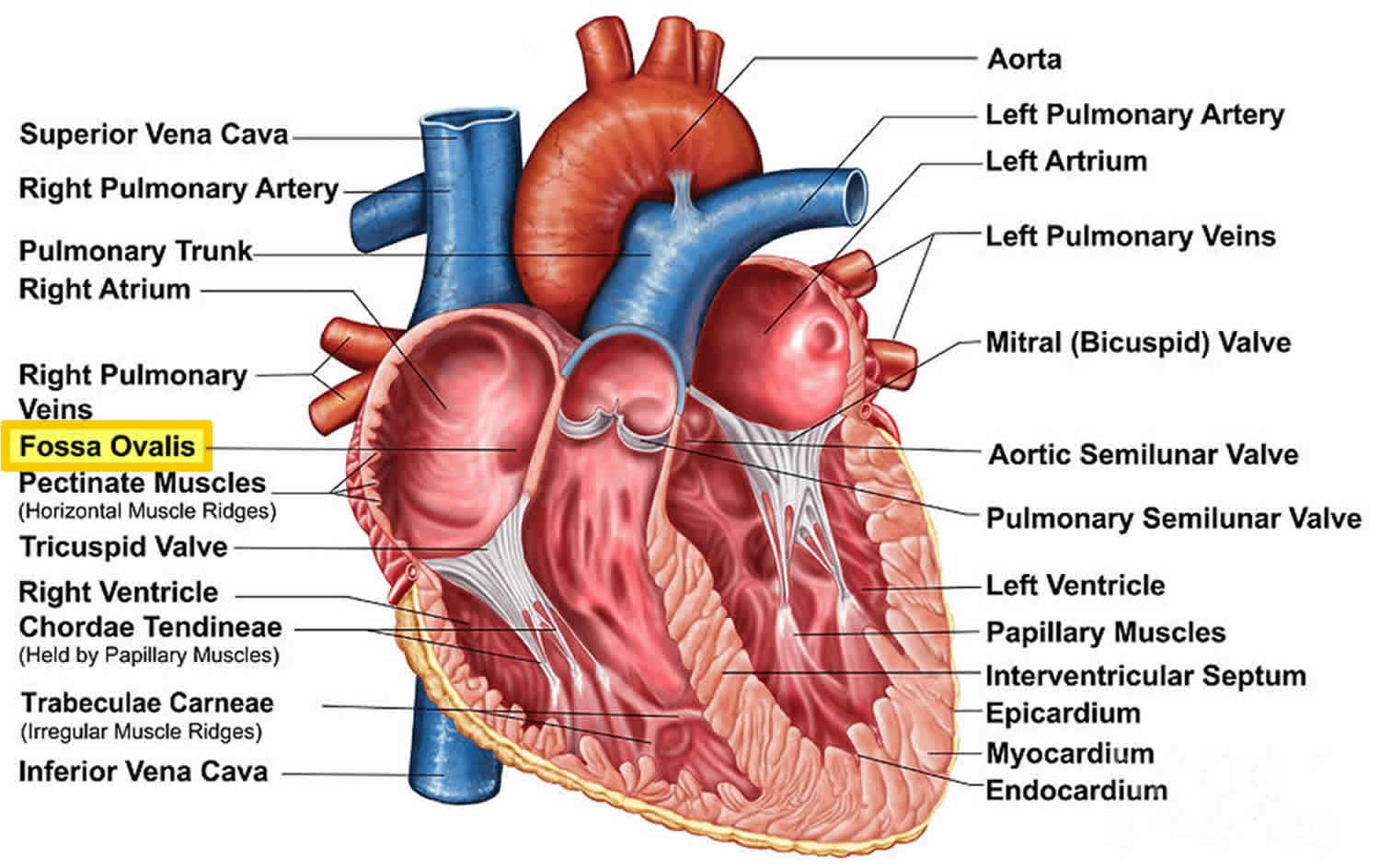 Figure 2. Formation of annulus fossa ovalis
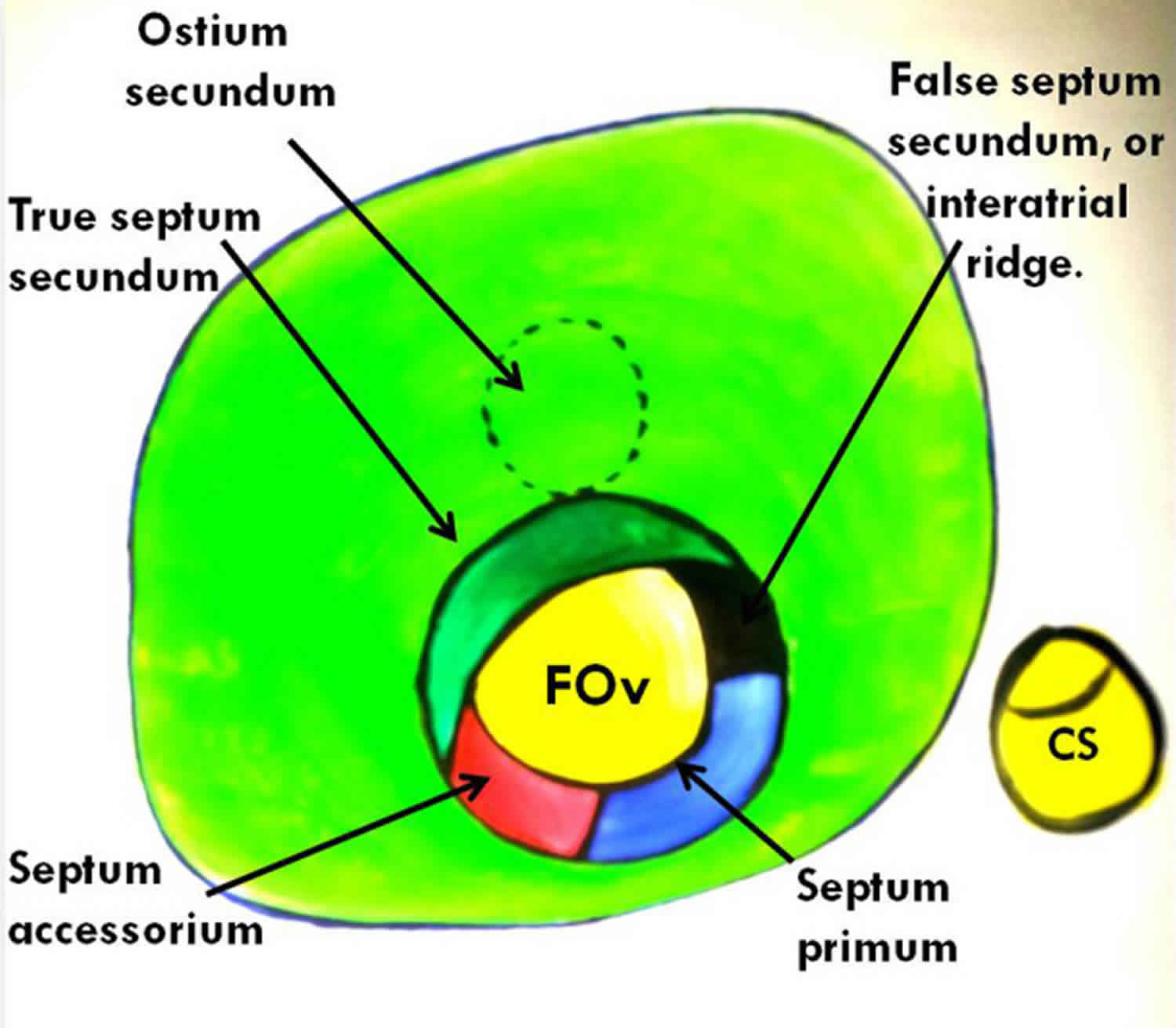
Abbreviations: FOv = fossa ovalis; CS = coronary sinus ostium.
[Source 6 ]Figure 3. Shapes of fossa ovalis
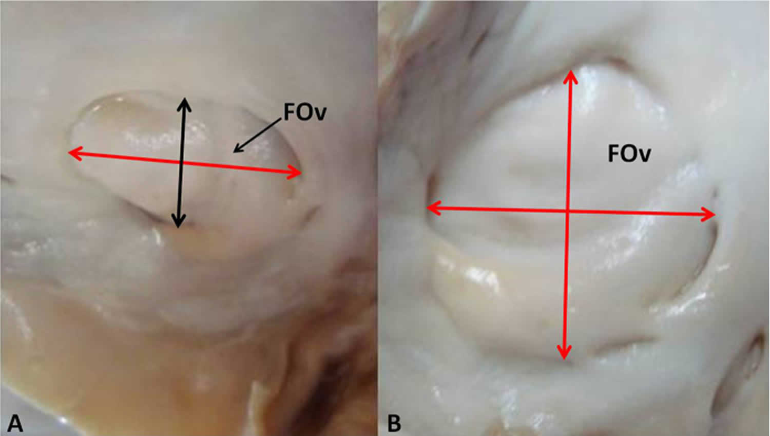
Footnote: (A) Oval and (B) circular. Double-headed red arrow indicates the long axis.
Abbreviation: FOv = fossa ovalis.
[Source 6 ]Figure 4. Annulus fossa ovalis or limbus fossa ovalis was found to be flat or raised at the margin. Annulus was raised in 46 cases (92%) and flat in 4 cases (8%)
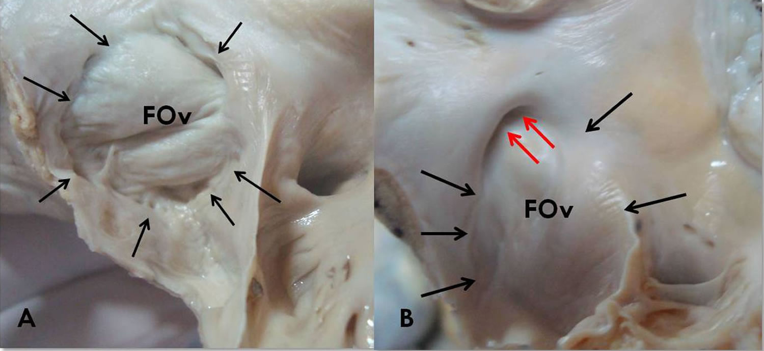
Footnote: (A) The annulus is raised along the whole circumference (black arrows). The fossa ovalis show redundancy. (B) The annulus is flat except in its upper part which shows a prominent recess (red arrows). Fossa ovalis is aneurysmal bulging towards right atrium.
Abbreviation: FOv = fossa ovalis.
[Source 6 ]Fossa ovalis function
In the normal heart, the fossa ovalis serves to prevent blood flow, i.e., shunting of blood, across the interatrial septum 13. During fetal development, the foramen ovale allows blood to pass from the right atrium to the left atrium, bypassing the nonfunctional fetal lungs while the fetus obtains its oxygen from the placenta 14. A flap of tissue called the septum primum and septum secundum form a one-way flap valve over the foramen ovale during that time, that allows shunting of blood from right atrium to the left atrium 4. The septum primum which is on the left atrial side forms the floor of the fossa ovalis, whereas the septum secundum derived from an infolding of the roof of the atrium forms the limbus of the fossa ovalis (annulus ovalis) which is the prominent oval margin of the fossa ovalis in the right atrium 3. After birth, the introduction of air into the lungs causes the pressure in the pulmonary circulatory system to drop. Physiologic increase in the left heart pressure at the time of birth allows the septum primum to create a tight seal with the septum secundum, creating a depression called the fossa ovalis 15, This fusion of the septum primum with the septum secundum and subsequent formation of the fossa ovalis occurs in 75% of cases, failure of which leads to a patent foramen ovale. Depending on the circumstances, a patent foramen ovale may be completely asymptomatic, or may require surgery 16. Also, the fossa ovalis undergoes thinning in utero 17.
The limbus of the fossa ovalis (annulus ovalis) represents the most direct anatomical landmark for atrial septal puncture for various procedures and may be located using the His bundle electrogram locating method 18. The following are significant procedures that require location of the fossa ovalis for transseptal puncture 5:
- Patent foramen ovale and atrial septal defect repair
- Right heart catheterization
- Percutaneous balloon valvuloplasty
- Radiofrequency catheter ablation
- Pulmonary vein isolation
- Left atrial appendage closure
- Catheter-based mitral valve repair
- Hemodynamic assessment of the mitral valve
- Paravalvular leak closure
- Alternative access to the left ventricle in the presence of a prosthetic aortic valve
Intracardiac echocardiography and fluoroscopy are useful for identifying the fossa ovalis, a critical landmark for electrophysiological procedures 19.
Fossa ovalis clinical significance
The fossa ovalis may be a frequent site of cardiac pathology. Known pathologies involving the fossa ovalis include:
- Premature closure of the foramen ovale and patent foramen ovale (PFO)
- Atrial septal defect (ASD)
- Rheumatic heart disease
- Amyloidosis
- Tumors: Prichard’s structures, myxomas, lipomatous hypertrophy of cardiac interatrial septum)
- Fossa ovalis membrane aneurysm
- Cardiac arrhythmias
Patent foramen ovale and atrial septal defect represent functionally similar pathologies; however, they differ in their etiology as well as clinical severity. Atrial septal defects are the result of the failure to form either of the interatrial septa (septum primum, septum secundum) or sinus venosus, whereas patent foramen ovales result when, in approximately 25% of the population, there is a failure of closure of the foramen ovale created by the septa 15. Both diseases are characterized clinically by the presence of migraines, paradoxical emboli and strokes, and decompression sickness 1. The size of a patent foramen ovale largely correlates with its clinical severity; larger lesions correlate with more severe illness and a higher risk of paradoxical emboli 20.
Atrial septal defects may occur in isolation or as part of a syndrome. Several genes have been implicated in the development of atrial septal defects. NKX2.5 gene is known to produce secundum type defects, TBX5 gene mutations result in Holt-Oram syndrome which features atrial septal defect 3. Atrial septal defects may undergo spontaneous closure depending on its initial size at the time of diagnosis, found to be the best predictor of the progression of such defects. Holes in the fossa ovalis measuring less than 8 mm and/or containing aneurysms may diminish in size or resolve spontaneously 21. However, failure of closure usually leads to enlargement of the defect and the need for surgical closure 21.
The structure and dimensions of the fossa ovalis are altered in the course of rheumatic heart disease 22. Rheumatic heart disease is characterized by extensive scarring of valves and other structures within the heart 23. The fossa ovalis may assume a more horizontal orientation in addition to a larger surface area; hemodynamic alterations are also prominent in this setting 22.
Amyloidosis, characterized by diffuse thickening of heart valves, does not spare the interatrial septum. Thickening of the interatrial septum is 100% specific for amyloidosis 24.
The fossa ovalis is a frequent site of atrial myxomas. Atrial myxomas are true neoplasms, appearing as pedunculated friable masses attached to the interatrial septum 25. These masses present with a wide range of symptoms ranging from dyspnea to hemiplegia 26.
Prichard’s structures are also benign structures derived from mature endothelial cells, they are more frequently found in individuals beyond 60 years of age and are unrelated to cardiac myxomas 27. These structures may be subendothelial or located within the atrial cavity, commonly on the left side of the fossa ovalis and may be the result of hemodynamic alterations in blood flow within the heart 28.
Lipomatous hypertrophy is fat accumulation greater than 2cm located on the interatrial septum. The mass characteristically spares the fossa ovalis. Arrhythmias and sudden cardiac death may be accompanying complications 29. Obstruction of the vena cava ostium may be present in larger lesions 30. The occurrence of lipomatous hypertrophy of the septum has correlations with obesity, older age, female gender, steroid use 30.
The fossa ovalis is found to be aneurysmal in some cases but is present with increased incidence in patients with stroke 3. Fossa ovalis membrane aneurysms present as focal bulging of the interatrial septum, with displacement towards the left or right atrium 31. Fossa ovalis membrane aneurysm carries a significant risk of fibrosis and thrombus formation within its wall with related complications, as well as the formation of an atrial septal defect (ASD) from a preexisting patent foramen ovale (PFO) leading to increased intracardiac shunting. It is often the result of elevated intracardiac pressures arising from secondary cardiac pathologies, most commonly ischemic heart disease, aortic and mitral valvular diseases 31. Transesophageal echocardiography represents the best method of visualizing fossa ovalis membrane aneurysms in patients suspected of cardiogenic emboli, although the emboli may arise from a different source 32.
Electrophysiological studies may reveal increased automaticity in the region of the limbus fossa ovalis. Such automaticity may manifest as reversible focal atrial tachycardia and chaotic atrial rhythm and is responsive to focal radiofrequency ablation of the limbus with the restoration of cardiac function 33.
References- Malik SB, Kwan D, Shah AB, Hsu JY. The right atrium: gateway to the heart–anatomic and pathologic imaging findings. Radiographics. 2015 Jan-Feb;35(1):14-31.
- Oduah MTA, Brown KN. Anatomy, Thorax, Heart Fossa Ovalis. [Updated 2019 Feb 7]. In: StatPearls [Internet]. Treasure Island (FL): StatPearls Publishing; 2019 Jan-. Available from: https://www.ncbi.nlm.nih.gov/books/NBK538432
- Joshi SD, Chawre HK, Joshi SS. Morphological study of fossa ovalis and its clinical relevance. Indian Heart J. 2016 Mar-Apr;68(2):147-52.
- Barik R. The three-dimensional fossa ovalis. Indian Heart J. 2016 Mar-Apr;68(2):211-2.
- Klimek-Piotrowska W, Hołda MK, Koziej M, Piątek K, Hołda J. Anatomy of the true interatrial septum for transseptal access to the left atrium. Ann. Anat. 2016 May;205:60-4.
- Joshi SD, Chawre HK, Joshi SS. Morphological study of fossa ovalis and its clinical relevance. Indian Heart J. 2016;68(2):147–152. doi:10.1016/j.ihj.2015.08.001 https://www.ncbi.nlm.nih.gov/pmc/articles/PMC4867950
- Davison P., Clift P.F., Steeds R.P. The role of echocardiography in diagnosis, monitoring closure and post-procedural assessment of patent foramen ovale. Eur J Echocardiogr. 2010;11:i27–i34.
- Rana BS, Shapiro LM, McCarthy KP, Ho SY. Three-dimensional imaging of the atrial septum and patent foramen ovale anatomy: defining the morphological phenotypes of patent foramen ovale. Eur J Echocardiogr. 2010 Dec;11(10):i19-25.
- Lam YY, Yu CM, Zhang Q, Yan BP, Yip GW. Enhanced detection of patent foramen ovale by systematic transthoracic saline contrast echocardiography. Int. J. Cardiol. 2011 Oct 06;152(1):24-7.
- Rana B.S., Shapiro L.M., McCarthy K.P. Three-dimensional imaging of the atrial septum and patent foramen ovale anatomy: defining the morphological phenotypes of patent foramen ovale. Eur J Echocardiogr. 2010;11:i19–i25.
- Boppana VS, Castaño A, Avula UMR, Yamazaki M, Kalifa J. Atrial Coronary Arteries: Anatomy And Atrial Perfusion Territories. J Atr Fibrillation. 2011 Sep-Nov;4(3):375.
- Sokolov VV, Brezhnev FF, Kharlamov EV. [Sources of the vascularization of the human interatrial septum of the heart with different variants of the atrial blood supply]. Arkh Anat Gistol Embriol. 1986 Jul;91(7):29-34.
- Terpenning S, White CS. Imaging pitfalls, normal anatomy, and anatomical variants that can simulate disease on cardiac imaging as demonstrated on multidetector computed tomography. Acta Radiol Short Rep. 2015 Jan;4(1):2047981614562443.
- Yee K, Lui F. Anatomy, Thorax, Heart Foramen Ovale. [Updated 2019 Aug 10]. In: StatPearls [Internet]. Treasure Island (FL): StatPearls Publishing; 2019 Jan-. Available from: https://www.ncbi.nlm.nih.gov/books/NBK545203
- Kanaganayagam GS, Malik IS. Modern management of a patent foramen ovale. JRSM Cardiovasc Dis. 2012 Oct 31;1(7).
- Patent foramen ovale: current pathology, pathophysiology, and clinical status. J Am Coll Cardiol. 2005 Nov 1;46(9):1768-76. Epub 2005 Sep 29. https://doi.org/10.1016/j.jacc.2005.08.038
- Terpenning S, White CS. Imaging pitfalls, normal anatomy, and anatomical variants that can simulate disease on cardiac imaging as demonstrated on multidetector computed tomography. Acta Radiol Short Rep. 2015 Jan;4(1):2047981614562443
- Shen J. [An application of His bundle electrogram to locate fossa ovalis during percutaneous transseptal balloon valvuloplasty]. Zhonghua Xin Xue Guan Bing Za Zhi. 1991 Jun;19(3):153-4, 197.
- Hanaoka T, Suyama K, Taguchi A, Shimizu W, Kurita T, Aihara N, Kamakura S. Shifting of puncture site in the fossa ovalis during radiofrequency catheter ablation: intracardiac echocardiography-guided transseptal left heart catheterization. Jpn Heart J. 2003 Sep;44(5):673-80.
- Kerut EK, Norfleet WT, Plotnick GD, Giles TD. Patent foramen ovale: a review of associated conditions and the impact of physiological size. J. Am. Coll. Cardiol. 2001 Sep;38(3):613-23.
- Demir T, Oztunç F, Eroğlu AG, Saltik L, Ahunbay G, Kutluğ S, Güzeltaş A, Altun G. Outcome for patients with isolated atrial septal defects in the oval fossa diagnosed in infancy. Cardiol Young. 2008 Feb;18(1):75-8.
- Chaudhari RG, Shinde SV, Deshpande JR. Morphometric analysis of fossa ovalis in rheumatic heart disease. Indian Heart J. 2005 Nov-Dec;57(6):662-5.
- Roberts WC, Ko JM. Some observations on mitral and aortic valve disease. Proc (Bayl Univ Med Cent). 2008 Jul;21(3):282-99.
- Munjewar C, Agrawal R, Sharma S. Cardiac amyloidosis: a report of two cases. Indian Heart J. 2014 Jul-Aug;66(4):473-6.
- Cohen R, Singh G, Mena D, Garcia CA, Loarte P, Mirrer B. Atrial Myxoma: A Case Presentation and Review. Cardiol Res. 2012 Feb;3(1):41-44.
- Onubogu U, West B, Orupabo-Oyan B. Atrial myxoma: a rare cause of hemiplegia in children. 2017 Sep/Oct 23Cardiovasc J Afr. 28(5):e1-e3.
- Acebo E, Val-Bernal JF, Gómez-Román JJ. Prichard’s structures of the fossa ovalis are not histogenetically related to cardiac myxoma. Histopathology. 2001 Nov;39(5):529-35.
- Val-Bernal JF, Martino M, Mayorga M, Garijo MF. Prichard’s structures of the fossa ovalis are age-related phenomena composed of nonreplicating endothelial cells: the cardiac equivalent of cutaneous senile angioma. APMIS. 2007 Nov;115(11):1234-40.
- Xanthopoulos A, Giamouzis G, Alexopoulos N, Kitai T, Triposkiadis F, Skoularigis J. Lipomatous Hypertrophy of the Interatrial Septum: A Case Report and Review of the Literature. CASE (Phila). 2017 Oct;1(5):182-189.
- Ribeiro RNF, Ribeiro BNF, Martins WA, Antunes LO, Marchiori E. Lipomatous hypertrophy of the interatrial septum. Radiol Bras. 2018 Mar-Apr;51(2):130-131.
- Topaz O, Edwards JE, Bojack-Mackey S, Titus JL. Aneurysm of fossa ovalis in adults: a pathologic study. Cardiovasc. Pathol. 2003 Jul-Aug;12(4):219-25.
- Ilercil A, Meisner JS, Vijayaraman P, Gentilucci M, Metveyeva P, Hla A, Strom JA, Chang CJ, Shirani J. Clinical significance of fossa ovalis membrane aneurysm in adults with cardioembolic cerebral ischemia. Am. J. Cardiol. 1997 Jul 01;80(1):96-8.
- Di Pino A, Caruso E, Gitto P. The limbus of the fossa ovalis: an unusual location for incessant focal atrial tachycardia in children. Europace. 2016 Aug;18(8):1251.


