Sneddon Wilkinson disease
Sneddon Wilkinson disease also called subcorneal pustular dermatosis, is a rare benign skin disease in which pus-filled pimples or blisters (pustules) form under the top (subcorneal) layer of the skin 1. Sneddon Wilkinson disease is not infectious or contagious and it is not cancerous 2. Sneddon Wilkinson disease is most common in middle-aged adults over the age of forty particularly affects women more often than men, but can develop in children 3. Pustules usually appear over a few hours and grow together to form round or wavy patterns 3. Subcorneal pustular dermatosis most often form in areas where the skin may touch or rub together, such as the groin area, underarms, inside the elbows, and behind the knees 4. The pustules may be mildly itchy or painful, but despite being pus filled, are not infected 3. The diagnosis of subcorneal pustular dermatosis is made based on the appearance of the pustules and the results of a skin biopsy (histologic findings) 3.
According to some authors, subcorneal pustular dermatosis can be classified as one of the neutrophilic dermatoses together with pyoderma gangrenosum, Sweet syndrome, and erythema elevatum diutinum 5, while others classify it within the group of autoinflammatory pustular neutrophilic diseases, together with pustular psoriasis variants 6. In addition, it must be noted that autoimmune intercellular IgA dermatosis with autoantibodies to desmocollin is almost indistinguishable from classic subcorneal pustular dermatosis, which makes the nosological classification of this entity remain controversial 7.
The cause of subcorneal pustular dermatosis is not known 3. There is currently no evidence it is inherited (no familial cases have been reported) and it is not contagious 2. Subcorneal pustular dermatosis may be associated with other diseases or health problems including several autoimmune diseases, blood (hematologic) diseases, infections, and cancers. Subcorneal pustular dermatosis has been described in association with IgA monoclonal gammopathies 8, multiple myeloma 9 and inflammatory diseases such as rheumatoid arthritis 10, ulcerative colitis 11 and Crohn disease 12. Rarely, subcorneal pustular dermatosis has been associated with taking certain medications (drug-induced subcorneal pustular dermatosis) 3.
Subcorneal pustular dermatosis may be treated with is oral dapsone, which often improves symptoms within one month. Alternative tablets are sulphapyridine and sulphamethoxypryidazine. However, the pustules may return when treatment is stopped 13. Often, intermittent treatment for months or years with a low dose is required to keep the skin clear. Other therapies have been tried with mixed results 3. Steroid creams or tablets may not clear the rash fully, other treatments, for example acitretin, colchicine, tetracycline antibiotics, immunosuppressive medication or biological treatments are sometimes helpful. Some patients respond to hospital treatment with ultra-violet light. The potential side effects of these treatments needs to be carefully balanced against the impact of subcorneal pustular dermatosis, which is a relatively harmless condition. While subcorneal pustular dermatosis may cause discomfort and cosmetic concerns, it typically does not affect overall health 4.
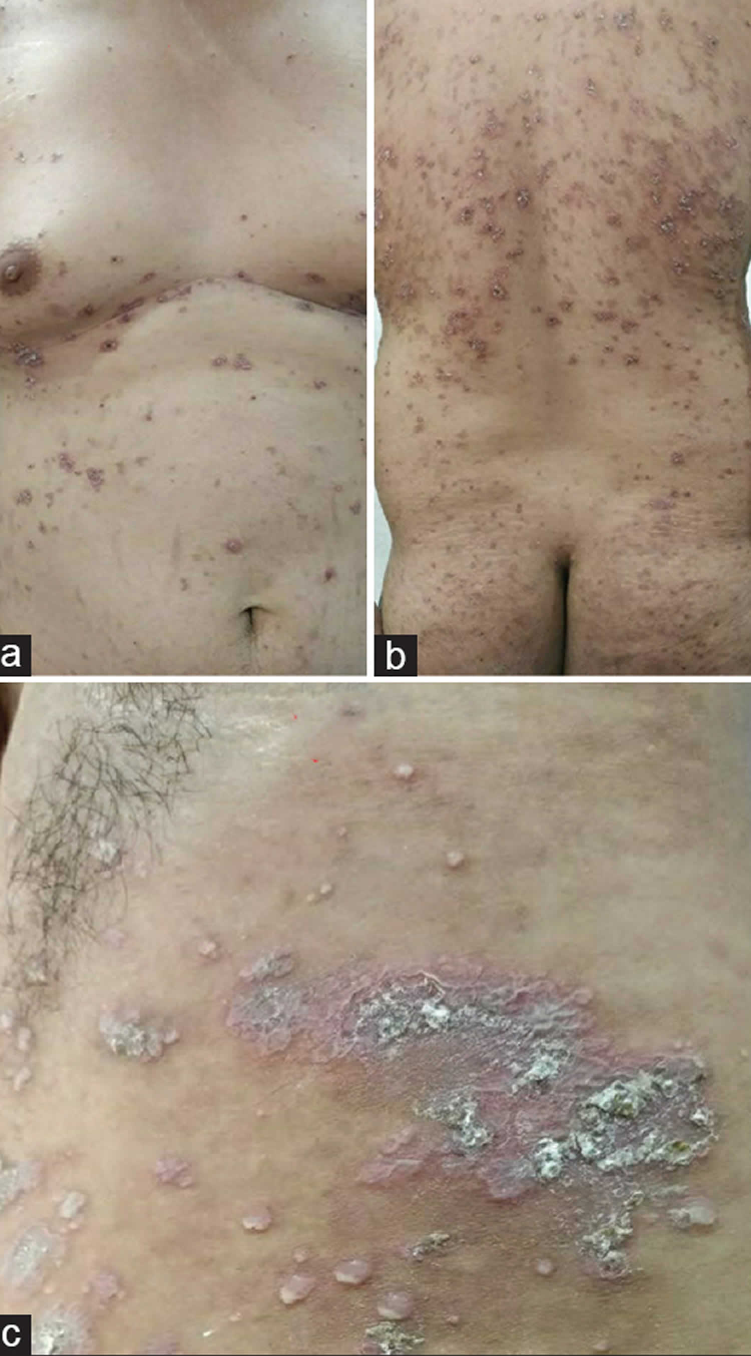
Figure 1. Sneddon Wilkinson disease
Footnote: A 28-year-old Caucasian man presented with 6-year duration of recurrent, erythematous, crusted, asymptomatic papules, scaly plaques, and grouped pustules affecting the neck, trunk, flexural aspects of extremities, and the groin. Review of systems was unremarkable and his family and social histories were noncontributory. On initial presentation, there were multiple pustules and papules as well as annular and serpiginous pink plaques with central scales and crusts and surrounding deep erythema. These lesions were symmetrically distributed on the groin and proximal upper and lower extremity flexor aspects.
[Source 14 ]Is subcorneal pustular dermatosis hereditary?
No, subcorneal pustular dermatosis does not run in families.
Can subcorneal pustular dermatosis be cured?
Presently there is no cure, but subcorneal pustular dermatosis can usually be controlled with medication.
Subcorneal pustular dermatosis causes
The cause of subcorneal pustular dermatosis is unknown. Sneddon Wilkinson disease is not infectious or contagious and it is not cancerous. Most often it occurs on its own, but has been linked to a variety of other diseases, for example inflammatory bowel disease, arthritis, thyroid disease and blood disorders. Some cases of subcorneal pustular dermatosis have been considered a variant of pustular psoriasis. Subcorneal pustular dermatosis can be very difficult to distinguish from generalised pustular psoriasis and acute generalized exanthematous pustulosis. They appear to be closely related disorders.
Other cases of subcorneal pustular dermatosis are argued to be a rare variant of pemphigus, known as subcorneal pustular dermatosis type IgA pemphigus. This subgroup of patients shows positive immunofluorescence with epidermal intercellular IgA deposits. The positive immunofluorescence can develop years after the initial diagnosis of subcorneal pustular dermatosis. Unlike pemphigus, a predominance of neutrophils and an absence or moderate acantholysis is observed; additionally, the condition is usually responsive to dapsone.
Subcorneal pustular dermatosis is associated with some other conditions. The most frequent are:
- IgA monoclonal gammopathy (accumulation of abnormal proteins in the blood) 15
- Multiple myeloma 9.
- Pyoderma gangrenosum 8.
Other less frequent associated conditions include rheumatoid arthritis, lupus erythematosus, hyperthyroidism and hypothyroidism, polycythemia rubra vera, and SAPHO syndrome (synovitis, acne, pustulosis, osteitis).
Subcorneal pustular dermatosis pathophysiology
The exact pathophysiology of subcorneal pustular dermatosis is unknown. The accumulation of neutrophils in the subcorneal layer suggests the presence of chemoattractants in the uppermost epidermis, but the stimulus for these chemoattractants was not found. Interleukin (IL)‒1 beta, IL-6, IL-8, IL-10, leukotriene B4, and complement fragment C5a are neutrophil chemoattractants that have been found at increased levels in scale extracts of patients with subcorneal pustular dermatosis compared with that of controls. Tumor necrosis factor (TNF)‒alpha levels have been found to be significantly elevated in the serum and blister fluid of patients with subcorneal pustular dermatosis 16. However, TNF-blockers were not found to be effective in all patients, although there are reports on successful treatment with this class of drugs 17. A case of a TNF-alpha-inhibitor–induced subcorneal pustular dermatosis has also been described 18.
Immunofluorescence studies are negative in the subcorneal pustular dermatosis of Sneddon-Wilkinson. However, a rare subtype of subcorneal pustular dermatosis has been reported to have a positive immunofluorescence with IgA deposition restricted to the upper epidermis and directed against desmocollin. As noted above, the clinical and histopathological characteristics have led experts to classify this variant as a variant of IgA pemphigus resembling subcorneal pustular dermatosis. In a large 2016 series of 49 patients with intercellular IgA dermatosis and 13 cases with subcorneal pustular dermatosis, it was confirmed that the subcorneal pustular dermatosis type of intercellular IgA dermatosis is clinically and histopathologically indistinguishable from classic subcorneal pustular dermatosis without immunoreactants 7.
Additionally, despite many attempts, no infectious agent or other immunogenic trigger has yet been identified in subcorneal pustular dermatosis patients. The eruption is regarded as sterile, although it may sometimes become secondarily infected with Staphylococcus aureus or streptococcal species. Preceding Mycoplasma pneumoniae infection was implicated in one report, but this case had an acute presentation that responded to 3 months of dapsone without relapse 19.
Subcorneal pustular dermatosis signs and symptoms
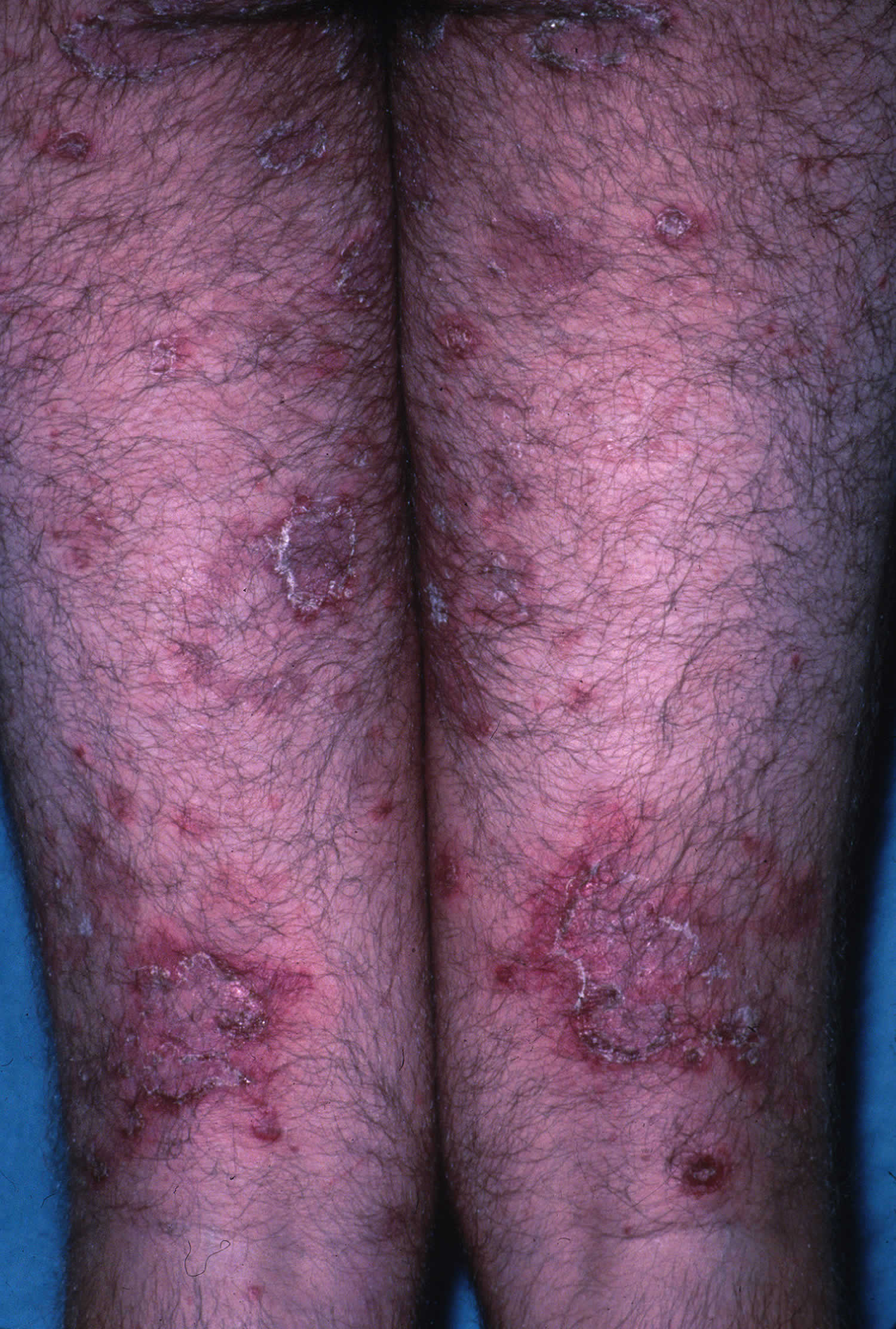
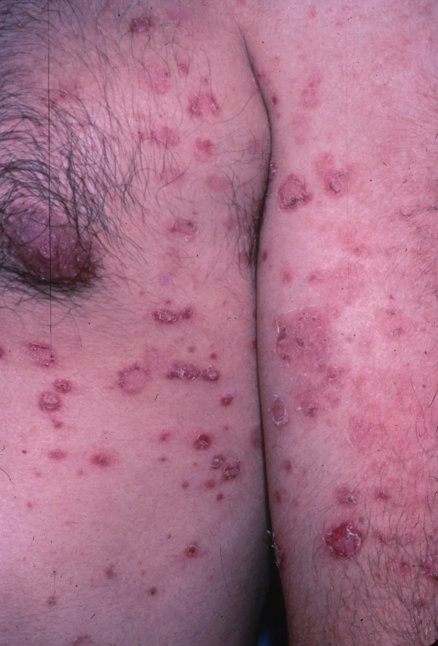
Subcorneal pustular dermatosis is characterized by numerous soft pustules at the skin surface. Individual pustular lesions arise within a few hours. They usually appear on the trunk, particularly in the skin folds such as the armpits and groin. They may appear on otherwise normal skin, but there are often within a red patch. The pustules resolve over a few days and are replaced by fine scale before there is another relapse and new pustules form again. The lesions are sometimes itchy, but generally the patient is otherwise well. It may flare-up for a few weeks and then clear up for months or years before reappearing. Systemic and toxic symptoms are not associated with acute episodes. However, malaise, fever, arthralgias, abnormalities of hepatic enzymes, and sclerosing glomerulonephritis have been reported in several cases 3. Patients typically do not have any symptoms or signs of mucosal involvement. The process may be repeated over many years.
Patients may present with histories notable for monoclonal gammopathies such as immunoglobulin A (IgA) more often than immunoglobulin G (IgG) 15; lymphoproliferative disorders especially multiple myeloma 9; pyoderma gangrenosum 8; and other inflammatory diseases such as rheumatoid arthritis 20, systemic lupus erythematosus (SLE) 21, Sjögren syndrome 22, diffuse scleroderma 23, ulcerative colitis 11 and Crohn disease 12. These conditions are well-recognized associations with subcorneal pustular dermatosis developing both before and after the diagnosis of subcorneal pustular dermatosis 24. Further associations were found with other dermatoses characterized by skin infiltration with neutrophils, such as pyoderma gangrenosum and SAPHO (synovitis, acne, pustulosis, osteitis) syndrome 25. Other anecdotally associated conditions include aplastic anemia 26, Mycoplasma pneumoniae infection 27, Coccidioides immitis infection 28, hyperthyroidism 29, APUDoma (amine precursor uptake and decarboxylation cell–derived tumor) 30 and thymoma 31.
Patients should be queried about a personal and family history of psoriasis, because differentiating subcorneal pustular dermatosis from pustular psoriasis can be difficult. Similarly, patients should be questioned about recent drug exposure because acute generalized exanthematous pustulosis is also in the differential diagnosis 32.
Subcorneal pustular dermatosis diagnosis
Sometimes subcorneal pustular dermatosis diagnosis is made just by looking at the skin, in particular if there are ‘half and half blisters’. The diagnosis is not always easy, as other blistering conditions, reactions to medicines and also a form of psoriasis (pustular psoriasis) can look similar.
Often, a skin biopsy i.e. a punch biopsy needs to be taken under a local anaesthetics for histology to confirm the diagnosis.
Skin swabs or scrapings may be taken to rule out a skin infection, and blood tests to look for an underlying illness.
Blood tests are likely to include a general screen such as blood count, calcium levels and liver function tests, and protein electrophoresis to look for a gammopathy.
Subcorneal pustular dermatosis treatment
Treatment is aimed at preventing complications. Dapsone is often successful, with the lesions resolving over a month. Ongoing maintenance with a lower dose is sometimes needed.
Other treatment options include:
- Acitretin
- Sulfapyridine or sulfamethoxypyridazine
- Phototherapy including UVB and psoralen with UVA (PUVA)
- Colchicine
- Ciclosporin or other immune suppressants such as mycophenolate mofetil
- Biological response mediators including infliximab and adalimumab
Miscellaneous other agents have been successfully used for individual patients. Systemic steroids are generally ineffective and may in fact precipitate a flare-up of subcorneal pustular dermatosis.
Patients need long term follow-up with periodic evaluation of serum protein electrophoresis and immunophoresis because paraproteinemia or myeloma may develop after several years.
Dapsone
Dapsone is the treatment of choice. The response is slower than that seen with dermatitis herpetiformis, with resolution usually occurring in about 4 weeks. Once disease control has been established, the dose should be tapered to the lowest dose needed to maintain control. Sulfapyridine and sulfamethoxypyridazine may also be used, but only a few isolated reports support their effectiveness.
Acitretin
Acitretin (etretinate) has been used to successfully treat subcorneal pustular dermatosis and should be considered as an alternative or additional treatment for those who are intolerant of, or unresponsive to, dapsone. Once disease control has been established, the dose should be tapered to the lowest dose needed to maintain control. Isotretinoin at 0.5 mg/kg/d appears to be ineffective.
Phototherapy
Phototherapy with psoralen with UVA (PUVA) 33, broadband UVB, and narrowband UVB alone or in combination with dapsone and/or retinoids can be successful at controlling subcorneal pustular dermatosis 34. Long-term maintenance regimens may be needed 35.
Additional therapies
Anecdotal case reports support the use of infliximab, tacalcitol 36, maxacalcitol 37, mizoribine 38, ketoconazole 39, tetracycline, minocycline, benzylpenicillin, vitamin E 40, azithromycin 41, cyclosporine 42, colchicine, pentoxifylline 43, intravenous immunoglobulins 44 and adalimumab with mycophenolate mofetil 45. Antimyeloma treatment should be considered in cases of subcorneal pustular dermatosis associated with IgA monoclonal gammopathy of undetermined significance, refractory to other therapies 46.
Systemic and topical corticosteroids are generally ineffective but may provide some control. They have been used in combination with dapsone to treat associated conditions such as pyoderma gangrenosum and multiple myeloma. A good response to systemic corticosteroids is atypical and is suggestive of a diagnosis of pustular psoriasis.
Long-term monitoring
Long-term follow-up is recommended. Periodic evaluations with serum protein electrophoresis and direct immunofluorescence should be performed every few years.
Paraproteinemia, myeloma, intraepidermal IgA staining, and pustular psoriasis may develop several years after the initial presentation of subcorneal pustular dermatosis. Identifying these conditions, as well as other associated diagnoses, can improve the understanding of the etiology and pathogenesis of subcorneal pustular dermatosis, clarify its relationship with IgA pemphigus and pustular psoriasis, and help define its nosologic classification.
Subcorneal pustular dermatosis prognosis
Subcorneal pustular dermatosis or Sneddon Wilkinson disease, is chronic and relapsing but benign. Subcorneal pustular dermatosis usually clears over a period of about 4 weeks when treated with a tablet called dapsone. The association of subcorneal pustular dermatosis with paraproteinemia or lymphoproliferative disorders, especially multiple myeloma, may alter the prognosis.
References- Subcorneal Pustular Dermatosis. https://emedicine.medscape.com/article/1124252-overview
- Subcorneal pustular dermatosis. https://www.bad.org.uk/for-the-public/patient-information-leaflets/subcorneal-pustular-dermatosis
- Watts PJ, Khachemoune A. Subcorneal Pustular Dermatosis: A Review of 30 Years of Progress. Am J Clin Dermatol. December, 2016; 17(6):653-671. https://www.ncbi.nlm.nih.gov/pubmed/27349653
- Subcorneal pustular dermatosis. https://www.uptodate.com/contents/subcorneal-pustular-dermatosis
- Cohen PR. Neutrophilic dermatoses: a review of current treatment options. Am J Clin Dermatol. 2009. 10 (5):301-12.
- Naik HB, Cowen EW. Autoinflammatory pustular neutrophilic diseases. Dermatol Clin. 2013 Jul. 31 (3):405-25.
- Hashimoto T, Teye K, Ishii N. Clinical and immunological studies of 49 cases of various types of intercellular IgA dermatosis and 13 cases of classical subcorneal pustular dermatosis examined at Kurume University. Br J Dermatol. 2016 Jun 3.
- Puechguiral-Renaud I, Carpentier O, Piette F, Delaporte E. Subcorneal pustulosis and Pyoderma gangrenosum associated with a biclonal gammopathy. Eur J Dermatol. 2006 Nov-Dec. 16(6):687-90.
- Takata M, Inaoki M, Shodo M, Hirone T, Kaya H. Subcorneal pustular dermatosis associated with IgA myeloma and intraepidermal IgA deposits. Dermatology. 1994. 189 Suppl 1:111-4.
- Butt A, Burge SM. Sneddon-Wilkinson disease in association with rheumatoid arthritis. Br J Dermatol. 1995 Feb. 132(2):313-5.
- Wargo JJ, Adams M, Trevino J. Subcorneal pustular dermatosis and episcleritis associated with poorly controlled ulcerative colitis. BMJ Case Rep. 2017;2017:bcr2016218123. Published 2017 Jan 30. doi:10.1136/bcr-2016-218123
- García-Salces I, Baldellou R, Hörndler C, Zubiri ML. Subcorneal pustular dermatosis with pathergy phenomenon in a patient with Crohn’s disease. J Eur Acad Dermatol Venereol. 2009 Mar. 23 (3):349-50.
- Cheng S, Edmonds E, Ben-Gashir M, Yu RC. Subcorneal pustular dermatosis: 50 years on. Clin Exp Dermatol. May 2008; 33(3):229-233. https://www.ncbi.nlm.nih.gov/pubmed/18355359
- Khachemoune, A., & Blyumin, M. L. (2003). Sneddon-Wilkinson disease resistant to dapsone and colchicine successfully controlled with PUVA. Dermatology Online Journal, 9(5). Retrieved from https://escholarship.org/uc/item/1vn304vn
- Kasha EE Jr, Epinette WW. Subcorneal pustular dermatosis (Sneddon-Wilkinson disease) in association with a monoclonal IgA gammopathy: a report and review of the literature. J Am Acad Dermatol. 1988 Nov. 19(5 Pt 1):854-8.
- Ono S, Otsuka A, Miyachi Y, Kabashima K. Subcorneal Pustular Dermatosis Exhibiting a High Serum TARC/CCL17 Level. Case Rep Dermatol. 2013 Jan. 5(1):38-42.
- Bonifati C, Trento E, Cordiali Fei P, Muscardin L, Amantea A, Carducci M. Early but not lasting improvement of recalcitrant subcorneal pustular dermatosis (Sneddon-Wilkinson disease) after infliximab therapy: relationships with variations in cytokine levels in suction blister fluids. Clin Exp Dermatol. 2005 Nov. 30(6):662-5.
- Sauder MB, Glassman SJ. Palmoplantar subcorneal pustular dermatosis following adalimumab therapy for rheumatoid arthritis. Int J Dermatol. 2013 May. 52 (5):624-8.
- Papini M, Cicoletti M, Landucci P. Subcorneal pustular dermatosis and mycoplasma pneumoniae respiratory infection. Acta Derm Venereol. 2003. 83(5):387-8.
- Iobst W, Ingraham K. Sneddon-Wilkinson disease in a patient with rheumatoid arthritis. Arthritis Rheum. 2005 Dec. 52(12):3771.
- Saulsbury FT, Kesler RW. Subcorneal pustular dermatosis and systemic lupus erythematosus. Int J Dermatol. 1984 Jan-Feb. 23 (1):63-4.
- Tsuruta D, Matsumura-Oura A, Ishii M. Subcorneal pustular dermatosis and Sjögren’s syndrome. Int J Dermatol. 2005 Nov. 44(11):955-7.
- Mokhtari F, Poostiyan N. Subcorneal Pustular Dermatosis: A Case Report of a Patient with Diffuse Scleroderma. Adv Biomed Res. 2018. 7:83.
- Subcorneal Pustular Dermatosis Clinical Presentation. https://emedicine.medscape.com/article/1124252-clinical
- Scarpa R, Lubrano E, Cozzi R, Ames PR, Oriente CB, Oriente P. Subcorneal pustular dermatosis (Sneddon-Wilkinson syndrome): another cutaneous manifestation of SAPHO syndrome?. Br J Rheumatol. 1997 May. 36 (5):602-3.
- Park BS, Cho KH, Eun HC, Youn JI. Subcorneal pustular dermatosis in a patient with aplastic anemia. J Am Acad Dermatol. 1998 Aug. 39 (2 Pt 1):287-9.
- Lombart F, Dhaille F, Lok C, Dadban A. Subcorneal pustular dermatosis associated with Mycoplasma pneumoniae infection. J Am Acad Dermatol. 2014 Sep. 71 (3):e85-6.
- Iyengar S, Chambers CJ, Chang S, Fung MA, Sharon VR. Subcorneal pustular dermatosis associated with Coccidioides immitis. Dermatol Online J. 2015 Aug 15. 21 (8).
- Taniguchi S, Tsuruta D, Kutsuna H, Hamada T. Subcorneal pustular dermatosis in a patient with hyperthyroidism. Dermatology. 1995. 190 (1):64-6.
- Villey MC, Ehrsam E, Marrakchi S, Colombel JF, Thomas P. Apudoma and subcorneal pustular dermatosis (Sneddon-Wilkinson disease). Dermatology. 1992. 185 (4):269-71.
- Agarwal A, Shivaswamy KN, Barani R et al. Subcorneal pustular dermatosis and thymoma:an association or a coincidence?. Indian J Dermatol. 2006. 51:272-4.
- Tajiri K, Nakajima T, Kawai K, Minemura M, Sugiyama T. Sneddon-Wilkinson disease induced by sorafenib in a patient with advanced hepatocellular carcinoma. Intern Med. 2015. 54 (6):597-600.
- Bauwens M, De Coninck A, Roseeuw D. Subcorneal pustular dermatosis treated with PUVA therapy. A case report and review of the literature. Dermatology. 1999. 198(2):203-5.
- Orton DI, George SA. Subcorneal pustular dermatosis responsive to narrowband (TL-01) UVB phototherapy. Br J Dermatol. 1997 Jul. 137(1):149-50.
- Marliere V, Beylot-Barry M, Beylot C, Doutre M. Successful treatment of subcorneal pustular dermatosis (Sneddon-wilkinson disease) by acitretin: report of a case. Dermatology. 1999. 199(2):153-5.
- Kawaguchi M, Mitsuhashi Y, Kondo S. A case of subcorneal pustular dermatosis treated with tacalcitol (1alpha,24-dihydroxyvitamin D3). J Dermatol. 2000 Oct. 27(10):669-72.
- Hoshina D, Tsujiwaki M, Furuya K. Successful treatment of subcorneal pustular dermatosis with maxacalcitol. Clin Exp Dermatol. 2016 Jan. 41 (1):102-3.
- Kono T, Terashima T, Oura H, Ishii M, Taniguchi S, Muramatsu T. Recalcitrant subcorneal pustular dermatosis and bullous pemphigoid treated with mizoribine, an immunosuppressive, purine biosynthesis inhibitor. Br J Dermatol. 2000 Dec. 143(6):1328-30.
- Verma KK, Pasricha JS. Ketoconazole as a therapeutic modality in subcorneal pustular dermatosis. Acta Derm Venereol. 1997 Sep. 77(5):407-8.
- Ayres S Jr, Mihan R. Letter: Subcorneal pustular dermatosis controlled by vitamin E. Arch Dermatol. 1974 Jun. 109(6):914.
- Bliziotis I, Rafailidis P, Vergidis P, Falagas ME. Regression of subcorneal pustular dermatosis type of IgA pemphigus lesions with azithromycin. J Infect. 2005 Aug. 51(2):E31-4.
- Karadogan SK, Aydogan K, Baskan EB, Tunali S. A case of subcorneal pustular dermatosis treated successfully with a combination of cyclosporin and prednisolone. J Eur Acad Dermatol Venereol. 2007 Apr. 21(4):536-7.
- Falcone LM, Pilcher MF, Kovach RF, Powers R. Pentoxyfilline as a Treatment for Subcorneal Pustular Dermatosis. Dermatol Ther. 2019 Jan 14. e12818.
- Kundak S, Bağ Ö, Gülez N, Ergin M. A child with subcorneal pustular dermatosis responded to IVIG treatment (Sneddon-Wilkinson disease). Reumatologia. 2017. 55 (6):323-327.
- Howell SM, Bessinger GT, Altman CE, Belnap CM. Rapid response of IgA pemphigus of the subcorneal pustular dermatosis subtype to treatment with adalimumab and mycophenolate mofetil. J Am Acad Dermatol. 2005 Sep. 53(3):541-3.
- von dem Borne PA, Jonkman MF, van Doorn R. Complete remission of skin lesions in a patient with subcorneal pustular dermatosis (Sneddon-Wilkinson disease) treated with antimyeloma therapy: association with disappearance of M-protein. Br J Dermatol. 2017 May. 176 (5):1341-1344.


