Stab wound
Stab wounds are incised wounds where the length of injury on the surface is less than the depth of penetration into the body, and are the result of a thrusting action, where the force is delivered along the long axis of a narrow, pointed object. The force of impact is concentrated at the tip of the implement, and the sharper the tip, the easier it will penetrate the skin. Stab wound is a specific form of penetrating trauma to the skin that results from a knife or a similar pointed object 1. The weapons of choice in the majority of assaults – both domestic and ‘on-the-street’ include lock knives, sheaf knives and kitchen knives. While stab wounds are typically known to be caused by knives, they can also occur from a variety of implements, including broken bottles and ice picks. Most stabbings occur because of intentional violence or through self-infliction 2. A stab wound usually causes a small opening at the skin, but may go very deep. As a result, nerves, tendons, blood vessels, and organs can be injured. Signs and symptoms of stab wound depends on various factors, including the type of penetrating weapon or object, the range from which the injury occurred, which organs may be injured, and the location and number of wounds. Stab wound treatment is dependent on many different variables such as the anatomical location and the severity of the injury.
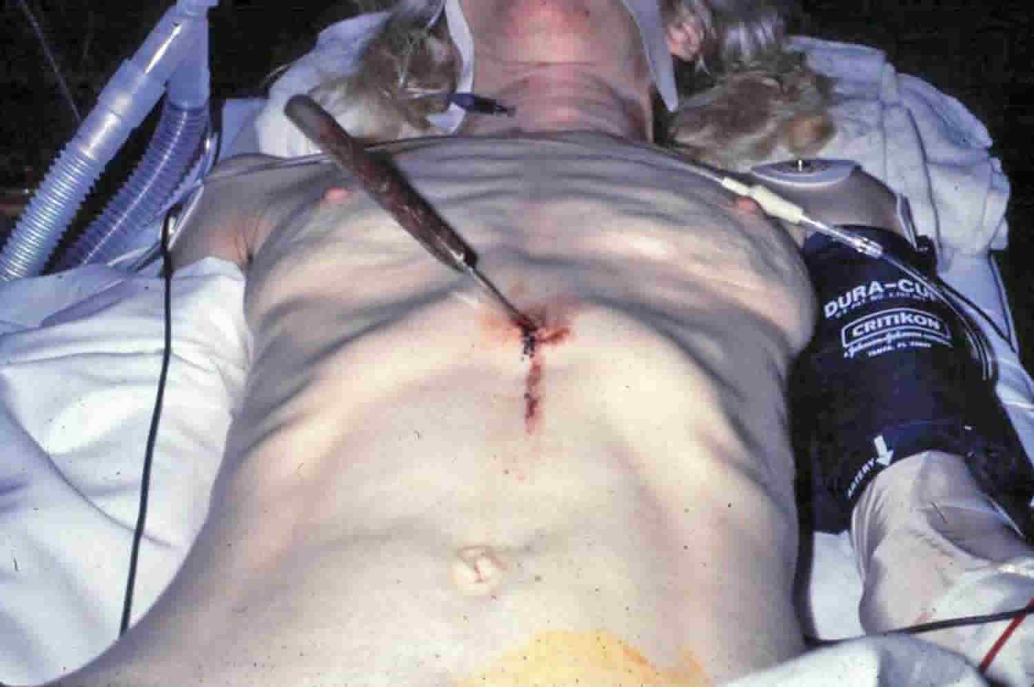
Characteristics of stab wounds:
- Clean cut edges
- One or both ends pointed
- Non-pointed end may be squared off or split (fish tail or boat shaped defect)
- Often gape (related to skin elasticity and Langer’s lines)
- Cross section of weapon may be illustrated when edges of wounds opposed
- Underlying bone may be scored by blade
- Abrasions may be present
- Frequently shows notching or a change in direction (caused by relative movement of the knife and body)
Stab wounds usually result from a thrusting mechanism with an object (e.g. knife or scissors) that penetrates the skin and underlying tissue and may extend deeper to breach anatomical cavities (e.g. peritoneal or pleural). The location of the injury, trajectory of the object, size of the object and depth of injury all affect the potential damage that can be inflicted.
Depending on the location vascular injury (e.g. aortic, carotid) and injury to highly vascular organs (e.g. spleen) may be acutely concerning. Injuries that may compromise breathing mechanics (e.g. pneumothorax) and airway patency (e.g. trachea) are also concerning. Visceral injury (e.g. intestinal perforation) is also significant.
A short (and non-exhaustive) list of locations and their potential injuries is given 3:
- limbs
- neurovascular injury
- tendon injury
- head and neck
- nerve injury
- arterial injury (e.g. carotid artery transection/dissection)
- bony injury (e.g. facial fracture)
- direct brain injury
- globe rupture
- tracheal injury
- esophageal injury
- chest
- nerve injury
- arterial injury (e.g. aortic injury)
- cardiac injury (e.g. ventricular laceration, traumatic shunt formation)
- diaphragmatic injury
- pneumothorax
- hemothorax
- hemopneumothorax
- abdomen and pelvis
- arterial injury
- venous injury
- bladder injury
- liver injury
- spleen injury
- kidney injury
- viscus perforation
- spine
- cord injury (e.g. complete cord transection, spinal cord herniation)
- meningeal injury (e.g. dural tear)
- arterial injury (e.g. epidural hematoma)
- muscle injury
Stab wounds should always be treated with suspicion, as a small entrance wound can hide injuries to deeper structures or foreign bodies. Stab wounds to the lower chest are associated with abdominal visceral injury in 15% of cases, whereas gunshot wounds to the lower chest are associated with abdominal visceral injury in nearly 50% of cases 4. The lower chest is defined as the area between the nipple line (fourth intercostal space) anteriorly, the tip of the scapula (seventh intercostal space) posteriorly, and the costal margins inferiorly. Because the diaphragm reaches the fourth intercostal space during expiration, the abdominal organs are at risk of injury even after what appears to be an “isolated chest” wound. Thus, wounds to the lower chest should also be managed as abdominal wounds to rule out intraabdominal injury. Occult injury to the diaphragm must be ruled out in patients with stab wounds to the lower chest. Patients undergoing diagnostic peritoneal lavage evaluation have different laboratory value cut-offs than standard anterior abdominal stab wounds. A red blood cell count of more than 10,000/μL is considered positive, and an indication for laparotomy whereas patients with a diagnostic peritoneal lavage red blood cell count between 1,000/μL and 10,000/μL should undergo laparoscopy or thoracoscopy.
Stab wounds are said by some authors to ‘gape open’ depending upon their anatomical location. Lines of tension in the skin are determined by the relative orientation of elastic and collagen fibers, and the cleavage lines of Langer correspond to body surface creases. Cox 5 reviewed, and ‘mapped’ (by making 22,600 puncture wounds in cadavers) the cleavage lines, and noted that it was Dupuytren who first observed (in 1834) that there was a disparity between the shape of an instrument and the shape of the skin wound produced by it. Wounds made with a conical instrument were linear, not circular, in shape and that the direction of these wounds differed in different parts of the body. The lines of cleavage, however, were first published by Langer in 1861.
It was postulated that the lines of cleavage were due to the intrinsic arrangement of collagen fibres in the skin – the majority running parallel to the long axis and the remainder interlacing. The orientation of these fibres was further investigated by Haut 6, examining the tensile strength (and failure) of rat skin. The tensile failure (giving rise to lacerations) depended upon location, orientation, age and strain rate.
Tensile strength of skin oriented circumferentially (along the lines of cleavage) exceeded that oriented longitudinally (i.e. parallel with the body axis).
Surgeons are well aware of the effect of cutting across Langer’s lines – the wound gapes open, as opposed to cutting in the direction of these elastic fibres under the skin surface – the resulting wound does not gape.
However, Hunt 7 adds a word of caution – experimental mapping of Langers lines on stab victims revealed a disparity between the orientation of stab wounds and gaping. The gaping wounds did not align ‘correctly’, and so limited information can be gleaned from this phenomenon. Indeed, he also notes that in addition to Langer’s lines, there are 33 other types of lines of tension described in the literature.
Byard et al 8 describe 3 cases in which wound morphology was substantially altered by their relationship with lines of skin cleavage.
A knife that has not fully entered the skin will only produce a wound of a size corresponding to that part of the blade that has penetrated – this will not necessarily represent the maximum dimensions of the knife, and any interpretation of knife wounds must take this into account. Where there are several wounds, measurements taken from each wound may go some way towards building up a composite picture of the true dimensions of the weapon.
Call your local emergency services number right away if any of these occur:
- Bleeding not controlled by direct pressure
- Wound bleeding for longer than 24 hours
For neck, chest, back, or abdomen wounds, call your local emergency services number if you have:
- Shortness of breath
- Painful breathing
- Back or abdomen pain that gets worse
- Blood in the stool or urine
- Weakness, dizziness, or fainting
Signs of infection:
- Increasing pain in the wound
- Fever of 100.4ºF (38ºC) or higher, or as directed by your healthcare provider
- Redness or swelling of the wound, or pus coming from the wound
Stab wound diagnosis
The approach to patients with penetrating trauma depends on type of penetrating weapon or object, the range from which the injury occurred, which organs may be injured, and the location and number of wounds and the patient’s hemodynamic status. Stab wounds are associated with a significantly lower incidence of intra-abdominal injuries; therefore, expectant management is indicated in hemodynamically stable patients.
Patients with penetrating abdominal trauma generally require complete laboratory profiles in case of need for emergent operation. Many imaging modalities can be useful in the evaluation of a patient with penetrating abdominal trauma. The imaging needs of each patient differ, depending on hemodynamic stability and associated injuries.
- Initial examination (primary survey, or ABCDEs) in patients with penetrating abdominal trauma includes assessment of the following:
- Airway, breathing, circulation (ABCs): Includes vital signs
- Level of consciousness (D, disability): To detect neurologic deficits
- Location(s) of the wound(s) (E, exposure): Inspect all body surfaces, and document all penetrating wounds
- Type of penetrating weapon or object
- Amount of blood loss
The secondary survey is a complete head-to-toe physical examination in hemodynamically stable patients and includes external inspection with respect to anatomic landmarks; abdominal evaluation for tympany, dullness to percussion, bowel sounds, and/or distention; and a digital rectal and genitourinary evaluation. In patients with life-threatening injuries, the secondary survey may be delayed for operative therapy.
Immediate surgical exploration is warranted for evidence of significant intra-abdominal injury, especially vascular trauma, such as the following:
- Hypotension (with or without abdominal distention)
- Narrow pulse pressure
- Tachycardia
- High or low respiratory rate
- Signs of inadequate end organ perfusion
- Peritoneal signs (eg, pain, guarding, rebound tenderness) and/or peritonitis
- Diffuse and poorly localized pain that fails to resolve
Laboratory testing
In case emergent operation is necessary, all patients with penetrating abdominal trauma should undergo certain basic laboratory testing, as follows:
- Blood type and cross-match
- Complete blood count (CBC)
- Electrolyte levels
- Blood urea nitrogen (BUN) and serum creatinine level
- Glucose level
- Prothrombin time (PT)/activated partial thromboplastin time (aPTT)
- Venous or arterial lactate level
- Calcium, magnesium, and phosphate levels
- Arterial blood gas (ABG)
- Urinalysis
- Serum and urine toxicology screen
Imaging studies
The following imaging studies may be used to evaluate patients with penetrating abdominal trauma:
- Chest radiography: To rule out penetration of the chest cavity
- Abdominal radiography in 2 views (anterior-posterior, lateral)
- Chest and abdominal ultrasonography: Focused assessment with sonography for trauma (FAST); includes 4 views (pericardial, right and left upper quadrants, pelvis)
- Abdominal CT scanning (including triple-contrast helical CT): Most sensitive and specific study in identifying and assessing liver or spleen injury severity [1]
Other radiologic studies that may be useful include the following:
- Skeletal survey: To detect any associated fractures
- Brain CT scanning: To detect any coincident head injuries
- Retrograde urethrogram/cystogram: To detect any urethral or bladder injury
- Intraoperative intravenous pyelography: To assess contralateral renal function in patients with kidney damage necessitating nephrectomy
Procedures
The following may be diagnostic and/or therapeutic procedures in patients with penetrating abdominal trauma:
- Gastric decompression in intubated patients: To prevent aspiration
- Foley catherization: To monitor fluid resuscitation
- Peritoneal lavage (open or closed): To identify hollow viscus or diaphragmatic injury
- Tube thoracostomy: To relieve hemothorax/pneumothorax
- Local wound exploration: Diagnostic aid to determine the track of penetration through the tissue layers
- Laparoscopy: To evaluate and treat intra-abdominal injuries, including stab wounds to the anterior abdomen or those with uncertain peritoneal penetration
Stab wound treatment
The approach to patients with penetrating stab wound depends on the following factors:
- Mechanism and location of injury
- Hemodynamic and neurologic status of the patient
- Associated injuries
- Institutional resources
In general, consultation with a general or trauma surgeon should be undertaken for victims of penetrating trauma.
At some centers, trauma surgeons perform the majority of operative repair, while at others, consultants may be involved as individual injuries are identified. For example, a vascular surgeon may repair major arterial and venous injuries or a urologist may address injuries to the bladder, kidneys, and ureters. Trauma surgeons, even if not directly performing care, should oversee the patient’s care and postoperative course.
Prehospital care
The scope of care that paramedics deliver at the scene of the injury has evolved in parallel with the changing standard of care in the hospital setting. Because most deaths occurred from exsanguination associated with prehospital hypotension, trauma system response has been designed to minimize care in the field and expedite transport to the emergency department (ED) and to reduce the time to definitive care.
Aggressive intravenous fluid administration to maintain or reach normotension is discouraged in patients with penetrating injury unless the patient manifests severe shock or prolonged transport is expected. Military data suggest that prehospital fluid administration can be guided by the patient’s mentation and the character of the radial pulse. Similarly, the recent military experience in the Middle East has led to a resurgence of interest in the use of tourniquets to control extremity hemorrhage in the prehospital setting.
The following medications may be used in the management of patients with penetrating abdominal trauma:
- Analgesics (eg, morphine sulfate, fentanyl citrate)
- Anxiolytics (eg, lorazepam, midazolam hydrochloride)
- Antibiotics (eg, cefotetan, metronidazole hydrochloride, gentamicin sulfate, vancomycin hydrochloride, ampicillin sodium-sulbactam sodium)
- Neuromuscular blocking agents (eg, succinylcholine, vecuronium bromide)
- Immune enhancement (eg, tetanus toxoid adsorbed or fluid)
Initial Emergency Department Care
A team leader should direct resuscitation and coordinate all care. Depending on the institution, it may be an emergency physician, trauma surgeon, or one of their supervised residents. Given the potential for significant injury, a junior level physician should not lead care without direct oversight.
When the patient arrives in the emergency department, advanced trauma life support (ATLS) protocols are initiated 9. The ABCDEs (airway, breathing, circulation, disability, exposure/environment) are assessed. The patient should be placed on a cardiac monitor, pulse oximeter, and 100% nonrebreather oxygen mask. Airway protection and ventilatory support are followed by circulatory resuscitation with fluid infusion.
Antibiotics should be administered to patients undergoing exploration.
Airway
Patients with severe shock or loss of ability to control their airway should be intubated to ensure appropriate oxygenation or ventilation. In general, occult cervical spine injury in penetrating trauma is highly unlikely. Unless there are clear deficits or associated blunt injury, cervical collars are rarely necessary and may hinder resuscitation.
Breathing
Tube thoracostomy or needle decompression should be undertaken immediately for patients with obvious pneumothorax. A patient who is otherwise stable, should have a chest radiograph performed in the trauma room. An upright positioned radiograph during expiration may provide the best evidence of pneumothorax. Ultrasonography for pneumothorax (as part of the Extended Focused Assessment With Sonography for Trauma [eFAST] or Focused Assessment With Sonography for Trauma [FAST] examination) has been shown to be highly accurate and may be used as the initial test, but it should be followed by a radiograph at some point.
Circulation
Resuscitation of the patient with penetrating abdominal trauma begins immediately upon arrival. Fluids should be administered rapidly. Normal saline or lactated Ringer solution can be used for crystalloid resuscitation.
Patients who present with hypotension are already in class III shock (30-40% blood volume loss) and should receive blood products as soon as possible; the same is true of patients with obvious significant bleeding. Consideration should be given to the early activation of massive transfusion protocols and damage control resuscitation in appropriate patients. Specific triggers for each remain to be better elucidated. Arterial access for continuous blood pressure monitoring is standard. Efforts should be made to limit hypothermia, including warm blankets and prewarmed fluids.
The route of intravenous access is important. Large-bore peripheral intravenous catheters (at least 2) in the upper extremities are the resuscitation lines of choice. These allow for rapid volume/blood infusion versus a central line where the infusion rate is slower.
Extensive debate exists in the literature on the amount and end points for resuscitation prior to definitive control of hemorrhage. Animal data and several studies in humans have suggested that “permissive hypotension”—actively or passively allowing the blood pressure to remain in the hypotensive range (ie, systolic pressure less than 90 mm Hg)—may prevent disruption of clot and dilution of clotting factors while maintaining adequate blood viscosity.
While no definite consensus exists, prevailing thought seems to promote limited resuscitation with avoidance of attempting to raise blood pressure to normal or near-normal levels until hemorrhage is definitively controlled.
Disability and exposure
A rapid and brief evaluation for neurologic deficits should be conducted.
All patients with penetrating trauma should be fully undressed. Complete exposure and head-to-toe visualization of the patient is mandatory in a patient with penetrating abdominal trauma. This includes the buttocks, posterior part of the legs, scalp, posterior part of the neck, and perineum. There is little to be gained by practicing spinal immobilization unless spinal injury is obvious.
Further intervention
Depending on the initial assessment, and in all seriously injured patients, a Foley catheter should be placed if possible to monitor urine output and to check for hematuria. In addition, a nasogastric tube (NGT) or orogastric tube (OGT) should be inserted to evaluate for intragastric blood and to decompress the stomach so as to reduce aspiration risk. Appropriate laboratory specimens should be immediately sent to the laboratory for evaluation.
After the initial evaluation, further evaluation depends on the hemodynamics and mechanism of wounds.
Expectant management
Although surgical management has generally been the standard of care for penetrating abdominal injuries, a study in 90 children by Cigdem et al 10 concluded that in the absence of hemodynamic instability or signs of hollow viscus perforation, the majority of abdominal stab wounds and many gunshot wounds in children can initially be managed nonoperatively.
In this study, patients with hemodynamic instability or signs of bowel perforation underwent immediate laparotomy; the remainder were followed with serial clinical examinations, radiologic evaluation, and hemoglobin levels. Of the 39 children who were managed surgically, 6 (15%) had no significant organ injury found during surgery; of the 51 patients who initially received conservative therapy, 2 children (3.9%) required surgery 10.
Patients with blood on rectal examination who are otherwise being managed expectantly (mostly stab wounds) should undergo rigid sigmoidoscopy to rule out rectal injury.
Emergency Department Thoracotomy
Victims of penetrating abdominal trauma who lose vital signs or who present with exsanguinating hemorrhage that is not controllable with direct external pressure are candidates for an emergency department left anterolateral-left thoracotomy. The purpose of this procedure is to relieve cardiac tamponade, control cardiac bleeding, obtain proximal aortic control, and provide open cardiac massage to improve cardiopulmonary cerebral resuscitation efforts.
This procedure is performed only in extremely selected circumstances, since survival from abdominal injury requiring a resuscitative ED thoracotomy is rare. It is much more effective if the arrest is due to cardiac injury with thoracoabdominal trauma. Patients who may be considered for thoracotomy are those who had vital signs on arrival or en route, with or without pulseless electrical activity on the cardiac monitor. Thoracotomy is rarely successful in blunt trauma.
The surgical procedure is as follows:
- After rapidly preparing and draping the entire chest, make a curvilinear incision from the left sternal border of the fifth intercostal space to the table, paralleling the course of the underlying rib.
- Divide all tissues above the rib with the scalpel.
- Halt respirations.
- Using a finger or Kelly clamp, pierce the intercostal muscle bundle above the rib, then divide with a curved Mayo scissor for the length of the incision.
- Reinflate the lungs.
- Insert a rib spreader with a ratchet mechanism placed laterally.
- Open the pericardium longitudinally to avoid injury to the pericardiacophrenic vessels and the phrenic nerve.
- Subluxing the heart into the left chest allows for open massage.
- Retract the left lung superiorly using a moist laparotomy pad, and divide the inferior pulmonary ligament using Metzenbaum scissors 11.
- The tissues overlying and just lateral to the vertebral bodies contain the aorta, esophagus, thoracic duct, and countless nerves. Usually, blunt dissection frees the aorta enough to place a Satinsky or long, curved DeBakey clamp. In certain circumstances, the aorta is not identified easily, and the aorta and esophagus must be clamped en masse in a patient who is in extremis.
- Warm saline is essential to prevent cooling of the heart, and pressor support usually is needed as well 11.
Surgical intervention
The indications for operative intervention include the following:
- Development of hemodynamic instability
- Development of increasing pain, peritoneal findings (eg, point tenderness, involuntary guarding, rebound tenderness)
- Diffuse and poorly localized pain that fails to resolve
Unstable patients or those with clear-cut peritonitis should undergo exploratory laparotomy.
Stable patients may have local wound exploration to ascertain whether the peritoneum was violated. If this cannot be performed or if flank or thoracoabdominal wounds are present, other methods must be used.
Diagnostic peritoneal lavage (DPL) remains an option, but it is currently being used less frequently. A positive focused assessment with sonography for trauma (FAST) examination result has a high positive predictive value for a therapeutic laparotomy, but a negative FAST examination result cannot be relied upon to rule out injury.
In patients with thoracoabdominal injury, a chest radiograph should be obtained. If no signs of diaphragmatic injury are present, laparoscopy is usually advocated; although some surgeons will elect not to perform this on patient with a right-sided wound, given the low likelihood of delayed complications.
The use of CT scan is still controversial; some centers use it as a screening test in patients with anterior stab wounds, while others feel the cost-benefit ratio is not justified. A triple contrast CT should be performed on patients with penetrating flank wounds.
Essentially all nonoperative patients, except those who have a wound that clearly does not penetrate the abdomen, should be observed for serial examinations. The literature is beginning to support a shortened time frame of 12 hours, but most centers use about 24 hours.
Surgical intervention begins with preparation of the patient in the operating room, as follows:
- The patient is placed in the supine position with arms extended
- The entire chest, abdomen, and pelvis, including the upper thighs, are prepped and draped
- Fluids and blood products should be readily available (and administered via warm lines)
- Warming devices should be placed on the patient’s upper and/or lower extremities
- Entering the abdominal cavity can release tamponade, resulting in a precipitous drop in blood pressure, so the anesthesia team must be informed when the midline incision is made
Intraoperative details
Essential components to the trauma laparotomy include the following:
- Control of bleeding
- Identification of injuries
- Control of contamination
- Reconstruction (if possible)
Procedure
- Initial control of bleeding is accomplished with 4-quadrant packing using laparotomy pads
- The abdominal wall is retracted, the falciform ligament is taken down, and packs are placed above the liver and the spleen and in both sides of the pelvis after the bowel is swept cephalad
- Once anesthesia has been given time to catch up with fluid resuscitation, the packs are removed one quadrant at a time, starting away from the sites of apparent bleeding
- All areas are examined for injuries; each solid organ and the entire bowel are inspected
- Contamination is controlled with the use of clamps, staples, or suture closures
- Depending on the character of the defect(s), resection may be necessary
- If the patient is stable enough to continue the operation, reconstruction may then be performed
Occasionally, patients with penetrating abdominal trauma develop such significant metabolic acidosis and coagulopathy that proceeding with the reconstruction phase of the laparotomy is not possible. In these cases, the operation is considered damage-control surgery, and the abdomen is closed rapidly.
Often, a temporary closure with an intravenous fluid bag or mesh (occasionally with a vacuum dressing) is used, as the patient has undergone massive fluid resuscitation and the bowel has become quite edematous, precluding primary closure of the abdomen. The patient is then transported to the intensive care unit for continued resuscitation and warming. Reconstruction then takes place upon return to the operating room in 24-48 hours.
Colon injuries
Primary repair of colonic injuries may be considered if the patient is hemodynamically stable and if the injury is fairly small with minimal fecal contamination. A diverting colostomy should be performed if the patient has any of the following:
- Multiple injuries
- Requirement for significant blood product resuscitation
- Acidosis, hypothermia, and coagulopathy
- A large defect (>50% of the circumference) and considerable fecal spillage
Other organ injuries
- Diaphragm – Lower-grade injuries may be repaired either via laparotomy or with laparoscopic or thoracoscopic techniques
- Liver – The key rules are gaining adequate exposure and obtaining hemostasis
- Spleen – On the basis of the patient’s hemodynamic status, comorbidities, and operative access, the surgeon will plan for splenorrhaphy or splenectomy
- Kidney – If at all possible, the kidney is salvaged with renography, using pledgeted sutures and wrapping, and capsular reapproximation; if nephrectomy is deemed necessary because of the severity of injury or instability of the patient, an intraoperative intravenous pyelogram is performed to confirm function of the contralateral kidney
- Stomach – Exposure and thorough inspection is necessary, facilitated by opening of the gastrocolic ligament; injuries extending into the lumen may be repaired quickly with a stapling device
- Diaphragm – For exploration, the Kocher maneuver is used to mobilize the duodenum, along with the pancreatic head and distal common bile duct; primary repair of injury is the goal, with protection of the repair using closed-suction drainage; diversion procedures are often used for protection
- Pancreas – Pancreatic duct status and injury location are determinants in the management; lacerations or contusions without ductal injury can be treated conservatively, while more severe injuries may require partial or complete pancreatectomy
Damage-control surgery
Damage control surgery involves abbreviated laparotomy after control of surgical hemorrhage and enteric spill, with physiologic resuscitation in the intensive care unit and staged abdominal reconstruction 12.
Damage-control techniques include the following:
- Perihepatic or intra-abdominal packing and towel clip closure of the abdomen
- Therapeutic decompressive celiotomy
- Prophylactically leaving open the abdominal fascia after laparotomy
Postoperative details
Patients should be monitored closely in the surgical intensive care unit after trauma laparotomy. Many patients will remain intubated and require ventilatory support. Attention should be paid to the following:
- Warming the patient
- Continuing fluid and blood product resuscitation
- Replacing electrolytes
- Monitoring drain outputs
- Patients with evidence of ongoing bleeding may benefit from angiographic evaluation for possible embolization; some require reexploration for control of hemorrhage
- Patients who have undergone damage-control procedures or have temporary abdominal closures must return to the operating room within 24-48 hours for definitive repair
Complications
Prevention is important for the following complications:
- Deep vein thrombosis and pulmonary embolism
- Stress ulceration and bleeding
- Pressure ulcers
- Atelectasis
- Ventilator-associated pneumonia
- Catheter-related sepsis
- ICU psychosis
Early postoperative complications include the following:
- Ongoing bleeding
- Coagulopathy
- Abdominal compartment syndrome
Later complications include the following:
- Acute respiratory distress syndrome
- Pneumonia
- Sepsis
- Intra-abdominal fluid collections
- Wound infections
- Enterocutaneous fistulae
- Small bowel obstruction
- Incisional hernias.
- Emergency! Stab wound. Am J Nurs. 1998 Sep;98(9):49. doi:10.2307/3471869
- Sugrue M, Balogh Z, Lynch J, Bardsley J, Sisson G, Weigelt J (August 2007). “Guidelines for the management of haemodynamically stable patients with stab wounds to the anterior abdomen”. ANZ Journal of Surgery. 77 (8): 614–20. doi:10.1111/j.1445-2197.2007.04173.x
- de Vries CS, Africa M, Gebremariam FA, van Rensburg JJ, Otto SF, Potgieter HF. The imaging of stab injuries. (2010) Acta radiologica (Stockholm, Sweden : 1987). 51 (1): 92-106. doi:10.3109/02841850903225198
- Abernathy’s Surgical Secrets 6th Edition, 2009. ISBN 978-0-323-05711-0. https://doi.org/10.1016/B978-0-323-05711-0.X0001-8
- Cox HT. The cleavage lines of the skin. Br J Surg 1941;29:234-40.
- Haut RC (1989), ‘The effects of orientation and location on the strength of dorsal rat skin in high and low speed tensile failure experiments’, Journal of biomechanical engineering’, 111 (2):136-146
- Hunt A.C. (2003), ‘Morphology of knife wounds’, Presentation to the British Association in Forensic Medicine Winter Meeting, Cardiff, Wales 29th November 2003
- Byard RW, Gehl A, Tsokos M (2005), ‘Skin tension and cleavage lines (Langer’s lines) causing distortion of ante- and post mortem wound morphology’, Int J Legal Med 119:226-230
- Richards CF, Mayberry JC. Initial management of the trauma patient. Crit Care Clin. 2004 Jan. 20(1):1-11.
- Cigdem MK, Onen A, Siga M, Otcu S. Selective nonoperative management of penetrating abdominal injuries in children. J Trauma. 2009 Dec. 67(6):1284-6; discussion 1287.
- Roberts JR, Hedges JR. Clinical Procedures in Emergency Medicine. Resuscitative thoracotomy. Philadelphia, Pa: WB Saunders; 2004. Vol 4: 18.
- Morris JA Jr, Eddy VA, Rutherford EJ. The trauma celiotomy: the evolving concepts of damage control. Curr Probl Surg. 1996 Aug. 33(8):611-700.


