Stomatitis
Stomatitis is an inflammation of the mucous lining of any structure in your mouth, which may involve the cheeks, gums, tongue, the inner aspect of the lips, throat, and/or the roof or floor of the mouth. Stomatitis is a type of mucositis. It can be acute or chronic, mild or serious. The inflammation can be the result of conditions within the mouth itself, such as poor oral hygiene, poorly fitted dentures, or mouth burns from hot food or drinks. It may also be caused by conditions that affect the entire body, such as medications, allergic reactions, or infections. A form of stomatitis known as stomatitis nicotina can be caused by smoking cigars, cigarettes, and/or pipes, and is characterized by small red bumps on the roof of the mouth.
When stomatitis also involves an inflammation of the gingiva, it is called gingivostomatitis. Inflammation of the vermilion of the lips is known as cheilitis, inflammation of the tongue is glossitis, inflammation of the gums is gingivitis, and inflammation of the back of the mouth is pharyngitis. Irritation and fissuring in the corners of the lips is termed angular stomatits or angular cheilitis. In children, a common cause of angular stomatitis is repeated lip-licking; in adults, it may be a sign of underlying iron deficiency anemia, or vitamin B deficiencies (e.g., B2-riboflavin, B9-folate, or B12-cobalamins), which in turn may be evidence of poor dietary habits or malnutrition (e.g., celiac disease).
Stomatitis key points
- Isolated stomatitis in patients with no other symptoms and signs or risk factors for systemic illness is usually caused by a viral infection or recurrent aphthous stomatitis (recurrent aphthous ulcers or canker sores).
- Extraoral symptoms, rash, or both suggest more immediate need for diagnosis.
Stomatitis causes
Stomatitis can be due to injury, infection, allergy, systemic or skin disease. Most commonly, stomatitis is due to:
- Dry mouth / xerostomia
- Dehydration
- Viral infection e.g., herpes simplex and herpes zoster
- Candida albicans infection (oropharyngeal candidiasis)
- Trauma including surgery
- Smoking tobacco
- Toxicity of chemotherapy drugs – including methotrexate used for psoriasis and other skin disorders
- Therapeutic radiation e.g. for oral cancer
- Dentures
- Folate deficiency
- Drugs
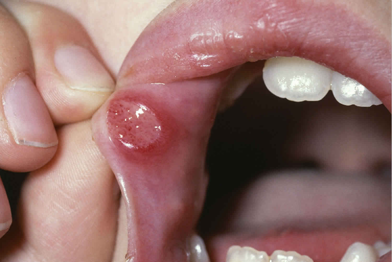
- Recurrent aphthous stomatitis also called recurrent aphthous ulcers
- Vitamin B12 deficiency
Life-threatening causes include conditions which may result in death or permanent disability within 24 hours if left untreated.
- Ebola
Some of the causes of stomatitis are listed below.
Causes of stomatitis
- Infectious
- Bacterial infection
- Necrotising periodontal disease
- Mycoplasma infection
- Syphilis
- Gonorrhea
- Rarely: actinomycosis, tuberculosis, Prevotella, H. pylori, Bartonella
- Fungal infection
- Candida albicans infection (oral candidiasis, thrush)
- Rarely: Blastomycosis, cryptococcosis, zygomycosis, histoplasma capsulatum (histoplasmosis)
- Viral infection
- Herpes simplex virus infection
- Varicella or herpes zoster infection
- Enterovirus or Coxsackie virus infection – hand foot and mouth, or herpangina
- Epstein-Barr virus infection – infectious mononucleosis
- Measles – Koplik spots
- Bovine papular stomatitis virus
- Cytomegalovirus infection
- Bacterial infection
- Non-infectious
- Systemic disorder
- Malnutrition including iron deficiency and vitamin c deficiency (scurvy)
- Inflammatory bowel disease
- Behçet disease
- Kawasaki disease
- Erythema multiforme
- Stevens-Johnson syndrome / toxic epidermal necrolysis
- Drugs
- Nicotine stomatitis
- Toxicity of chemotherapy drugs
- Methotrexate-induced stomatitis
- Lichenoid drug eruption
- Physical irritation
- Thermal burns from hot foot or drink
- Denture stomatitis
- Contact stomatitis
- Contact stomatitis due to irritants (acidic or sharp food) and allergies, e.g. to sesquiterpene lactones in food, toothpaste or propolis
- Immunobullous disesase
- Immunobullous disorders
- Pemphigus vulgaris
- Paraneoplastic pemphigus
- Linear IgA bullous dermatosis
- Bullous pemphigoid
- Mucous membrane pemphigoid
- Pemphigoid gestationis
- Other
- Chronic ulcerative stomatitis
- Recurrent aphthous ulceration
- Geographic tongue / migratory glossitis
- Erosive lichen planus
- Lupus erythematosus
- Autoimmune progesterone dermatitis
- Oral leukoplakia (precancerous state)
- Systemic disorder
Stomatitis symptoms
Stomatitis results in pain, stinging and soreness. It can present with:
- Red patches
- Mouth ulcers
- Blisters
- Peeling
- Swelling
- Oral dysaesthesia (abnormal unpleasant sensation felt when touched such as numbness)
- Burning mouth syndrome – soreness despite normal appearance
These can lead to dehydration and malnutrition.
Stomatitis diagnosis
History of present illness should ascertain the duration of symptoms and whether the patient ever had them previously. Presence and severity of pain should be noted. The relation of symptoms to food, drugs, oral hygiene materials (eg, toothpaste, mouth rinses), and other substances (particularly occupational exposure to chemicals, metals, fumes, or dust) is sought.
Review of systems seeks symptoms of possible causes, including chronic diarrhea and weakness (inflammatory bowel disease, celiac disease); genital lesions (Behçet disease, syphilis); eye irritation (Behçet disease); and weight loss, malaise, and fever (nonspecific chronic illness).
Past medical history should ascertain known conditions that cause oral lesions, including herpes simplex, Behçet disease, inflammatory bowel disease, and risk factors for oral lesions, including immunocompromised state (eg, cancer, diabetes, organ transplant, use of immunosuppressants, HIV infection). Whether chemotherapy or radiation therapy has ever been used to manage cancer needs to be determined. Drug history should note all recent drugs used. History of tobacco use should be noted. Social history should include sexual contact, particularly oral sex, unprotected sex, and sex with multiple partners.
Physical examination
Vital signs are reviewed for fever. The patient’s general appearance is noted for lethargy, discomfort, or other signs of significant systemic illness.
The mouth is inspected for the location and nature of any lesions.
The skin and other mucosal surfaces (including the genitals) are inspected for any lesions, rash, petechiae, or desquamation. Any bullous lesions are rubbed for the Nikolsky sign (upper layers of epidermis move laterally with slight pressure or rubbing of skin adjacent to a blister).
Red flags
The following findings are of particular concern:
- Fever
- Cutaneous bullae
- Ocular inflammation
- Immunocompromise
Relevant investigations depend on the likely cause of stomatitis and whether it is accompanied by other symptoms internally or skin rashes.
Testing may include:
- Bacterial swabs and culture
- Viral swabs and culture
- Tissue scrapings for mycology
- Biopsy for histology and direct immunofluorescence
- Blood tests
- Patch tests to identify contact allergy
- Biopsy
Patients with acute stomatitis and no symptoms, signs, or risk factors for systemic illness probably require no testing.
If stomatitis is recurrent, viral and bacterial cultures, complete blood count (CBC), serum iron, ferritin, vitamin B12, folate, zinc, and endomysial antibody (for sprue) are done. Biopsy at the periphery of normal and abnormal tissue can be done for persistent lesions that do not have an obvious etiology.
Systematically eliminating foods from the diet can be useful, as can changing brands of toothpaste, chewing gum, or mouthwash
Interpretation of findings
Occasionally, causes are obvious in the history (eg, cytotoxic chemotherapy; significant occupational exposure to chemicals, fumes, or dust). Recurrent episodes of oral lesions occur with recurrent aphthous stomatitis, herpes simplex, and Behçet disease. History of diabetes, HIV infection or other immunocompromise, or recent antibiotic use should increase suspicion of Candida infection. Recent drug use (particularly sulfa drugs, other antibiotics, and antiepileptics) should increase suspicion of Stevens-Johnson syndrome.
Some causes typically have extraoral, noncutaneous findings, some of which suggest a cause. Recurrent gastrointestinal symptoms suggest inflammatory bowel disease or celiac disease. Ocular symptoms can occur with Behçet disease and Stevens-Johnson syndrome. Genital lesions may occur with Behçet disease and primary syphilis.
Some causes usually also have extraoral, cutaneous findings.
Cutaneous bullae suggest Stevens-Johnson syndrome, pemphigus vulgaris, or bullous pemphigoid. Prodrome of malaise, fever, conjunctivitis, and generalized macular target lesions suggests Stevens-Johnson syndrome. Pemphigus vulgaris starts with oral lesions, then progresses to flaccid cutaneous bullae. Bullous pemphigoid has tense bullae on normal-appearing skin. The Nikolsky sign is usually positive in Stevens-Johnson syndrome and pemphigus vulgaris.
Cutaneous vesicles are typical with chickenpox or herpes zoster. Unilateral lesions in a band along a dermatome suggest herpes zoster. Diffuse, scattered vesicular and pustular lesions in different stages suggest chickenpox.
Kawasaki disease usually has a macular rash, desquamation of hands and feet, and conjunctivitis; it occurs in children, usually those < 5 year. Oral findings include erythema of the lips and oral mucosa.
Other cutaneous lesions may implicate erythema multiforme, hand-foot-and-mouth disease (resulting from coxsackievirus), or secondary syphilis.
Some causes have isolated oral findings, including recurrent aphthous stomatitis, most viral infections, acute necrotizing ulcerative gingivitis, primary syphilis, gonorrhea, and Candida.
Location of oral lesions may help identify the cause. Interdental ulcers occur with primary herpes simplex or acute necrotizing ulcerative gingivitis. Lesions on keratinized surfaces suggest herpes simplex, recurrent aphthous stomatitis, or physical injury. Physical injury typically has an irregular appearance and occurs near projections of teeth, dental appliances, or where biting or an errant toothbrush can injure the mucosa. An aspirin burn next to a tooth and pizza burn on the palate are common.
Primary herpes simplex infection causes multiple vesicular lesions on the intraoral mucosa on both keratinized and nonkeratinized surfaces and always includes the gingiva. These lesions rapidly ulcerate. Clinical manifestation occurs most often in children. Subsequent reactivations (secondary herpes simplex, cold sore) usually appear starting in puberty on the lip at the vermilion border and, rarely, on the hard palate.
Acute necrotizing ulcerative gingivitis causes severe inflammation and punched-out ulcers on the dental papillae and marginal gingivae. A severe variant called noma (gangrenous stomatitis) can cause full-thickness tissue destruction (sometimes involving the lips or cheek), typically in a debilitated or malnourished patient. It begins as a gingival, buccal, or palatal (midline lethal granuloma) ulcer that becomes necrotic and spreads rapidly. Tissue sloughing may occur.
Isolated oral gonorrhea very rarely causes burning ulcers and erythema of the gingiva and tongue, as well as the more common pharyngitis. Primary syphilis chancres may appear in the mouth. Tertiary syphilis may cause oral gummas or a generalized glossitis and mucosal atrophy. A common sign of HIV becoming AIDS is hairy leukoplakia (vertical white lines on the lateral border of the tongue).
Candida albicans and related species, which are normal oral flora, can overgrow in people who have taken antibiotics or corticosteroids or who are immunocompromised, such as patients with AIDS. Candida albicans can cause whitish, cheesy plaques that leave erosions when wiped off. Sometimes only flat, erythematous areas appear (erosive form of Candida).
Stomatitis treatment
Treatment for stomatitis depends on the cause. If it is due to allergy to a medication, the medication must be promptly stopped. However, it may be necessary to continue a causative medication when stomatitis arises as an expected adverse reaction to chemotherapy.
Infections may require specific treatment such as antibiotics for streptococcal pharyngitis, topical antifungal or oral antifungal agent for candida infection.
Nutritional deficiencies should be identified and corrected, for example, folic acid can reduce methotrexate-induced stomatitis.
Immunobullous diseases may be treated with systemic corticosteroids or other immunosuppressive treatments.
Symptomatic treatment may include numerous topical treatments alone or in combination, are used to ease symptoms:
- Antiseptic mouthwash
- Protective pastes
- Local anesthetic mouthwash or spray
- Sucralfate plus aluminum-magnesium antacid rinse
- Oral analgesics (pain killers)
- Topical corticosteroids.
For topical anesthesia of discomfort that may interfere with eating and drinking, the following may be effective:
- Lidocaine rinse
- Sucralfate plus aluminum-magnesium antacid rinse
A 2-minute rinse is done with 15 mL (1 tbsp) 2% viscous lidocaine every 3 hour as required; patient expectorates when done (no rinsing with water and no swallowing unless the pharynx is involved). A soothing coating may be prepared with sucralfate (1-g pill dissolved in 15 mL water) plus 30 mL of aluminum-magnesium liquid antacid; the patient should rinse with or without swallowing. Many institutions and pharmacies have their own variation of this formulation (magic mouthwash), which sometimes also contains an antihistamine.
If the physician is certain the inflammation is not caused by an infectious organism, the patient can:
- Rinse and expectorate after meals with dexamethasone elixir 0.5 mg/5 mL (1 tsp)
- Apply a paste of 0.1% triamcinolone in an oral emollient
- Wipe amlexanox over the ulcerated area with the tip of a finger
Chemical or physical cautery can ease the pain of localized lesions. Silver nitrate sticks are not as effective as low-power (2- to 3-watt), defocused, pulsed-mode CO2 laser treatments, after which pain relief is immediate and lesions tend not to recur locally.


