Subinvolution
Subinvolution of placental site also known as placental site vascular subinvolution or subinvolution of uteroplacental arteries, is an important contributor to secondary postpartum bleeding 1. Subinvolution of the placental site refers to delayed or inadequate physiologic closure and sloughing of the superficial modified spiral arteries at the placental attachment site. Subinvolution can be identified and documented by the typical clinical features and the histologic findings in postpartum endometrial curettings and hysterectomy specimens. Although detailed studies have been performed by several authors to determine the normal and abnormal anatomy and histology of the uterine placental site 2, there is a relative lack of practice-oriented literature on the topic of subinvolution. Subinvolution of the placental site is commonly associated with delayed post partum or post abortal hemorrhage. It tends to occur in the older group of obstetrical patients and in multiparae 3.
Normal involution of the uteroplacental arteries
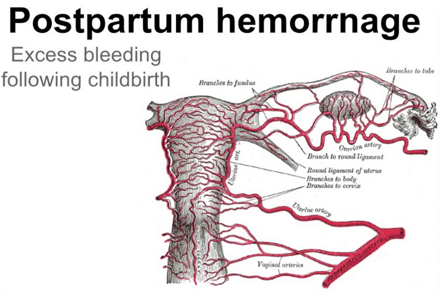
After delivery, a physiologic mechanism of uteroplacental arterial involution is required to eliminate these altered vessels. In the third trimester, this process begins modestly as the endovascular extravillous cytotrophoblasts are replaced by maternal-derived endothelial cells. The exact temporal relationships are not fully elucidated, but several involution events occur within the uteroplacental vascular bed in the few days after delivery. These changes include occlusive fibrointimal thickening, endarteritis, thrombosis, regeneration of the internal elastic lamina, and disappearance of both endovascular and interstitial extravillous cytotrophoblasts. Although the histomorphology of involutional events is well described, the exact molecular basis that triggers these changes remains poorly understood 4. Necrosis and sloughing of the decidua and superficial endomyometrium occur in tandem with the involutional vascular changes. Contraction of the uterine smooth muscle also contributes to mechanical shrinkage and involution of the placental site as a whole 5. Taken together, one very important consequence of these changes is to limit the loss of blood at the placental site after delivery.
Subinvolution causes
The cause of subinvolution is not known, but this process may be a manifestation of an abnormal interaction between fetal-derived trophoblasts and maternal tissue. Subinvolution has also been reported to cause bleeding after induced abortion 6 and after molar pregnancy evacuation 7. Clinical associations cited in the literature include multiparity and older age 8.
The extravillous cytotrophoblasts play an important role in the development of adequate uteroplacental arterial flow, and an abnormal interaction between these cells and the maternal tissue is proposed as the mechanism for subinvolution 8. An immunologic basis for this type of altered fetomaternal interaction has been suggested and is an especially attractive idea when compared to the findings in preeclampsia. In preeclampsia, poor investment and remodeling of the spiral arteries by extravillous cytotrophoblasts is found. Mounting epidemiologic and molecular evidence suggests that poor interaction between extravillous cytotrophoblasts and maternal decidual tissue may be due to an abnormal immunologic recognition process 9. The respective pathophysiology of preeclampsia and subinvolution may represent opposite ends of a spectrum of abnormal cellular interactions at the fetomaternal tissue interface. Other evidence supporting an abnormal immunologic basis for subinvolution includes the observations by Andrew et al 10 of differential immunoglobulin and complement deposition in involuted and subinvoluted vessels. In this study, the deposition of immunoglobulins (IgG, IgA, IgM) and complement proteins (C1q, C3d, C4) in the walls of normally involuted vessels is described; these components are absent in subinvoluted vessels. Another insight into the mechanism of subinvolution is related to extravillous cytotrophoblast apoptosis. While the extravillous cytotrophoblasts in preeclampsia appear to lose expression of the antiapoptosis protein Bcl-2, increased expression of this protein has been reported in subinvolution 11. Perhaps persistent expression of this antiapoptosis protein plays a role in maintaining the uteroplacental vessels in a state similar to gestation in cases of subinvolution. Despite these interesting observations, there remains no uniform consensus as to a defined pathophysiologic sequence of events in subinvolution.
The diagnosis of subinvolution can be made by pathologic examination of postpartum endometrial curettage material 1. Vascular subinvolution may be detected in the presence of rare degenerating chorionic villi, which is a valuable observation when the amount of villous tissue present does not account for bleeding 1. Therefore, the diagnosis of subinvolution should be carefully excluded in all cases, and the temptation to only record the presence or absence of villous tissue should be avoided.
A number of microscopic features have been described in the literature 10. The essential findings identifiable on hematoxylin-eosin staining include large, patent, and dilated superficial myometrial vessels with pink hyaline material replacing the media and partial intravascular thrombosis of variably aged thrombi 1. These abnormal vessels are typically clustered with little intervening myometrial tissue and may be located adjacent to normally involuted vessels 1. Extravillous interstitial and/or endovascular trophoblasts should also be identified. The cytologic features of extravillous cytotrophoblast cells include polygonal shape, abundant amphophilic cytoplasm, and vesicular nuclei. Immunohistochemistry may be helpful to identify these cells, as they stain readily with commercially available antibodies directed at low-molecular-weight cytokeratin, inhibin-α, Mel-CAM, and human placental lactogen 12. Rare cells may also stain with β human chorionic gonadotrophin. Other, though less constant, microscopic features have been described in subinvolution. These include lack of internal elastic lamina duplication, partial or complete absence of a true endothelial lining, and immunohistochemical expression of the antiapoptosis protein Bcl-2. Histochemical stains for elastin (elastic von Gieson or elastochrome) can be very helpful in delineating the vessel wall anatomy. Immunoperoxidase staining for vascular endothelial markers may also be helpful in defining the extent of endothelial regeneration, particularly anti-CD31. The constellation of histologic findings in cases of subinvolution likely reflects the nature of this process as a biologic continuum of changes, lying somewhere between appropriate involution and complete lack of it. The extent of subinvolution varies from case to case, and the diagnostic findings may be quantitatively limited in any given specimen. In cases where the complete set of essential findings is not observed, subinvolution may be suggested as a diagnostic possibility. A diagnosis of subinvolution should only be rendered in the appropriate clinical setting, which is secondary (delayed) postpartum bleeding. Physiologic patency of uteroplacental arteries will be seen in the immediate (<24 hour) postpartum period and should not be unequivocally interpreted as subinvolution.
Subinvolution symptoms
Subinvolution symptoms include:
- Postpartum hemorrhage
- Prolonged lochial discharge
- Larger than normal uterus
- Boggy uterus (occasionally)
Delayed postpartum bleeding should always raise the possibility of subinvolution. Although subinvolution may cause bleeding anytime between 1 week and several months postpartum, the most common reported timeline for presentation is within the second week after delivery 8. Some patients presented within the second week postpartum, some presented between the third and seventh weeks postpartum. The severity of the bleeding is also variable, but typically there is an abrupt onset of increased uterine bleeding, which prompts the patient to seek medical attention.
Subinvolution treatment
Subinvolution treatment depends on the cause. Endometrial curettage may not be curative, and continued brisk bleeding may require hysterectomy in spite of fluid and coagulation maintenance.
References- Jamie A. Weydert and Jo Ann Benda (2006) Subinvolution of the Placental Site as an Anatomic Cause of Postpartum Uterine Bleeding: A Review. Archives of Pathology & Laboratory Medicine: October 2006, Vol. 130, No. 10, pp. 1538-1542.
- Brosens, J. J. , R. Pijnenborg , and I. A. Brosens . The myometrial junctional zone spiral arteries in normal and abnormal pregnancies: a review of the literature. Am J Obstet Gynecol 2002. 187:1416–1423.
- OBER WB, GRADY HG. Subinvolution of the placental site. Bull N Y Acad Med. 1961;37(10):713–730. https://www.ncbi.nlm.nih.gov/pmc/articles/PMC1804714
- Andrew, A. , J. B. Bulmer , and L. Morrison . et al. Subinvolution of the uteroplacental arteries: an immunohistochemical study. Int J Gynecol Pathol 1993. 12:28–33.
- Anderson, W. R. and J. Davis . Placental site involution. Am J Obstet Gynecol 1968. 102:23–33
- Lee, E. T. , N. J. Marley , and J. R. Bevan . A rare late complication of first trimester induced abortion requiring hysterectomy—subinvolution of the placental bed: case report. Br J Obstet Gynaecol 1986. 93:777–781.
- Khong, T. Y. and M. M. Chin . Subinvolution of the placental bed after molar pregnancy. J Reprod Med 1996. 41:352–354.
- Andrew, A. C. , J. B. Bulmer , and M. Wells . et al. Subinvolution of the uteroplacental arteries in the human placental bed. Histopathology 1989. 15:395–405.
- Redman, C. W. and I. L. Sargent . Latest advances in understanding preeclampsia. Science 2005. 308:1592–1594.
- Andrew, A. , J. B. Bulmer , and L. Morrison . et al. Subinvolution of the uteroplacental arteries: an immunohistochemical study. Int J Gynecol Pathol 1993. 12:28–33
- DiFederico, E. , O. Genbacev , and S. J. Fisher . Preeclampsia is associated with widespread apoptosis of placental cytotrophoblasts within the uterine wall. Am J Pathol 1999. 155:293–301
- Santos, L. D. , J. L. Yong , and X. J. Wu . Applications of monoclonal antihuman inhibin alpha subunit in endometrial curettings. Pathology 2000. 35:217–223.


