What is tinea barbae
Tinea barbae is the name used for fungal infection of the beard and moustache areas of the face with a dermatophyte fungus. Tinea barbae is less common than tinea capitis and generally affects only adult men.
Tinea barbae often presents with marked inflammation and aggregated pustules, exudation and crusting. Affected hairs tend to come away quite easily. Tinea barbae is frequently misdiagnosed as a bacterial infection although it may be distinguished from boils secondary to bacteria by the relative lack of pain and the ease with which the hairs come away. Milder cases need to be differentiated from bacterial folliculitis, pseudofolliculitis, acne or rosacea
The cause of tinea barbae is most often a zoophilic (animal) fungus:
- Trichophyton verrucosum (originating from cattle)
- Trichophyton mentagrophytes var. equinum (originating from horses)
Tinea barbae is usually due to infection of coarse facial hair with an ectothrix pattern (spores on the outside). In ectothrix infections, the fungal filaments (hyphae) and spores (arthroconidia) cover the outside of the hair.
It is extremely difficult to totally get rid of tinea barbae with only topical medications; oral antifungal medications are usually required. However, if the infection has just started, you might try one of the following over-the-counter antifungal creams or lotions:
- Terbinafine
- Clotrimazole
- Miconazole
Apply the cream to each lesion and to the normal-appearing skin 2 cm beyond the border of the affected skin for at least 2 weeks until the areas are completely clear of lesions. Remember, you will probably not be able to totally get rid of the tinea barbae with topical creams.
Stop shaving the affected area until you start treatment. If you must shave, use a new disposable razor each time you shave.
Since people often have tinea infections on more than one body part, examine yourself for other ringworm infections, such as in the groin (tinea cruris), on the feet (tinea pedis, athlete’s foot), and anywhere else on the body (tinea corporis).
Have any household pets or farm animals evaluated by a veterinarian to make sure they do not have a fungal (ie, dermatophyte) infection. If the veterinarian discovers an infection, be sure to have the animal treated.
Is tinea barbae contagious?
Yes, tinea barbae is contagious and is passed from person to person, animal to person, and from contaminated objects (such as towels and pillows) to person. It would be possible for beard ringworm in one person to be passed as a facial, body, or scalp ringworm in another person because all the infections are caused by the same fungi.
The infection is spread through close contact with an infected person, or by sharing combs, hairbrushes, hats, clothing, towels, beds and other furniture with someone who is infected. It’s also possible to catch ringworm from infected animals such as dogs, cats, horses or farm animals.The fungus can live for long periods of time in the environment and therefore infection can occur many months later.
Avoid sharing combs, hairbrushes, hats, towels, pillowcases, or helmets with other people. Fungus can live in combs, hairbrushes, and hair accessories, so clean them with simple bleach or purchase new ones. Do not visit the hairdressers or barbers until the infection is clear.Wash all bedding, towels and hats at 140 °F (60°C).
Tinea barbae causes
Tinea barbae is caused by an infection with a type of fungus called a dermatophyte. Dermatophytes are found in humans, animals and in the environment. Hair can be infected with Trichophyton (abbreviated as “T”.) and Microsporum (“M”.) fungi. Lesions are probably triggered by autoinoculation, which is usually observed after local trauma caused by scratch or, more commonly, by shaving razors 1.
The cause of tinea barbae is most often a zoophilic (animal) fungus 2, 3, 4, 5, 6, 7:
- Trichophyton verrucosum (originating from cattle)
- Trichophyton mentagrophytes var. equinum (originating from horses)
- Trichophyton rubrum
Other causative organisms that were documented are 7:
- Trichophyton violaceum,
- Trichophyton megninii,
- Trichophyton schoenleinii,
- Trichophyton tonsurans,
- Trichophyton interdigitale,
- Trichophyton ernacei,
- Microsporum canis,
- Microsporum nanum,
- Mycroscporum gypseum,
- Epidermophyton floccosum.
Until 2000, most of the tinea barbae cases that were documented in the literature were caused by zoophilic dermatophytes from infected farm animals and domestic animals, especially dairy cows (Trichophyton verrucosum), sheep, pigs (Trichophyton ernacei), horses, dogs, and cats (Microsporum canis) 7, 8. More recently, infections with anthropophilic (direct contact with infected humans) Trichophyton rubrum have been a frequently reported causative organism, as noted in multiple individual case reports worldwide and case series published from Portugal and Mexico 7, 4, 5, 2, 6. Human to human transmission of tinea barbae is rare but possible. Individual case reports in the recent past have documented autoinoculations with T. rubrum in patients with tinea pedis 4, 5, 2.
Almost anyone with facial hair can get tinea barbae, though it is most commonly seen in men who shave and in women who have coarse facial or neck hair. It also tends to occur more often in people who live in warm, humid climates and people who work with farm animals. Tinea barbae is more commonly seen in warmer, more humid climates. It is most frequently passed to humans from animals, so agricultural workers are the most commonly infected people with beard ringworm.
Tinea barbae symptoms
Tinea barbae most often affects farmers and farmworkers and is due to direct contact with an infected animal. It is rarely passed from one person to another.
The most common locations for tinea barbae infection include the following:
- Chin
- Cheeks
- Neck
- Upper lip
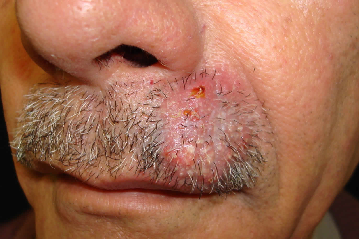
Tinea barbae may affect either the outer surface (superficial) or the deep portion of the skin that holds shafts of hair (hair follicles) 9. If the infection is superficial, tinea barbae appears as a pink-to-red scaly patch ranging in size from 1 to 5 cm. Alternatively, small pus-filled bumps (pustules) may be seen around hair follicles in the affected skin. In deeper forms of tinea barbae, you may see firm red nodules covered with pustules or scabs that may ooze blood and pus. The hairs can be pulled out easily. Surprisingly, it is not excessively itchy or painful.
Tinea barbae is usually itchy. Deeper forms of tinea barbae may be accompanied by fever and swollen lymph glands.
Clinically, tinea barbae may be inflammatory or non-inflammatory, depending on the fungus and the patient’s immune response 1. Deep folliculitis is inflammatory; its clinical lesions are follicular pustules and fistula on the skin surface 4. The non-inflammatory subtype is superficial and similar to tinea corporis or superficial folliculitis.
Tinea barbae can result in an “id reaction” also known as ‘dermatophytid’, especially just after starting antifungal treatment. A itchy rash, papular ‘id eruption’, particularly around the outer helix of the ear or the palms and soles, may accompany treatment initiation, but should not be confused with a drug reaction 10. These eruptions represent a cell mediated host response to the dermatophyte after effective therapy has been initiated and do not warrant cessation of systemic antimycotic therapy. Topical (or occasionally, if very severe, oral) corticosteroids may provide symptomatic relief.
Tinea barbae diagnosis
The diagnosis of tinea barbae is confirmed by microscopy and culture of skin scrapings and hair pulled out by the roots. The samples are looked at under the microscope and cultured to confirm that a fungal infection is the cause. Fungus grows slowly therefore the culture results can take up to 6 weeks.
If you have many pus-filled lesions or if deeper lumps are present, your physician may wish to perform a procedure to grow out the fungus (fungal culture) in order to discover the particular organism that may be causing the infection. The procedure involves:
- Penetrating the pus-filled lesion with a needle, scalpel, or lancet.
- Rubbing a sterile cotton-tipped applicator across the skin to collect the pus.
- Sending the specimen away to a laboratory.
The fungal culture can take up to 6 weeks to produce final results.
Sometimes, diagnosis is made on skin biopsy showing characteristic histopathological features of tinea barbae.
Tinea barbae treatment
Topical antifungal agents may be adequate for mild cases of tinea barbae, but it is usually treated with oral antifungal medicines 11, 12, 13, 14, 15, 16.
Since tinea barbae usually requires oral antifungal pills in order to get rid of the infection completely, your physician will likely recommend one of the following oral medications:
- Terbinafine
- Itraconazole
- Griseofulvin
- Fluconazole
- Ketoconazole
Recommended dosages for the available antifungal agents for tinea barbae are 15:
- Terbinafine 125 mg to 250 mg once a day
- Ketoconazole 200 to 400 mg a day
- Fluconazole 200 mg once a day
- Itraconozole 100 mg once a day
Tinea barbae should go away within 4–6 weeks after using effective treatment. Even though four weeks of treatment is sufficient, some authors have suggested continuing treatment for 2 to 3 weeks after the resolution of the lesions 15.
Oral antifungal agents that have proven effective in the management of tinea barbae are terbinafine, azoles, and griseofulvin 16. Griseofulvin used to be the preferred drug, but it is rarely used now due to its side effects, drug resistance, need for prolonged therapy, and increased relapse rates due to the rapid clearance of the drug from the skin 3, 17. In the past, tinea barbae was treated for 12 weeks with griseofulvin. Recent case reports have demonstrated complete resolution of the tinea barbae with 4-6 weeks of therapy with both terbinafine and azoles 1, 4, 5, 2, 18.
Common side effects for terbinafine and azoles include nausea, abdominal pain, diarrhea, rash, elevated transaminases, and visual disturbances 3. When on oral antifungal therapy, periodic monitoring of liver, kidney, and blood function is recommended. Significant drug interactions can happen when liver metabolized drugs are taken along with azoles 3. The role of treatment with steroids and other topical agents like selenium sulfide is not well defined, and it is the prescriber’s discretion on a case to case basis 15. Oral or systemic antibiotics are warranted when a secondary bacterial infection is suspected. Surgery is rarely indicated, and incision and drainage of kerion are not recommended 16.
Tinea barbae prognosis
Tinea barbae is a rare dermatophytosis infection with only a few hundred cases reported in the literature 15. Most of the reported cases of tinea barbae in the literature were either single case reports or small case series. Though the data available is limited, it has been very reassuring, as almost all cases responded well to oral antifungal therapy with complete resolution and no significant complications have been reported in the literature 15.
References- Furlan KC, Kakizaki P, Chartuni JC, Valente NY. Sycosiform tinea barbae caused by trichophyton rubrum and its association with autoinoculation. An Bras Dermatol. 2017 Jan-Feb;92(1):160-161. doi: 10.1590/abd1806-4841.20174802
- A. Kawada, Y. Aragane, A. Maeda, T. Yudate, T. Tezuka, M. Hiruma, Tinea barbae due to Trichophyton rubrum with possible involvement of autoinoculation, British Journal of Dermatology, Volume 142, Issue 5, 1 May 2000, Pages 1064–1065, https://doi.org/10.1046/j.1365-2133.2000.03510.x
- Rutecki GW, Wurtz R, Thomson RB. From Animal to Man: Tinea Barbae. Curr Infect Dis Rep. 2000 Oct;2(5):433-437. doi: 10.1007/s11908-000-0073-1
- Szepietowski, J.C. and Matusiak, Ł. (2008), Trichophyton rubrum autoinoculation from infected nails is not such a rare phenomenon. Mycoses, 51: 345-346. https://doi.org/10.1111/j.1439-0507.2007.01481.x
- Yin, X., Du, X. and Zhang, H. (2011), A case of tinea barbae due to Trichophyton rubrum infection by autoinoculation from the infected fingernails. Mycoses, 54: e864-e866. https://doi.org/10.1111/j.1439-0507.2011.02012.x
- Duarte, B, Galhardas, C, Cabete, J. Adult tinea capitis and tinea barbae in a tertiary Portuguese hospital: A 11-year audit. Mycoses. 2019; 62: 1079– 1083. https://doi.org/10.1111/myc.12991
- Bonifaz, A., Ramírez-Tamayo, T. and Saúl, A. (2003), Tinea Barbae (Tinea Sycosis): Experience with Nine Cases. The Journal of Dermatology, 30: 898-903. https://doi.org/10.1111/j.1346-8138.2003.tb00345.x
- R. U. Sidwell, I. Chan, N. Francis, C. B. Bunker, Trichophyton erinacei kerion barbae from a hedgehog with direct osculatory transfer to another person, Clinical and Experimental Dermatology, Volume 39, Issue 1, 1 January 2014, Pages 38–40, https://doi.org/10.1111/ced.12197
- Ringworm, Beard (Tinea Barbae). https://skinsight.com/skin-conditions/tinea-barbae-ringworm-of-beard
- lkit M, Durdu M, Karakas M. Cutaneous id reactions: a comprehensive review of clinical manifestations, epidemiology, etiology,and management. Crit Rev Microbiol 2012;38: 191–202
- Reiss E, Shadomy HJ, Lyon GM., III . Fundamental Medical Mycology. Hoboken, NJ: Wiley-Blackwell; 2012. Dermatophytosis; pp. 527–66.
- lkit M, Durdu M, Karakaş M. Majocchi’s granuloma: a symptom complex caused by fungal pathogens. Med Mycol. 2012;50:449–57. doi: 10.3109/13693786.2012.669503
- Feng WW, Chen HC, Chen HC. Majocchi’s granuloma in a 3-year-old boy. Pediatr Infect Dis J. 2006 Jul;25(7):658-9. doi: 10.1097/01.inf.0000224312.87417.fc
- Ely JW, Rosenfeld S, Seabury Stone M. Diagnosis and management of tinea infections. Am Fam Physician. 2014 Nov 15;90(10):702-10. https://www.aafp.org/pubs/afp/issues/2014/1115/p702.html
- Kuruvella T, Pandey S. Tinea Barbae. [Updated 2022 Sep 26]. In: StatPearls [Internet]. Treasure Island (FL): StatPearls Publishing; 2023 Jan-. Available from: https://www.ncbi.nlm.nih.gov/books/NBK563204
- Drake LA, Dinehart SM, Farmer ER, Goltz RW, Graham GF, Hordinsky MK, Lewis CW, Pariser DM, Skouge JW, Webster SB, Whitaker DC, Butler B, Lowery BJ, Elewski BE, Elgart ML, Jacobs PH, Lesher JL Jr, Scher RK. Guidelines of care for superficial mycotic infections of the skin: tinea capitis and tinea barbae. Guidelines/Outcomes Committee. American Academy of Dermatology. J Am Acad Dermatol. 1996 Feb;34(2 Pt 1):290-4. doi: 10.1016/s0190-9622(96)80137-x
- Hainer BL. Dermatophyte infections. Am Fam Physician. 2003 Jan 1;67(1):101-8. https://www.aafp.org/pubs/afp/issues/2003/0101/p101.html
- Duarte, B, Galhardas, C, Cabete, J. Adult tinea capitis and tinea barbae in a tertiary Portuguese hospital: A 11-year audit. Mycoses. 2019; 62: 1079– 1083. https://doi.org/10.1111/myc.12991


