What is toxic epidermal necrolysis
Toxic epidermal necrolysis (T.E.N.) is characterized by fever (>100.4 °F or >38 °C), widespread tender redness (erythema) affecting >30% skin surface associated with mucosal involvement. Redness (erythema) is followed by extensive full thickness cutaneous and mucosal necrosis and denudation within 2 or 3 days. Similar symptoms and signs involving less than 10% of the body surface are classified as Stevens-Johnson syndrome; if 10-30% of body surface area is affected it is classified as T.E.N. or Stevens-Johnson syndrome overlap.
The prodromal rash may be non-specific or there may be target lesions. Later, fluid filled blisters spread with lateral pressure (Nikolsky’s sign) and peel off to reveal bright red oozing dermis. Hair and nails may be shed.
The mechanism is related to cell-mediated cytotoxic reaction against epidermal cells by activated lymphocytes and their cytokines.
T.E.N. toxic epidermal necrolysis is a medical emergency with significant mortality and should be managed in a burns unit or intensive care facility. Luckily it is rare, with an annual incidence of one per million.
Drugs are responsible for more than 80% of cases of toxic epidermal necrolysis., most often begun one to three weeks prior to presentation. A patient with a history of toxic epidermal necrolysis is at serious risk of recurrence if exposed to the same drug or another drug in the table below. Family members are also at increased risk compared to the normal population.
Acute graft versus host disease may also result in toxic epidermal necrolysis and is usually fatal.
SCORTEN is an illness severity score that has been developed to predict mortality in Stevens-Johnson syndrome and toxic epidermal necrolysis cases. One point is scored for each of seven criteria present at the time of admission. These criteria are:
- Age >40 years
- Presence of a malignancy
- Heart rate >120
- Initial percentage of epidermal detachment >10%
- Serum urea level >10 mmol/L
- Serum glucose level >14 mmol/L
- Serum bicarbonate level <20 mmol/L
Predicted mortality
- SCORTEN 0-1 > 3.2%
- SCORTEN 2 > 12.1%
- SCORTEN 3 > 35.3%
- SCORTEN 4 > 58.3%
- SCORTEN 5 or more > 90%
Who gets toxic epidermal necrolysis?
Toxic epidermal necrolysis and Stevens-Johnson syndrome is a very rare complication of medication use (estimated at 1–2/million each year for Stevens-Johnson syndrome, and 0.4–1.2/million each year for toxic epidermal necrolysis).
- Anyone on medication can develop Stevens-Johnson syndrome/toxic epidermal necrolysis unpredictably.
- It can affect all age groups and all races.
- It is slightly more common in females than in males.
- It is 100 times more common in association with human immunodeficiency virus infection (HIV).
Genetic factors are important.
- There are HLA associations in some races to anticonvulsants and allopurinol.
- Polymorphisms to specific genes have been detected (e.g., CYP2C coding for cytochrome P450 in patients reacting to anticonvulsants).
More than 200 medications have been reported in association with Stevens-Johnson syndrome/toxic epidermal necrolysis.
- It is more often seen with drugs with long half-lives compared to even a chemically similar related drug with a short half-life. A half-life of a medication is the time that half of the delivered dose remains circulating in the body.
- The medications are usually systemic (taken by mouth or injection) but toxic epidermal necrolysis has been reported after topical use.
- No drug is implicated in about 20% of cases
- Stevens-Johnson syndrome/toxic epidermal necrolysis has rarely been associated with vaccination and infections such as mycoplasma and cytomegalovirus. Infections are generally associated mucosal involvement and less severe cutaneous disease than when drugs are the cause.
The drugs that most commonly cause Stevens-Johnson syndrome/toxic epidermal necrolysis are:
- Sulfonamides: cotrimoxizole
- Beta-lactam: penicillins, cephalosporins
- Anti-convulsants: lamotrigine, carbamazepine, phenytoin, phenobarbitone
- Allopurinol
- Paracetamol/acetominophen
- Nevirapine (non-nucleoside reverse-transcriptase inhibitor)
- Nonsteroidal anti-inflammatory drugs (NSAIDs) (oxicam type mainly).
Toxic epidermal necrolysis causes
Toxic epidermal necrolysis and Stevens-Johnson syndrome, is a rare and unpredictable reaction to medication. The mechanism has still not been understood and is complex.
Drug specific CD8+ cytotoxic lymphocytes can be detected in the early blister fluid. They have some natural killer cell activity and can probably kill keratinocytes by direct contact. Cytokines implicated include perforin/granzyme, Fas-L and tumor necrosis factor alpha (TNFα).
The most common drugs causing toxic epidermal necrolysis are:
- Allopurinol
- Sulphonamides
- Anticonvulsants: phenytoin, carbamazepine, lamotrigine
- Nonsteroidal anti-inflammatories drugs (NSAIDs)
Other causes:
- Mycoplasma pneumoniae and cytomegalovirus infections
- Vaccinations
- Cancer, especially hematological cancers
- Systemic diseases
- Contrast medium
- External chemical exposure
- Herbal medicines
- Foods
- Bone marrow transplantation
- Radiotherapy
Medicines (listed alphabetically)
This list of drugs known to cause toxic epidermal necrolysis and Stevens-Johnson syndrome is not exclusive:
- Abacavir
- Acetominophen
- Allopurinol
- Antibiotics
- Anticonvulsants
- Antifungals
- Antivirals
- Atenolol
- Azathioprine
- Beta-lactam antibiotics
- Captopril
- Carbamazepine
- Cephalosporins
- Clomipramine
- Dapsone
- Diltiazem
- Gold salts
- Ibuprofen
- Imidazole antifungals, eg ketoconazole, itraconazole, fluconazole
- Isoniazid
- Lamotrigine
- Mexiletine
- Minocycline
- Naproxen
- Nevirapine (non-nucleoside reverse-transcriptase inhibitor)
- Nonsteroidal anti-inflammatory drugs (NSAIDs)(oxicam type mainly)
- Paracetamol
- Penicillins
- Phenobarbitone
- Phenytoin
- Sulfasalazine
- Sulfonamides
- Trimethoprim
- Valproic acid
- Vemurafenib
There are probably two major pathways involved:
- Fas-Fas ligand pathway of apoptosis has been considered a pivotal step in the pathogenesis of toxic epidermal necrolysis. The Fas ligand (FasL), a form of tumor necrosis factor, is secreted by blood lymphocytes and can bind to the Fas ‘death’ receptor expressed by keratinocytes.
- Granule-mediated exocytosis via perforin and granzyme B resulting in cytotoxicity (cell death). Perforin and granzyme B can be detected in early blister fluid and it has been suggested that levels may be associated with disease severity.
Toxic epidermal necrolysis prevention
People who have survived Stevens Johnson syndrome/toxic epidermal necrolysis must be educated to avoid taking the causative drug or structurally related medicines as Stevens Johnson syndrome/toxic epidermal necrolysis may recur. Cross-reactions can occur between:
- The anticonvulsants carbamazepine, phenytoin, lamotrigine and phenobarbital
- Beta-lactam antibiotics penicillin, cephalosporin and carbapenem
- Nonsteroidal anti-inflammatory drugs (NSAIDs)
- Sulfonamides: sulfamethoxazole, sulfadiazine, sulfapyridine.
In the future, we may be able to predict who is at risk of Stevens Johnson syndrome/toxic epidermal necrolysis using genetic screening.
Allopurinol should be prescribed for good indications (e.g., gout with hyperuricemia) and commenced at low dose (100 mg/day), as Stevens Johnson syndrome/toxic epidermal necrolysis is more likely at doses > 200 mg/day.
Toxic epidermal necrolysis symptoms
Toxic epidermal necrolysis and Stevens-Johnson syndrome usually develops within the first week of antibiotic therapy but up to 2 months after starting an anticonvulsant. For most drugs the onset is within a few days up to 1 month.
Before the rash appears, there is usually a prodromal illness of several days duration resembling an upper respiratory tract infection or ‘flu-like illness.
Toxic epidermal necrolysis and Stevens-Johnson syndrome symptoms may include:
- Fever > 102.2 °F or > 39 °C
- Sore throat, difficulty swallowing
- Runny nose and cough
- Sore red eyes, conjunctivitis
- General aches and pains.
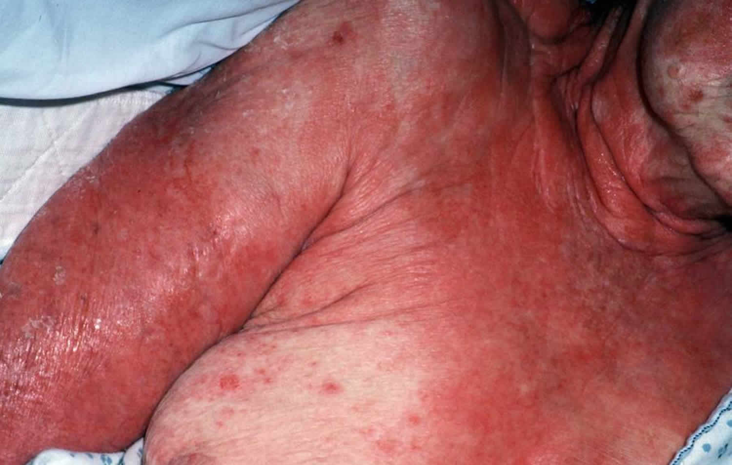
There is then an abrupt onset of a tender/painful red skin rash starting on the trunk and extending rapidly over hours to days onto the face and limbs (but rarely affecting scalp, palms or soles). The maximum extent is usually reached by 4 days.
The skin lesions may be:
- Macules — flat, red and diffuse (measles-like spots) or purple (purpuric) spots
- Diffuse erythema
- Targetoid — as in erythema multiforme
- Blisters — flaccid (i.e., not tense).
The blisters then merge to form sheets of skin detachment, exposing red, oozing dermis. The Nikolsky sign is positive in areas of skin redness. This means that blisters and erosions appear when the skin is rubbed gently.
Mucosal involvement is prominent and severe, although not forming actual blisters. At least 2 mucosal surfaces are affected including:
- Eyes (conjunctivitis, less often corneal ulceration, anterior uveitis, panophthalmitis) — red, sore, sticky, photosensitive eyes
- Lips/mouth (cheilitis, stomatitis) — red crusted lips, painful mouth ulcers
- Pharynx, esophagus — causing difficulty eating
- Genital area and urinary tract — erosions, ulcers, urinary retention
- Upper respiratory tract (trachea and bronchi) — cough and respiratory distress
- Gastrointestinal tract — diarrhea.
The patient is very ill, extremely anxious and in considerable pain. In addition to skin/mucosal involvement, other organs may be affected including liver, kidneys, lungs, bone marrow and joints.
Toxic epidermal necrolysis complications
Toxic epidermal necrolysis has significant morbidity as a result of dehydration, protein loss, thermoregulatory difficulties, renal tubular necrosis, eroded gastrointestinal tract, interstitial pneumonitis, neutropaenia, liver and heart failure. The mortality rate approaches 30%, most commonly from bacterial sepsis and toxic epidermal necrolysis is especially dangerous in those over 60.
During the acute phase, potentially fatal complications include:
- Dehydration and acute malnutrition
- Infection of skin, mucous membranes, lungs (pneumonia), septicemia (blood poisoning)
- Acute respiratory distress syndrome
- Gastrointestinal ulceration, perforation and intussusception
- Shock and multiple organ failure including kidney failure
- Thromboembolism and disseminated intravascular coagulopathy.
How is toxic epidermal necrolysis diagnosed?
Toxic epidermal necrolysis or Stevens-Johnson syndrome is suspected clinically and classified based on the skin surface area detached at maximum extent.
Toxic epidermal necrolysis with spots
- Detachment > 30% of body surface area
- Widespread purpuric macules or flat atypical targets
Toxic epidermal necrolysis without spots
- Detachment of > 10% of body surface area
- Large epidermal sheets and no purpuric macules
Stevens-Johnson syndrome
- Skin detachment < 10% of body surface area
- Widespread erythematous or purpuric macules or flat atypical targets
Overlap Stevens-Johnson syndrome/toxic epidermal necrolysis
- Detachment between 10% and 30% of body surface area
- Widespread purpuric macules or flat atypical targets
The category cannot always be defined with certainty on initial presentation. The diagnosis may therefore change during the first few days in hospital.
Investigations in toxic epidermal necrolysis
If the test is available, elevated levels of serum granulysin taken in the first few days of a drug eruption may be predictive of Stevens-Johnson syndrome/toxic epidermal necrolysis.
Skin biopsy is usually required to confirm the clinical diagnosis and to exclude staphylococcal scalded skin syndrome and other generalized rashes with blisters.
The histopathology shows keratinocyte necrosis (death of individual skin cells), full thickness epidermal/epithelial necrosis (death of an entire layer of skin), minimal inflammation (very mild lymphocytic infiltrate of the superficial dermis). The direct immunofluoresence test on the skin biopsy is negative, indicating the disease is not due to deposition of antibodies in the skin.
Blood tests do not help to make the diagnosis but are essential to make sure fluid and vital nutrients have been replaced, to identify complications and to assess prognostic factors (see below). Abnormalities may include:
- Anemia occurs in virtually all cases (reduced haemoglobin).
- Leucopenia (reduced white cells), especially lymphopenia (reduced lymphocytes) is very common (90%).
- Neutropenia (reduced neutrophils), if present, is a bad prognostic sign.
- Eosinophilia (raised eosinophil count) and atypical lymphocytosis (odd-looking lymphocytes) do not occur.
- Mildly raised liver enzymes are common (30%) and approximately 10% develop overt hepatitis.
- Mild proteinuria (protein leaking into urine) occurs in about 50%. Some changes in kidney function occur in the majority.
Patch testing rarely identifies the culprit in Stevens-Johnson syndrome/toxic epidermal necrolysis following recovery, and is not recommended.
Toxic epidermal necrolysis treatment
The most recently prescribed medications should be immediately discontinued and should never be readministered. The patient should wear a MedicAlert bracelet. Recurrent toxic epidermal necrolysis is more severe than the initial episode.
Management should be in an intensive care unit and may require:
- Expert supportive care (warm environment, fluid replacement, correction of electrolyte disturbances, administration of a high caloric enteral intake, prevention of sepsis, suction of oropharynx)
- Analgesics
- Intravenous antibiotics for sepsis
- Mucosal emollients
- Debridement of frankly necrotic skin
- Non-stick occlusive burns dressings
- Granulocyte colony stimulating factor for neutropaenia
- Intravenous immunoglobulins or ciclosporin to arrest the cytotoxic process
Systemic corticosteroids may increase mortality and in general should be avoided. Their use is controversial.
Recovery takes 10 days to 3 weeks and may be followed by irregular pigmentation and mucosal scarring. Eye complications are relatively common.
Care of a patient with Stevens Johnson syndrome/toxic epidermal necrolysis requires:
- Cessation of suspected causative drug(s) — the patient is less likely to die and complications are less if the culprit drug is on or before the day that blisters/erosions appear
- Hospital admission — preferably immediately to an intensive care and/or burns unit with specialist nursing care, as this improves survival, reduces infection and shortens hospital stay
- Consider fluidised air bed
- Nutritional and fluid replacement (crystalloid) by intravenous and nasogastric routes — reviewed and adjusted daily
- Temperature maintenance — as body temperature regulation is impaired, patient should be in a warm room (30–32C)
- Pain relief — as pain can be extreme
- Sterile handling and reverse isolation procedures.
Skin care
- Examine daily for extent of detachment and for infection (take swabs for bacterial culture).
- Topical antiseptics can be used (e.g., silver nitrate, chlorhexidine [but not silver sulfadiazine as it is a sulfa drug])
- Dressings such as gauze with petrolatum, non-adherent nanocrystalline-containing silver gauze or biosynthetic skin substitutes can reduce pain.
- Avoid using adhesive tapes and unnecessary removal of dead skin; leave the blister roof as a ‘biological dressing’.
Eye care
- Daily assessment by ophthalmologist
- Frequent eye drops/ointments (antiseptics, antibiotic, corticosteroid)
Mouth care
- Mouthwashes
- Topical oral anesthetic
Genital care
- If ulcerated, prevent vaginal adhesions using intravaginal steroid ointment, soft vaginal dilators.
Lung care
- Consider aerosols, bronchial aspiration, physiotherapy
- May require intubation and mechanical ventilation if trachea and bronchi are involved
Urinary care
- Catheter because of genital involvement and immobility
- Culture urine for bacterial infection
General
- Psychiatric support for extreme anxiety and emotional lability
- Physiotherapy to maintain joint movement and reduce risk of pneumonia
- Regular assessment for staphylococcal or gram negative infection
- Appropriate antibiotic should be given if infection develops; prophylactic antibiotics are not recommended and may even increase the risk of sepsis
- Consider heparin to prevent thromboembolism (blood clots).
The role of systemic corticosteroids (cortisone) remains controversial. Some clinicians prescribe high doses of corticosteroids for a short time at the start of the reaction, e.g., prednisone 1–2 mg/kg/day for 3–5 days. However concerns have been raised that they may increase the risk of infection, impair wound healing and other complications, and they have not been proven to have any benefit. They are not effective later in the course of the illness.
Case reports and small patient series have reported benefit from active adjuvant treatments delivered during the first 24–48 hours of illness. As Stevens Johnson syndrome/toxic epidermal necrolysis is fortunately a rare condition, controlled trials of therapies in large numbers of patients are difficult.
- Ciclosporin 3–5 mg/kg/day is reported to reduce mortality by 60% compared to patients with similar SCORTEN score on admission that were not treated with ciclosporin.
Other options include:
- Anti-TNFα monoclonal antibodies (eg, infliximab, etanercept)
- Cyclophosphamide
- Intravenous immunoglobulin (IVIG) 2–3 g/kg given over 2–3 days
- Plasmapheresis.
Thalidomide, trialed because of its anti-TNFα effect, increased mortality, and should not be used.
Toxic epidermal necrolysis prognosis
The acute phase of toxic epidermal necrolysis or Stevens Johnson syndrome lasts 8–12 days.
Repithelialization of denuded areas takes several weeks, and is accompanied by peeling of the less severely affected skin. Survivors of the acute phase have increased on-going mortality especially if aged or sick.
Long-term consequences include:
- Pigment change — patchwork of increased and decreased pigmentation
- Skin scarring, especially at sites of pressure or infection
- Loss of nails with permanent scarring (pterygium) and failure to regrow
- Scarred genitalia — phimosis (constricted foreskin which cannot retract) and vaginal adhesions (occluded vagina)
- Joint contractures
- Lung disease — bronchiolitis, bronchiectasis, obstructive disorders.
Eye problems can lead to blindness:
- Dry and/or watery eyes, which may burn and sting when exposed to light
- Conjunctivitis: red, crusted, or ulcerated conjunctiva
- Corneal ulcers, opacities and scarring
- Symblepharon: adhesion of conjunctiva of eyelid to eyeball
- Ectropion or entropion: turned-out or turned-in eyelid
- Trichiasis: inverted eyelashes
- Synechiae: iris sticks to cornea.
It may take weeks to months for symptoms and signs to settle.


