Triplane fracture
Triplane fracture also called triplanar fracture, is fracture of the distal tibia only occurring in adolescents (12 to 15 year old age group) 1. The incidence is slightly higher in boys than in girls 2. As the physiological closure of the physeal plate begins medially, the lateral (open) physis is prone to triplane fracture. Some doctors prefer to call triplane fracture an adolescent tibial triplane fracture because this term is more descriptive of the age of occurrence, location, and fracture pattern. It is also termed transitional injury because it occurs during the period of transition from skeletal immaturity to skeletal maturity. The name triplane fracture is due to the fact of the fracture expanding both in frontal and lateral as well as transverse planes. Most authors regard it as a type IV Salter-Harris fracture. As physeal closure has to begin at an end, triplane fractures have occasionally been reported in other sites too, e.g. distal radius 3, proximal tibia 4, distal femur 5.
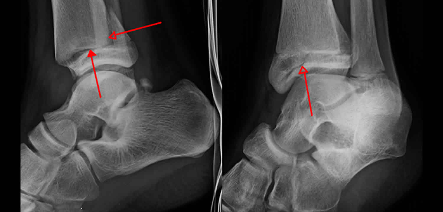
Triplane fracture comprises of:
- a vertical fracture through the epiphysis
- a horizontal fracture through the physis
- an oblique fracture through the metaphysis
Treatment of triplane fractures depends on the amount of displacement between the broken bones. Articular congruity at the ankle joint surface, not physeal arrest or growth retardation, is the major concern with triplane fractures. Therefore, minimally displaced fractures (less than 2 millimeters) and non-displaced fractures can be treated with a long-leg cast nonoperatively, but displaced fractures (greater than 2 millimeters) require anatomic reduction and internal fixation. Malunited fractures with more than 2 mm of intra-articular displacement are associated with poor outcomes.
Triplane fracture anatomy
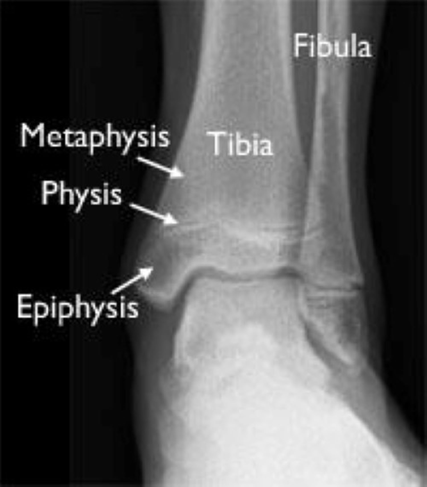
The tibia is the main weightbearing bone of the lower leg. The tibial metaphysis consists of the distal quarter of the tibia, excluding the tibial growth plate and epiphysis. The distal tibial physis, also called the growth plate, is located between the tibial metaphysis and the epiphysis. The distal tibial epiphysis is bordered proximally by the physeal growth plate and distally by its articulation with the articular surface of the talar dome. It contributes 50% of tibial growth and approximately 4-6 mm (0.25 in.) of longitudinal growth per year 6.
The long bones of the body do not grow from the center outward. Instead, growth occurs at each end of the bone around the growth plate. When a child becomes full-grown, the growth plates harden into solid bone.
Because growth plates are the last portion of bones to harden, they are vulnerable to fracture. In fact, the ligaments that attach the tibia and fibula to the talus bone are generally stronger than the growth plates. This is why an ankle twist that would result in a sprain in an adult is more likely to cause a growth plate fracture in a child.
Pediatric ankle fractures account for 9% to 18% of all growth plate fractures. In children age 10 to 15 years, only injuries to the wrist and hand are more common than ankle fractures. These older children are more likely to participate in strenuous sports activities, and their growth plates are not yet fully mature.
The fibula is situated laterally along the length of the tibia in the lower leg, giving stability to the lateral ankle joint and serving in a nonweightbearing role.
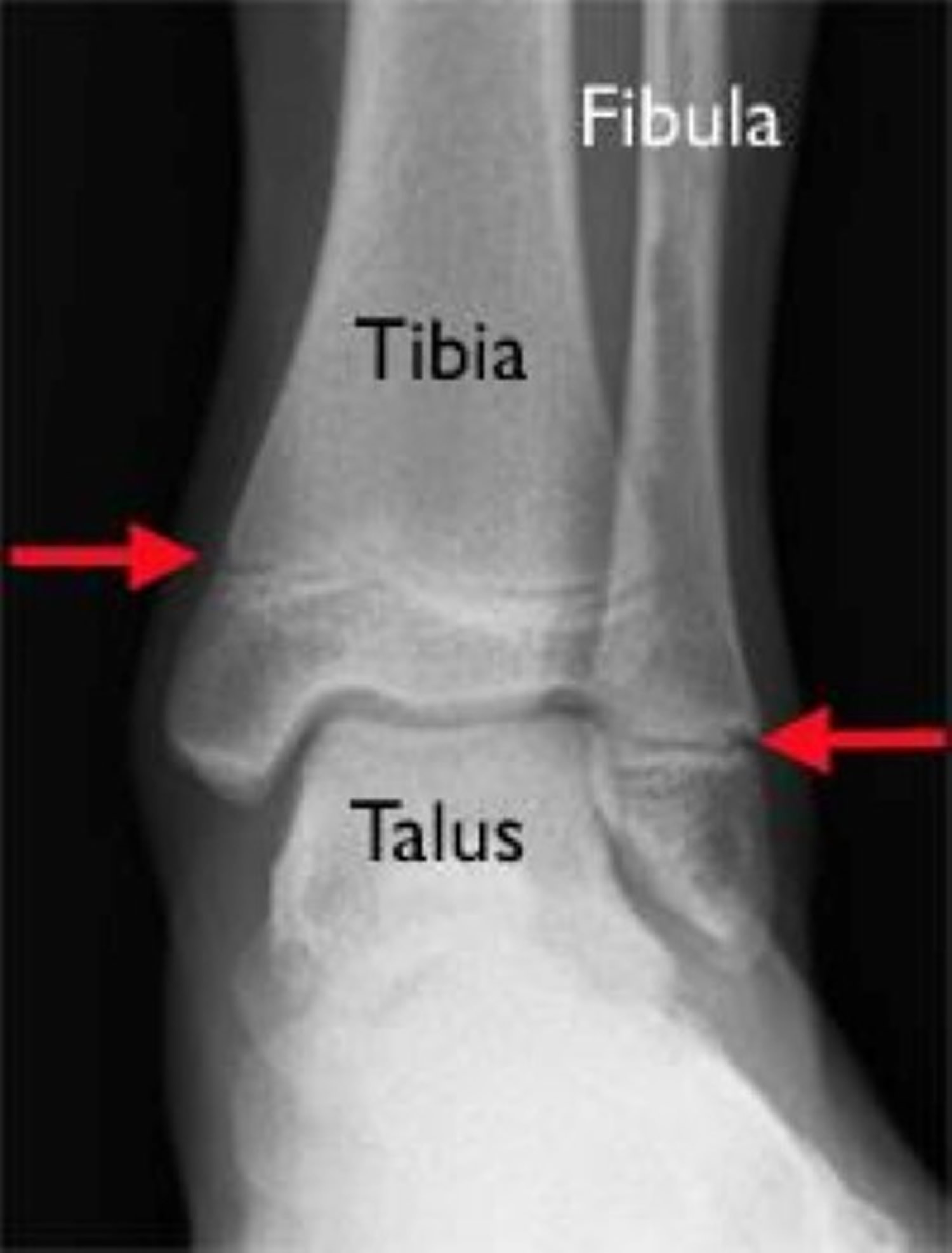
The ankle joint bears more weight per unit surface area than any other joint in the body. The ankle joint is formed by the fibula (laterally), the tibia (superiorly and medially), and the dome of the talus (inferiorly). The joint is saddle-shaped. The dome of the talus becomes wider anteriorly, such that when the foot is in dorsiflexion, the talus is situated more snugly in the tibiofibular saddle than when the foot is plantarflexed. Thus, plantarflexion (a position contributing to the triplane fracture) is a less stable position of the ankle than is dorsiflexion.
The only pure motions of the ankle joint are dorsiflexion and plantarflexion. Inversion and eversion of the ankle joint take place at the subtalar joint formed by the opposition of the talus and the inferiorly situated calcaneus. The talus always moves in the same direction as the calcaneus in normal gait.
Ligamentous support of the ankle is extensive. Ligaments situated laterally consist of anterior and posterior talofibular and tibiofibular ligaments. The strong deltoid ligament is located medially and is the only ligament of the ankle containing elastic fibers.
The tibia and fibula are joined by the anterior and posterior talofibular ligaments distally and the interosseous membrane more proximally.
Knowledge of the anatomic planes of the body is essential to understanding the lines, planes, and fragments produced in a triplane fracture. In the anatomic position (ie, with the person standing, palms forward), these two-dimensional planes are as follows:
- The horizontal plane passes horizontally through the body, dividing it into upper and lower segments
- The coronal plane passes through the body from one shoulder to the other, dividing it into front and back segments
- The sagittal plane passes through the body from front to back, dividing it into right and left segments
Motions of the ankle and foot are described by a number of interchangeable terms, including the following:
- Eversion – External rotation
- Inversion – Internal rotation
- Dorsiflexion – Extension
- Plantarflexion – Flexion
- Abduction – Lateral deviation of the foot on a longitudinal axis through the tibia
- Adduction – Medial deviation of the foot on a longitudinal axis through the tibia
- Supination – Adduction and inversion
- Pronation – Abduction and eversion
Neurovascular structures in the area of the ankle and foot include the following:
- Medially, both the posterior tibial artery and tibial nerve pass deep to the flexor retinaculum spanning between the distal tibia and the calcaneus
- Arterial pulses of the foot and ankle should be checked in any injury to the region and are readily palpable over the posterior tibial artery area medially and the dorsalis pedis artery on the dorsum of the foot between the bases of first and second metatarsals
Figure 1. Growth plates of the tibia and fibula (arrows)
Triplane fracture causes
Triplane fracture typically occur as a result of external rotation of the foot in the presence of the asymmetric physiologic epiphysiodesis characteristic for the distal tibial physis. In distal tibia, the triplane fracture occurs due to the asymmetrical closure of the distal tibial physis, from central to anteromedial to posteromedial and finishing with closure of the lateral margin of the physis 7.
Triplane fracture symptoms
Without an x-ray, it is often difficult to differentiate between an ankle sprain and a more serious ankle fracture. Initially, both sprains and fractures may cause pain and swelling.
A strong sign of a fracture, however, is when a child cannot put weight on the injured ankle. Possible signs include swelling, localized/referred pain, and/or deformity of the ankle.
Any fracture with an open skin wound is cause for significant concern, and the injured child should be taken to the nearest emergency room or urgent care facility as soon as possible.
Triplane fracture diagnosis
After discussing your child’s medical history and how the injury occurred, your doctor will do a careful examination. Your doctor will look for:
- Swelling and tenderness
- Bruising
- A deformed or crooked appearance of the ankle
- Tears or openings in the skin
Skin wounds are a sign of a potential open fracture. This type of fracture is particularly serious because once the skin is broken through, infection in both the wound and the bone can occur. To prevent infection, open fractures require immediate treatment, including irrigation to clear the wound of debris and bacteria, and surgery to repair the fracture.
During the physical examination, your doctor will feel for pulses in your child’s leg and foot. He or she will also check for sensation (feeling) and movement.
If your doctor suspects a triplane fracture, he or she will order additional tests to provide more information about your child’s injury.
- X-rays. The most common way to evaluate a fracture is with x-rays, which provide clear images of bone. X-rays will usually show whether a bone is intact or broken.
- Magnetic resonance imaging (MRI). If the physical examination suggests a fracture but the x-rays do not show it, your doctor may order a magnetic resonance imaging (MRI) scan. These tests provide high resolution images of both bones and soft tissues, like ligaments.
- Computed tomography (CT). This type of scan can create a cross-section image of the ankle. It is especially useful when the fracture extends into the ankle joint.
Triplane fracture treatment
Treatment options comprise surgery (i.e. open reduction and internal fixation [ORIF]) and conservative management (i.e. long leg cast immobilization). Physeal arrest leading to angular deformity may occur but is considered rare.
Patients with non-displaced triplane fractures (< 2 mm displacement) are treated with closed reduction and a non-weight-bearing long-leg cast (4-6 weeks), followed by a short-leg walking cast with crutches for additional 4 weeks. Serial radiographs are obtained at weekly intervals during the first 3 weeks to check for late displacement. For displaced fractures, closed reduction is attempted with general anesthesia. Following removal of the final cast, progressive return to normal activity is encouraged with ongoing physical therapy and range-of-motion (ROM) exercises.
If a closed reduction results in more than a 2-millimeter “step off” — meaning that the broken piece juts out along the bottom surface of the bone — then surgery is necessary. Open reduction and pins and/or screws will more accurately align the broken pieces and reduce damage to the articular cartilage that protects the end of the bone.
Open reduction and internal fixation
Open reduction and internal fixation (ORIF) is performed for a triplane fracture demonstrating 2 mm or more of displacement after attempted closed reduction. The surgical approach depends on the fracture planes and can be anterolateral for lateral fractures or anteromedial for medial fractures. Small stab incisions are often needed for the placement of screws, either solid or cannulated.
The strength of operative screws and pins has increased progressively, while the diameters of operative screws have decreased. Titanium-based materials of greater diameter may be replaced by composite materials of lesser diameter, thus lessening the trauma associated with their operative placement.
Reduction and fixation of the metaphyseal spike may be all the surgery that is needed. An alternative is the placement of epiphyseal screws parallel to the joint surface, avoiding the growth plate and the ankle joint. More than one screw is needed, and the primary goal is reduction of the physeal fracture and joint surface.
Preoperatively, it is essential to detect all other injuries and address them adequately, as well as other comorbidities and preexisting medical conditions and needs. In persons with open fractures, tetanus immunization should be updated preoperatively if needed, and prophylactic antistaphylococcal antibiotics should be administered.
Intraoperatively, portable or fixed overhead radiography or C-arm fluoroscopy is needed to evaluate the results of internal fixation (ie, to confirm that the fracture is reduced and that screw placement is satisfactory) before the patient leaves the operating room. Future intraoperative CT C-arm or navigation technology will be more readily available and will increase the accuracy of placement of screws and internal fixation in these complex fractures.
The anterolateral epiphyseal fragment of a three-part injury is reduced and held with either a screw or a Kirschner wire (K-wire). Before the patient leaves the operating room, a final set of postreduction radiographs is completed.
Arthroscopic reduction and internal fixation of two-part triplane fractures has been described as having advantages over traditional ORIF 8.
Postoperative care
Postoperatively, standard incision care and suture removal are performed as directed by the physician. An above-the-knee cast is used for 4-6 weeks, followed by a below-the-knee partial weightbearing cast. When internal fixation has been accomplished and early physical therapy or range-of-motion (ROM) exercises are desired, the short leg cast may be replaced by a removable air splint.
Complications
In general, young healthy adolescents do well after a triplane fracture 9, even though it is a serious injury. Potential complications include the following:
- Tibial length growth retardation or deformity around the ankle secondary to epiphyseal growth plate injury
- Posttraumatic arthritis
- Postoperative infection
- Osteomyelitis
- Pressure sores from the cast
- Fracture blisters
- Compartment syndrome.
- Lynn MD. The triplane distal tibial epiphyseal fracture. Clin Orthop. 1972;86:187.
- Patel S, Haddad F. Triplane fractures of the ankle. Br J Hosp Med (Lond). 2009 Jan. 70 (1):34-40.
- Mingo-Robinet J, Torres-Torres M, Gonzalez-Rodriguez M. Triplane fracture of distal radius treated surgically: case report and review of the literature. J Pediatr Orthop B. 2014;23 (3): 227-30. doi:10.1097/BPB.0000000000000011
- Kanellopoulos AD, Yiannakopoulos CK, Badras LS. Triplane fracture of the proximal tibia. Am J. Orthop. 2004;32 (9): 452-4.
- Masquijo JJ, Allende V. Triplane fracture of the distal femur: a case report. J Pediatr Orthop. 2011;31 (5): e60-3. doi:10.1097/BPO.0b013e318221096b
- Crawford AH. Ankle fractures in children. Instr Course Lect. 1995. 44:317-24.
- Dvonch V. M., Bunch W. H. Pattern of closure of the proximal femoral and tibial epiphyses in man. Journal of Pediatric Orthopaedics. 1983;3(4):498–501. doi: 10.1097/01241398-198309000-00015
- Jennings MM, Lagaay P, Schuberth JM. Arthroscopic assisted fixation of juvenile intra-articular epiphyseal ankle fractures. J Foot Ankle Surg. 2007 Sep-Oct. 46 (5):376-86.
- Tan AC, Chong RW, Mahadev A. Triplane fractures of the distal tibia in children. J Orthop Surg (Hong Kong). 2013 Apr. 21 (1):55-9.


