What is vasoconstriction
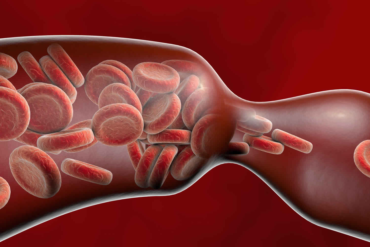
Vasoconstriction is the constriction or narrowing of a blood vessels by the smooth muscles in their walls. When blood vessels constrict, blood flow is slowed or blocked.
Vasoconstriction may be slight or severe. Vasoconstriction may result from disease, drugs, or psychological conditions. Medicines that cause vasoconstriction include:
- Decongestants, including pseudoephedrine
- Cough and cold combinations
- Caffeine
Medicines may be used to increase or reduce vasoconstriction in very ill people.
Does vasoconstriction increase blood pressure?
Yes. Vasoconstrictors are also called vasopressors, increase vasoconstriction, which leads to increased systemic vascular resistance. Increasing the systemic vascular resistance leads to increased mean arterial pressure (MAP) and increased perfusion to organs. The American College of Critical Care Medicine 1 guidelines recognize that a mean arterial pressure (MAP) of 60 to 65 mm Hg is required to perfuse organs. If after appropriate fluid resuscitation the mean arterial pressure (MAP) does not improve to about 60 mm Hg, it is recommended that vasopressors be initiated. Norepinephrine is recommended as the initial vasopressor per the Surviving Sepsis Campaign recommendations. Vasopressin or epinephrine are the 2 recommended vasopressors to add in addition to norepinephrine, although the evidence for these recommendations is considered weak 1.
Vasopressors act to increase cardiac output and systemic vascular resistance through increasing contractility and heart rate as well inducing vasoconstriction peripherally. The three main groups are catecholamine, smooth muscle, and dopaminergic receptors.
The most common catecholamine active medications are phenylephrine, norepinephrine, and epinephrine. Each of these three medications has varying activity on the alpha and beta receptors. Alpha receptors are peripheral vasoconstrictors to increase systemic vascular resistance. Beta-1 receptors have mostly positive chronotropic (heart rate) and inotropic (contractility) effects on the heart. Beta-2 receptors act as vasodilators in many organ systems.
Phenylephrine is a pure alpha-1 agonist, inducing peripheral arterial vasoconstriction. Reflex bradycardia may occur due to selective vasoconstriction and elevation of blood pressure. Blood pressure, mean arterial pressure, and systemic vascular resistance are increased.
Norepinephrine has mixed alpha-1 and beta activity (beta-1 greater than beta-2), with slightly more alpha-1 activity compared to beta activity. This leads to a more significant increase in blood pressure than increased heart rate. Blood pressure, mean arterial pressure, systemic vascular resistance, and cardiac output are increased with norepinephrine.
Epinephrine has essentially equivocal activity on alpha-1 and beta receptors. Epinephrine increases systemic vascular resistance, heart rate, cardiac output, and blood pressure.
Vasopressin acts on V-1 receptors to stimulate smooth muscle contraction of the vessels as well as V-2 receptors in the kidneys as an anti-diuretic. There are no inotropic or chronotropic effects. Only blood pressure and systemic vascular resistance is increased with vasopressin.
Dopamine is a precursor of norepinephrine and epinephrine which acts in a dose-dependent fashion on dopaminergic receptors as well as alpha and beta receptors. At low doses, dopaminergic receptors activate renal artery vasodilation. At doses 5 to 15 micrograms/kg/min, alpha, and beta-adrenergic activation increase renal blood flow, heart rate, contractility, and cardiac output. At higher doses greater than 15 micrograms/kg per minute, the main effects are on the alpha stimulation.
Vasoconstriction symptoms
The symptoms of vasoconstriction depends on its location. For example, in the arms or legs, vasoconstriction typically results in feelings of tightness, achiness, and numbness within a person’s arms and legs. It can range from mild in its effects to extremely uncomfortable.
Hypoxic pulmonary vasoconstriction
The pulmonary circulation differs from the systemic in several important aspects, the most important being that pulmonary arteries constrict to moderate physiological (∼20–60 mmHg PO2) hypoxia, whereas systemic arteries vasodilate 2. This phenomenon is called hypoxic pulmonary vasoconstriction and is responsible for maintaining the ventilation–perfusion ratio during localized alveolar hypoxia. Pulmonary vasoconstriction is a physiological phenomenon and mechanism in response to alveolar hypoxia or lower oxygen levels in the smaller, higher resistant pulmonary arteries and, to some extent, the pulmonary veins 3. The process of vasoconstriction of the pulmonary arteries serves to redirect blood flow within the vasculature away from poorly oxygenated parts of the lungs towards better-oxygenated portions in the lungs. Ventilation and perfusion matching is the physiological balancing process between air that reaches the alveoli and perfusion. Profusion is the blood reaching the capillaries of the alveoli and is predicated on the appropriate functioning of vasoconstriction of the pulmonary vasculature in lower-oxygen states. Poor oxygen availability to tissues has profound and overarching systemic ramifications manifesting in a plethora of pathologies starting within the lungs itself. Maintaining correct and appropriate oxygen homeostasis is a critical component for systemic stability and functioning, and it begins with the pulmonary vasculature and an intact pulmonary vasoconstriction reflex. Despite many years of study, the precise mechanisms underlying hypoxic pulmonary vasoconstriction remain unresolved 2. However, there is now overwhelming evidence that hypoxia can stimulate several pathways leading to a rise in the intracellular Ca2+ concentration in pulmonary artery smooth muscle cells. This rise in intracellular Ca2+ is consistently found to be relatively small and hypoxic pulmonary vasoconstriction seems also to require rho kinase-mediated Ca2+ sensitization. There is good evidence that hypoxic pulmonary vasoconstriction also has an as yet unexplained endothelium dependency.
Hypoxic pulmonary vasoconstriction is an issue with the pulmonary vasculature. However, inappropriate and chronic pulmonary vasoconstriction can lead to pulmonary hypertension which has systemic ramifications. The development of pulmonary hypertension creates an increased load on the right heart. Excess pressure within the pulmonary artery characterizes pulmonary hypertension. The right ventricle which propels poorly oxygenated venous return into the pulmonary artery will have to work excessively harder to overcome the greater pressures in the pulmonary circuit, leading to hypertrophy and muscle damage of the heart tissue on the right side. The grim consequences of pulmonary hypertension are heart failure manifesting with systemic cyanosis and edema, and or death.
It is well accepted that the elevation of vascular smooth muscle cell intracellular Ca2+ concentration is an early and critical element leading to hypoxic pulmonary vasoconstriction, and involves Ca2+ entry across the cell membrane. However, the Ca2+ channels and signal transduction pathways involved, as well as the oxygen sensor(s) responsible for their activation, are hotly debated. Currently, the best documented hypothesis for hypoxic pulmonary vasoconstriction proposes that Ca2+ entry is mediated primarily via voltage-dependent L-type channels 4, as a result of hypoxia-induced inhibition of voltage-gated K+ channels and consequent depolarization. According to this scheme, gated K+ channel inhibition is caused by a decrease in the ambient intracellular concentration of H2O2 which results when mitochondrial electron transport, and consequently the production of superoxide ion, falls due to the lack of O2. There is an enormous body of evidence supporting this hypothesis, which has been presented in a number of authoritative reviews 5.
However, others have presented evidence that hypoxia increases the levels of H2O2 and other reactive oxygen species in hypoxic pulmonary vasoconstriction 6. Morever, it is noteworthy that both in vivo studies of hypoxic pulmonary vasoconstriction in humans 7 and in vitro studies of hypoxic pulmonary vasoconstriction in isolated human pulmonary arteries 8 show that blockers of L-type Ca2+ channels, which according to this model should abolish hypoxic pulmonary vasoconstriction, produce a variable and in some cases rather small inhibition of this response. Work from our laboratory 9 and many others has similarly demonstrated the relative insensitivity of hypoxic pulmonary vasoconstriction to L-type Ca2+ channel blockers in animal models ranging from the perfused lung to isolated pulmonary artery and pulmonary artery smooth muscle cells 10. Moreover, a substantial and growing literature indicates that drugs which block non-voltage-gated Ca2+-elevating pathways such as the release of Ca2+ from intracellular stores and the opening of Ca2+-permeable cation channels, also attenuate, and in some cases abolish, hypoxic pulmonary vasoconstriction.
Key evidence that the release of intracellular Ca2+ stores plays an important role in hypoxic pulmonary vasoconstriction was presented by Jabr et al. 11 in isolated dog pulmonary artery. They found that hypoxic pulmonary vasoconstriction in these arteries was strongly inhibited by depletion of ryanodine- and caffeine-sensitive Ca2+ stores, yet was enhanced when Ca2+ stores were depleted with thapsigargin or cyclopiazonic acid. From this, they concluded that hypoxia was releasing ryanodine-sensitive Ca2+ stores, and that some of this Ca2+ was being buffered by a separate, cyclopiazonic acid-sensitive store. Similarly, Liu et al. 12 found that ryanodine pretreatment abolished hypoxic pulmonary vasoconstriction in isolated rabbit and pig intrapulmonary arteries, and Morio & McMurtry 13 showed that ryanodine suppressed hypoxic pulmonary vasoconstriction in perfused rat lung. Wilson et al. 14 have presented extensive evidence that cyclic ADP ribose, which acts on ryanodine receptors to stimulate Ca2+ release, is elevated by hypoxia in small pulmonary arteries. Moreover, the sustained (phase 2) contraction to hypoxia in these arteries was completely suppressed by the cyclic ADP ribose antagonist 8-Br-cADPR, a fascinating and potentially pivotal observation which, however, awaits independent verification by other laboratories.
The release of intracellular Ca2+ stores triggers store-operated Ca2+ entry in pulmonary artery smooth muscle cells as in many other types of cell and it would therefore be expected that hypoxia-induced Ca2+ release should activate store-operated Ca2+ entry. Snetkov et al. 15 have reported that contraction in rat intrapulmonary arteries appears to be more sensitive to store-operated Ca2+ entry activated by store depletion than it is in systemic arteries of similar size. This unique property of intrapulmonary arteries is also observed with regard to multiple agonists. Lin et al. 16 demonstrated the effect of diltiazem (10 μm), which blocks L-type Ca2+ channels, and La3+ (10 μm), which is mainly selective for store-operated Ca2+ entry, on contractions to 5-HT, angiotensin II, endothelin-1, and PGF2α in intrapulmonary arteries, mesenteric and femoral arteries of the rat. For each agonist, intrapulmonary arteries are much more susceptible to block by La3+ and less sensitive to diltiazem than are both types of systemic arteries, indicating the general importance of voltage-independent mechanisms in the excitation–contraction coupling in intrapulmonary arteries.
Theories of the vasoconstriction reflex
Hypoxia needs to be present for the vasoconstriction reflex to initiate. There are contrasting views as to where hypoxia is first identified in the vasculature. The classical understanding suggested that hypoxia is initially identified within the pulmonary artery, whereas a new concept submits that low oxygen levels are detected at the interaction of the capillaries and alveoli. This latter concept further suggests that from the capillary/alveoli junction, gap junctions embedded throughout the pulmonary vascular endothelium assist in transducing an electrical signal to the pulmonary arterioles, causing them to constrict.
Original understanding of the vasoconstriction reflex
In comparison to this newer understanding, the original mechanism is believed to involve voltage-gated potassium and calcium channels. These channels are located in the smooth muscle cells of the pulmonary arteries and are very sensitive to hypoxia. In addition to the critical roles of potassium and calcium in hypoxic pulmonary vasoconstriction, there are indications that there could be other ion channels contributing to hypoxic pulmonary vasoconstriction. These ion channels are the transient receptor potential vanilloid 4 (TRPV4) and the transient receptor potential canonical 6 (TRPC) 3.
Pulmonary hypertension pathophysiology
The complete pathophysiology of pulmonary hypertension is not entirely understood. The current causes of pulmonary hypertension are stratified into five groups.
- The first group is idiopathic pulmonary hypertension, most likely related to inheritable and intrinsic pulmonary arterial pathology.
- The second group is pulmonary hypertension due to left heart disease causing a non-flowing circulation circuit that allows pressure to build up excessively into the pulmonary vasculature.
- The third group is pulmonary hypertension due to pathology within or related to the lungs. This group includes several different diseases such as COPD, sleep apnea, inflammatory scarring as a result of interstitial lung disease, chronic exposure to a high altitude environment, and certain developmental abnormalities.
- The fourth group is associated with frequent occurrence of embolic deposition in the lung’s vessels or thrombosis known as chronic thromboembolic pulmonary hypertension (CTEPH).
- The fifth group is designated for any remaining unclear or unestablished etiologies and causes that are multifactorial. Examples of the multifactorial processes that could contribute to or cause pulmonary hypertension are metabolic disorders specifically the thyroid, hematological disorders, and systemic pathologies that involve the respiratory system such as sarcoidosis.
Idiopathic Pulmonary Hypertension
Of these groups, idiopathic pulmonary hypertension has the least defined mechanisms and is the group that is still is being understood. There are two prevailing concepts to the origin and progression of idiopathic pulmonary hypertension. The most prevalent understanding of what precipitates pulmonary hypertension was commonly believed to be derangement in the production and underproduction of local vasoconstrictors and dilators simply denoted as the vasoconstriction theory. The vasodilators include prostacyclin and nitric oxide, and the vasoconstrictors include thromboxane and endothelin.
Roles of Chemical and Molecular Agents in Idiopathic Pulmonary Hypertension
In the vasoconstriction theory, nitric oxide plays a critical role in maintaining appropriate vascular tone produced and regulated by two enzymes, nitric oxide synthase 2 and 3. Patients with idiopathic pulmonary hypertension are found to have diminished levels of nitric oxide (NO). Prostacyclin is a product of arachidonic metabolism, is an effective vasodilator systemically, and exerts effects on platelets as well. In patients with remodeling in the lungs vasculature, there are decreased levels of prostacyclin and the enzyme prostacyclin synthase, giving more validity to the correlation between the role of pulmonary arterial vasodilators and pulmonary hypertension. On the contrary, endothelin 1 is a systemic vasoconstrictor which is found in abnormally high quantities in idiopathic pulmonary hypertension.
Newer Understanding of Idiopathic Pulmonary Hypertension
The new understanding that is becoming more accepted is more centered on the concept of endothelial and smooth muscle cell activity in the context of arterial remodeling. The issue in remodeling the vessels is specifically involving the extracellular matrix that creates the architecture in the vessel that contributes to increased vascular resistance. The bone morphogenetic receptor type 2 (BMR2) is involved with cell growth, proliferation, and osteogenesis. It is believed that a mutation in bone morphogenetic receptor type 2 (BMR2) is directly involved in idiopathic pulmonary hypertension. Another process that can contribute to the pulmonary vasoconstriction is an inappropriately high sensitivity to depolarization in the smooth muscle cells within the vessels. This alteration in the resting potential set point is possibly altered due to potassium channel irregularities that cause increase availability of calcium.
Sleep apnea and pulmonary hypertension
A distinct relationship exists between obstructive sleep apnea (OSA) and the existence of pulmonary hypertension. The link of causation has not been established to define obstructive sleep apnea as the direct singular cause of pulmonary hypertension, however, an association is there. It is thought to believe that obstructive sleep apnea contributes to pulmonary hypertension by inducing hypoxia during sleep which creates a cycle of oxygen desaturation that leads to decreased nitric oxide synthase, endothelin derangements, and an increase of sympathetic tone.
Diagnosing Pulmonary Hypertension
Hypoxic pulmonary vasoconstriction manifests clinically as pulmonary hypertension. In order to fully properly diagnose pulmonary hypertension, tests must establish whether the clinical criteria has been met, what part of the vasculature is pathologic, the degree of pulmonary hypertension, and a potential cause. Diligent and appropriate investigations need to be conducted due to the ambiguous nature and usual lack of suspicion for pulmonary hypertension. Early detection is paramount to long-term morbidity and mortality. The initial process of diagnosis begins with detecting pulmonary hypertension in the clinical setting. A routine or situational ECG is one way that suspicion can be formed outside of a history and physical. Right hearted issues such as hypertrophy should be expected, regardless of whether the right atrium or the right ventricle are involved, overcompensating against the excess pressure of the pulmonary circuit.
Echocardiography
One of the preliminary tests performed is an echocardiogram to give credence to clinical suspicion and to legitimize further testing to confirm a diagnosis. Echocardiography will relate the physiological circumstances within the four chambers of the heart, specifically the right half, to provide insight to any excess pressure build up or inappropriate hypertrophy which is what is expected in pulmonary hypertension. In addition to the size of the right atrium and ventricle, the echocardiogram evaluates right ventricular ejection fraction through the tricuspid annular plane systolic excursion (TAPSE), another important variable in assessing for the presence of hypertension in the lung’s vasculature.
Right Heart Catheterization
If preliminary findings warrant further investigation, a clinician would conduct a right heart catheterization to confirm a diagnosis. A right heart catheterization will provide information regarding the mean pulmonary artery pressure, right atrial pressure, and pulmonary artery occlusion pressure. It also can provide empirical information that would be utilized to calculate a transpulmonary gradient and the pulmonary vascular resistance.
Different Variables and Calculations
In a healthy individual at rest, the mean pulmonary artery pressure (mPAP) is 14 +/- 3 mmHg while a resting (mPAP) of greater than 25 mmHg would meet the criteria for a diagnosis of pulmonary hypertension. Pulmonary artery occlusion pressure (PAOP) and mean pulmonary artery pressure (mPAP) are variables in the calculations for determining transpulmonary gradient and pulmonary vascular resistance.
- The equation to calculate pulmonary vascular resistance: (mPAP-PAOP)/(CO).
- The equation to calculate the transpulmonary gradient: mPAP-PAOP.
One of the most sensitive findings of right heart catheterization is the pulmonary artery occlusion pressure. It has been the most susceptible and prone incorrect measurement. It is import to keep in mind that there are five criteria delineated to make sure the interpretation of pulmonary artery occlusion pressure (PAOP) is valid.
Additional Testing
Additional testing needs to be completed to rule out other disease processes that could be contributing to the pathology. These include spiral CT scans or ventilation-perfusion lung scan in the case of renal impairment to dismiss or identify the presence of thromboembolic activity within the lung’s vasculature. Pulmonary function testing to identify and categorize the presence of any intrinsic lung disease that may be contributing. Additional high-resolution CT imaging can be helpful in finding the presence of disease in the parenchyma of the lungs. Potentially likely etiologies of pulmonary hypertension are connective tissue diseases and obstructive sleep apnea. They will be investigated with serological studies and polysomnography respectively. A frequent and quick test to obtain further information is the cardiopulmonary exercise test known as the 6-minute walk test. This basic test evaluates functional capacity which, in short, represents how much physical exertion a patient can make before feeling overwhelmed.
Hypoxic pulmonary vasoconstriction treatment
Nonsurgical Treatment Options
Therapy will be catered to the patient’s individual pathological profile. All pharmaceutical regimens are not always indicated for patients; however, if there are no contraindications, anticoagulation is recommended for all diagnoses of idiopathic pulmonary hypertension. If pharmacological therapy is indicated, agents such as phosphodiesterase-5 inhibitors, calcium channel blockers, and endothelin receptor antagonists can be utilized. If pulmonary vasoconstriction is refractory to pharmacological therapy or if the severity of disease is extreme, surgery can be an option. Patients with obstructive sleep apnea should be screened for pulmonary hypertension, and likewise, pulmonary hypertensive patients should be evaluated for sleep apnea because there could be an association. If a patient has sleep apnea, a continuous positive airway pressure (CPAP) mask is prescribed for the patient to wear during sleep.
Surgical Options
Surgical options entail creating an anatomical environment that is conducive to relieving pressures on the right side of the heart with a procedure called an atrial septostomy producing a right-left shunt at the level of the atria. Once the disease reaches a state where correction is unattainable, lung transplantation options can be explored. In this scenario, both lungs are usually transplanted to lessen the potential for postoperative complications.
Reversible cerebral vasoconstriction syndrome
Reversible cerebral vasoconstriction syndrome is a rare cerebrovascular disorder characterized by severe headaches with or without focal neurological deficits or seizures, and a reversible segmental and multifocal vasoconstriction of cerebral arteries 17. Reversible cerebral vasoconstriction syndrome occurs predominantly in females before the age of 50. The most common clinical feature of reversible cerebral vasoconstriction syndrome is severe acute headache. The major complication of reversible cerebral vasoconstriction syndrome is ischemic or hemorrhagic stroke.
Reversible cerebral vasoconstriction syndrome is characterized by recurrent severe thunderclap headaches with or without other neurological symptoms and diffuse segmental narrowing of the cerebral arteries which is reversible within 3 months 18. Reversible cerebral vasoconstriction syndrome may occur spontaneously but in over 50% of cases, it can be provoked by various precipitating factors, the most common being the post-partum state and exposure to various vasoactive medications or illicit drugs and selective serotonin-reuptake inhibitors (SSRIs). One third to a half of cases develop hemorrhagic or ischemic brain lesions or a combination of both. Posterior reversible encephalopathy syndrome (PRES) often occurs in association with reversible cerebral vasoconstriction syndrome and the conditions are likely to share a common pathophysiology 18.
The exact pathophysiology of reversible cerebral vasoconstriction syndrome remains unknown and the prevailing hypothesis involves a transient disturbance in the control of cerebral vascular tone with autonomic dysregulation, oxidative stress, and genetic predisposition being postulated. Significant differential diagnoses include subarachnoid hemorrhage (SAH) due to aneurysmal rupture, cervical artery dissection, and primary angiitis of the central nervous system. Although there is no proven treatment, calcium channel antagonists including nimodipine and verapamil have been administered with reported reduction of headache intensity but without effect on the time course of cerebral vasoconstriction. Glucocorticoids have been reported as an independent predictor of worse outcome and should be avoided. The cornerstone of reversible cerebral vasoconstriction syndrome management remains largely supportive with bed rest and analgesics and removal of precipitating factors. Invasive neurointerventional techniques should be reserved for severe deteriorating cases. The condition is usually benign and self-limited and the majority of patients have a favorable outcome but around 5-10% are left with permanent neurological deficits and rare cases may die.
Reversible cerebral vasoconstriction syndrome diagnostic criteria
The key diagnostic criteria for reversible cerebral vasoconstriction syndrome proposed by Calabrese et al. 19 have since been slightly modified by the International Headache Society (Table 1). Although these criteria have not been validated in any prospective study, they have proved very useful clinically to diagnose reversible cerebral vasoconstriction syndrome and to increase physician awareness of the disease.
Table 1. Diagnostic criteria for reversible cerebral vasoconstriction syndrome
- Severe, acute headaches, with or without additional neurologic signs or symptoms
- Uniphasic disease course with no new symptoms after 1 month of onset
- No evidence for aneurysmal subarachnoid hemorrhage (SAH)
- Normal or near-normal findings on CSF analysis (protein level, <80 mg/dL; leukocyte level, <10/mm3; normal glucose level)
- Multifocal segmental cerebral artery vasoconstriction demonstrated on either catheter angiography or indirectly on CTA/MRA
- Reversibility of angiographic abnormalities within 12 weeks after onset. If death occurs before the follow-up studies are completed, postmortem rules out such conditions as vasculitis, intracranial atherosclerosis, and aneurysmal subarachnoid hemorrhage (SAH), which can also manifest with headache and stroke
Potential Triggers of reversible cerebral vasoconstriction syndrome
Although the true incidence of reversible cerebral vasoconstriction syndrome remains uncertain, the syndrome does not appear rare on the basis of the rates of patient recruitment or presentation into prospective and retrospective studies 21. Furthermore, recent reports have suggested that the incidence of reversible cerebral vasoconstriction syndrome may be increasing, though it is unclear whether this reflects a true increase in the incidence of the syndrome versus a consequence of improved imaging techniques and physician awareness 22. Nevertheless, reversible cerebral vasoconstriction syndrome likely remains underdiagnosed and should be included in the differential diagnosis of young patients presenting with severe headache or cryptogenic stroke 23.
Reversible cerebral vasoconstriction syndrome commonly affects patients 20–50 years of age (mean, 42–45 years), though other age groups, including children and adolescents, can be affected 23. Most interesting, the mean age of men presenting with reversible cerebral vasoconstriction syndrome tends to be a decade younger than that of female patients (fourth decade) 24. There is a female predominance, with an average female/male ratio from 3 large series of patients of approximately 2.4:1 25. Reversible cerebral vasoconstriction syndrome does not appear to be limited to any single ethnic or racial group 21. As Ducros 21 highlighted in her review of reversible cerebral vasoconstriction syndrome, differences in patient characteristics in large published series could reflect either intrinsic differences in reversible cerebral vasoconstriction syndrome among various patient populations and/or differences in patient recruitment criteria.
Reversible cerebral vasoconstriction syndrome can occur spontaneously, without an obvious underlying cause, or can be secondary to an identifiable trigger (roughly 25%–60% of cases) 26. The delay in exposure to an exogenous trigger and the development of reversible cerebral vasoconstriction syndrome can be anywhere between a few days and several months 25. In cases in which medications act as the exogenous trigger for the syndrome, patients may be taking the drug on a regular basis or infrequently, either at recommended dosages or in excess 25. For those patients without an obvious precipitant, reversible cerebral vasoconstriction syndrome may be induced in vulnerable individuals due to spontaneous or evoked vascular and/or neuronal discharges 19.
A diverse group of possible exogenous triggers for secondary reversible cerebral vasoconstriction syndrome have been proposed, though the potential delay between exposure and development of the syndrome (in some cases weeks to months) and the ubiquity of some triggers (coughing, laughing, and so forth) raise the possibility that some of these associations may be coincidental 25. However, the association of reversible cerebral vasoconstriction syndrome with the most commonly reported triggers is more compelling, including the use of vasoactive drugs and the postpartum state, which together account for more than half of cases in most published series (approximately 50% and 9%–10% of cases respectively) 27. Sympathomimetic drugs commonly taken over the counter for upper respiratory tract infections, including phenylpropanolamine and pseudoephedrine, as well as antimigrainous medications, have historically been associated with subarachnoid hemorrhage and ischemic stroke in rare cases, which in retrospect likely reflects the sequelae of drug-induced reversible cerebral vasoconstriction syndrome 28. The association between reversible cerebral vasoconstriction syndrome and the postpartum state is thought to possibly reflect increased levels of both pro- and antiangiogenic factors, some of which have also been associated with eclampsia, such as placental growth factor.5 reversible cerebral vasoconstriction syndrome encountered in the postpartum period typically is encountered anywhere from 1 to 3 weeks following an uncomplicated pregnancy, though presentation as late as 6 weeks has been reported 29.
Reversible cerebral vasoconstriction syndrome is commonly associated with a history of migraine headaches (20%–40% of cases), which may, in part, be due to the known role of migraine medications as a trigger for the syndrome 30. Cervical arterial dissection has also been associated with reversible cerebral vasoconstriction syndrome, though it remains uncertain whether this represents a potential etiology or complication of the syndrome 31. In a prospective study identifying patients with reversible cerebral vasoconstriction syndrome or cervical arterial dissection, Mawet et al. 32 found that 12% of patients in the reversible cerebral vasoconstriction syndrome cohort (n = 173) had or developed cervical arterial dissection, while 7% of patients in the cervical dissection cohort (n = 285) developed reversible cerebral vasoconstriction syndrome. In rare cases, multiple cervical arterial dissections may be present 31. Finally, some published series have noted a significant association between reversible cerebral vasoconstriction syndrome and cannabis use 33.
Reversible cerebral vasoconstriction syndrome signs and symptoms
The thunderclap headache is a defining clinical feature of reversible cerebral vasoconstriction syndrome and is defined as a severe, throbbing headache that reaches peak intensity within 60 seconds of onset. In reversible cerebral vasoconstriction syndrome, the pain is often bilateral and diffuse, though it can originate in the occipital region 23. Thunderclap headache has been reported in 94%–100% of patients with reversible cerebral vasoconstriction syndrome and may be the sole presenting symptom in 70%–76% of cases 34. Often, there is significant delay between the onset of headache and patient presentation for medical care (average, 7 days) 34. The thunderclap headache can be associated with other symptoms, including nausea, emesis, diplopia, elevations in blood pressure, and photosensitivity 23. In patients with reversible cerebral vasoconstriction syndrome who have migraines, the thunderclap headache is typically described as differing in location, degree, and quality from their usual migraines 35. A minority of patients with reversible cerebral vasoconstriction syndrome may present with a more mild or subacute headache, though the complete absence of headache is rare 26.
Thunderclap headache is not specific for reversible cerebral vasoconstriction syndrome and can be associated with various other medical conditions, including aneurysmal subarachnoid hemorrhage, primary headache disorder, pituitary apoplexy, cerebral venous sinus thrombosis, unruptured cerebral aneurysm, cervical arterial dissection, and third ventricle colloid cyst, among others 36. In fact, prior reports suggest that reversible cerebral vasoconstriction syndrome will ultimately be diagnosed in less than half (45%) of patients presenting with a thunderclap headache 37. For example, Grooters et al. 37 found that only 8.8% of patients presenting to a single center with thunderclap headache and no evidence of aneurysmal subarachnoid hemorrhage were ultimately diagnosed with reversible cerebral vasoconstriction syndrome.
However, some characteristics of the thunderclap headache associated with reversible cerebral vasoconstriction syndrome may be more specific for the syndrome. For example, in contradistinction to patients with aneurysmal subarachnoid hemorrhage, the thunderclap headache associated with reversible cerebral vasoconstriction syndrome typically demonstrates a waxing and waning course, often completely resolving within 3 hours (range, minutes to days), only to recur repeatedly during 1–3 weeks 23. On average, the last episode occurs 7–8 days after symptom onset 21. In reversible cerebral vasoconstriction syndrome, the number of exacerbations may vary between 1 and 20 episodes and often are triggered by bathing, stress, sexual intercourse, change in position, exertion, and coughing 23. A more moderate headache may persist between the acute episodes 21.
The exact etiology of the thunderclap headache encountered in cerebral vasoconstriction syndrome remains uncertain. Some authors have postulated that cerebral vasoconstriction may be the cause because the cerebral vasculature receives innervation from the first division of the trigeminal nerve and the dorsal ganglion of the second cervical nerve 19. However, the time course of patient symptoms such as headache and cerebral vasoconstriction argues against a causal relationship. For example, although patients typically present acutely with thunderclap headache, cerebral vasoconstriction often does not become evident for a week or more following symptom onset. Furthermore, resolution of vasoconstriction may take weeks to months in some individuals, persisting long after the resolution of patient symptomatology 38.
Other clinical presentations and consequences of reversible cerebral vasoconstriction syndrome
Other clinical presentations, or sequelae, of reversible cerebral vasoconstriction syndrome include generalized seizures, encephalopathy, focal neurologic deficits, altered mental status, transient ischemic attacks, ischemic stroke, intracranial hemorrhage, cerebral edema, and PRES (posterior reversible encephalopathy syndrome) 25. In her meta-analysis of 3 large case series of patients with reversible cerebral vasoconstriction syndrome, Ducros 21 found that focal neurologic deficits were present in 8%–43% of patients, seizures in 1%–17%, cortical subarachnoid hemorrhage in 30%–34% (1 study had hemorrhage as an exclusion criterion and was not included), cerebral infarction in 6%–39%, and concomitant PRES (posterior reversible encephalopathy syndrome) in 9%–38%. This wide range in reported incidence of various sequelae of reversible cerebral vasoconstriction syndrome may reflect recruitment bias, with more ill patients being more likely to present for medical care; selection criteria; and the clinical context in which patients were encountered 21. For example, reported rates of ischemic infarct and intracranial hemorrhage in patients developing reversible cerebral vasoconstriction syndrome postpartum appear to be higher than those in series included by Ducros 39.
Although patients with reversible cerebral vasoconstriction syndrome may initially present with generalized seizure, seizures rarely persist and long-term antiepileptic therapy is generally not indicated 23. Hypertension is commonly encountered in patients with reversible cerebral vasoconstriction syndrome in the acute period; however, it is unclear whether high blood pressure is from pain associated with headache, a response to cerebral vasoconstriction, or some other manifestation of the syndrome 40. As previously described, cervical arterial dissections may be encountered in patients with reversible cerebral vasoconstriction syndrome and should be excluded in patients who present with neck pain and/or territorial cerebral infract 23.
Focal neurologic deficits encountered with reversible cerebral vasoconstriction syndrome include visual deficits, hemiplegia, dysarthria, aphasia, numbness, cortical blindness, or ataxia 19. Focal deficits of vision, sensory, sensation, and motor function are encountered in decreasing frequency 23. Focal neurologic deficits may be transient or permanent, often reflecting the sequelae of TIA or ischemic infarct resulting from severe segmental cerebral vasoconstriction, though some transient deficits may be due to a migraine-type aura phenomenon 23. Neurologic deficits lasting >24 hours are unlikely to improve and likely reflect the sequelae of ischemic infarct, which typically occur in bilateral watershed zones of the cerebral hemispheres 21. Cerebellar infarcts are also possible 21.
Risk factors for the development of intracranial hemorrhage in patients with reversible cerebral vasoconstriction syndrome include a history of migraines, older age, and female sex 41. Subarachnoid hemorrhage, the most common hemorrhagic complication of reversible cerebral vasoconstriction syndrome, is most often focal and localized in superficial cerebral sulci near the cerebral convexities 23. Given this distribution, subarachnoid hemorrhage associated with reversible cerebral vasoconstriction syndrome may be missed on imaging and CSF analysis, and its incidence in the syndrome consequently is underestimated 41. It has been postulated that vasoconstriction of small arterioles early in the course of reversible cerebral vasoconstriction syndrome, along with hypertension and breakdown of autoregulatory mechanisms, may precipitate the rupture of small pial vessels with resulting subarachnoid hemorrhage 30. Other patterns of intracranial hemorrhage encountered in reversible cerebral vasoconstriction syndrome include intraparenchymal hemorrhage and subdural hematomas 23. Intraparenchymal hemorrhage can be see in up to 6%–20% of patients and most often is unifocal and lobar in location 23.
The various sequelae of reversible cerebral vasoconstriction syndrome tend to occur at different times during the course of the syndrome 34. Hemorrhagic complications, such as subarachnoid and intraparenchymal hemorrhage and concomitant PRES and seizures, most often occur during the first week of illness 23. In contradistinction, ischemic events and their resulting focal neurologic deficits often arise later in reversible cerebral vasoconstriction syndrome, peaking between 1 and 2 weeks following patient presentation 34. Ischemic stroke can occur even later in the course of the syndrome, occasionally after resolution of symptoms such as headache, and presumably reflects the well-documented delay in resolution of cerebral vasoconstriction 42. Overall, Ducros et al. 34 found that ischemic events such as TIA and stroke occurred on average approximately 8 days later than hemorrhagic complications 34.
Association with posterior reversible encephalopathy syndrome
Reversible cerebral vasoconstriction syndrome and posterior reversible encephalopathy syndrome overlap significantly in their clinical and radiographic features, and the 2 entities are frequently encountered concurrently. posterior reversible encephalopathy syndrome is a clinical and radiographic syndrome characterized by headache, visual changes, seizure, and imaging findings, including cerebral edema affecting the cerebral cortex and underlying white matter, manifesting as areas of hyperintensity on T2 and FLAIR imaging, most often involving the occipital and posterior parietal lobes. However, other distribution patterns can be encountered with posterior reversible encephalopathy syndrome, including involvement of the frontal and temporal lobes, basal ganglia, deep white matter, and brain stem. While the areas of cerebral edema encountered in posterior reversible encephalopathy syndrome are often reversible, progression to cytotoxic edema and infarct can occur. Finally, like reversible cerebral vasoconstriction syndrome, the exact pathophysiologic etiology of posterior reversible encephalopathy syndrome remains unknown, though a breakdown in autoregulation of the cerebral vasculature leading to hyperperfusion is 1 possible mechanism.
Posterior reversible encephalopathy syndrome-like reversible cerebral edema is encountered in anywhere from 9% to 38% of patients with reversible cerebral vasoconstriction syndrome, while most patients with posterior reversible encephalopathy syndrome (>85%) demonstrate some element of reversible cerebral vasoconstriction syndrome-like cerebral vasoconstriction when conventional angiography is performed. When posterior reversible encephalopathy syndrome-like features are encountered in cases of reversible cerebral vasoconstriction syndrome, the anatomic distribution of cerebral edema is similar to posterior reversible encephalopathy syndrome encountered in other settings. Furthermore, posterior reversible encephalopathy syndrome and reversible cerebral vasoconstriction syndrome often arise concurrently as complications of various medical conditions, including intravenous immunoglobin therapy, Guillain-Barre syndrome, immunosuppression, stem cell transplantation, blood transfusions, and septic shock. Finally, both posterior reversible encephalopathy syndrome and reversible cerebral vasoconstriction syndrome demonstrate similar clinical features, including an acute, self-limited course and symptomatology, such as headache, confusion, seizure, and transient or permanent neurologic deficits. Given the significant overlap between the 2 entities, it is possible that reversible cerebral vasoconstriction syndrome and posterior reversible encephalopathy syndrome may represent a spectrum of potential clinical manifestations of a common underlying pathophysiology involving various degrees of altered cerebral vascular tone and endothelial dysfunction.
Reversible cerebral vasoconstriction syndrome treatment
The treatment of reversible cerebral vasoconstriction syndrome is based on observational data obtained from retrospective studies 23. Current treatment recommendations for reversible cerebral vasoconstriction syndrome include withdrawal of any suspected exogenous triggers, including vasoactive medications, and intensive care unit–level care, symptom relief with analgesics, blood pressure control, and seizure prophylaxis 23. Patients should avoid activities that are associated with the onset of symptoms/headache as much as possible. Calcium channel blockers, including nimodipine, have been administered to patients with reversible cerebral vasoconstriction syndrome via oral and intravenous routes and have been shown in prospective and retrospective studies to provide symptom relief, including headache 26. However, calcium channel blockers have not been shown to influence the evolution of cerebral vasoconstriction or the possible complications of reversible cerebral vasoconstriction syndrome, including intracranial hemorrhage and ischemic stroke 23.
Other vasodilators, such as phosphodiesterase inhibitors, have also been used with anecdotal success in case reports 43. However, vasodilators, including calcium channel blockers, must be used with caution because drops in systolic blood pressure may impair cerebral perfusion in patients with reversible cerebral vasoconstriction syndrome with severe cerebral vasoconstriction 25. Intra-arterial administration of vasodilators and balloon angioplasty have been performed in cases of severe reversible cerebral vasoconstriction syndrome-related vasoconstriction, though the indications and efficacy of these treatments remain unclear 23. Although reversible cerebral vasoconstriction syndrome vasoconstriction has been shown to improve following intra-arterial vasodilator therapy, recurrence of arterial narrowing has been reported, sometimes necessitating multiple treatment sessions 44. Glucocorticoid steroids have been administered to patients with reversible cerebral vasoconstriction syndrome, without improvement in either patient symptoms or sequelae of the disease. Some case series have even suggested that steroid therapy may be associated with worse outcomes in reversible cerebral vasoconstriction syndrome 45.
Reversible cerebral vasoconstriction syndrome prognosis
Fortunately, the prognosis for most patients with reversible cerebral vasoconstriction syndrome is very good. The syndrome typically follows a self-limiting, monophasic course, with resolution of symptoms by 3 weeks, and no new symptoms after 1 month 23. By definition, resolution of vasoconstriction should occur by 3 months. However, a minority of patients will demonstrate delayed clinical worsening in the first few weeks following symptoms onset, most often due to the development of an ischemic infarct 46. A more fulminant course of reversible cerebral vasoconstriction syndrome leading to permanent disability or death can be encountered in 5%–10% of patients 46. Recurrence of reversible cerebral vasoconstriction syndrome appears to be rare, though some patients may have chronic mild headaches and fatigue on follow-up 25.
Reversible cerebral vasoconstriction syndrome encountered in the postpartum period deserves special attention because it has been reported to be more likely to follow a fulminant course, with multifocal infarct, intracranial hemorrhage, extensive vasogenic edema, and death 23. When Fugate et al. 39 evaluated patients with postpartum angiopathy in a small retrospective series (n = 18), they found focal neurologic deficits in 50%, visual disturbances in 44%, encephalopathy in 33%, seizure in 28%, intracranial hemorrhage in 39%, vasogenic edema in 35%, and infarction in 35%. Somewhat unusual for reversible cerebral vasoconstriction syndrome, only slightly less than half of patients in this small series achieved a complete recovery, while the remaining patients either died or were left with significant neurologic deficits 39.
References- VanValkinburgh D, McGuigan JJ. Inotropes And Vasopressors. [Updated 2018 Oct 27]. In: StatPearls [Internet]. Treasure Island (FL): StatPearls Publishing; 2018 Jan-. Available from: https://www.ncbi.nlm.nih.gov/books/NBK482411
- Hypoxic pulmonary vasoconstriction: mechanisms and controversies. J Physiol. 2006 Jan 1; 570(Pt 1): 53–58. doi: 10.1113/jphysiol.2005.098855 https://www.ncbi.nlm.nih.gov/pmc/articles/PMC1464287/
- Khan M, Sharma S. Physiology, Pulmonary Vasoconstriction. [Updated 2018 Oct 27]. In: StatPearls [Internet]. Treasure Island (FL): StatPearls Publishing; 2018 Jan-. Available from: https://www.ncbi.nlm.nih.gov/books/NBK499962
- Inhibition of hypoxic pulmonary vasoconstriction by calcium antagonists in isolated rat lungs. McMurtry IF, Davidson AB, Reeves JT, Grover RF. Circ Res. 1976 Feb; 38(2):99-104.
- Hypoxic pulmonary vasoconstriction: role of ion channels. Mauban JR, Remillard CV, Yuan JX. J Appl Physiol (1985). 2005 Jan; 98(1):415-20.
- Hypoxic pulmonary vasoconstriction: redox events in oxygen sensing. Waypa GB, Schumacker PT. J Appl Physiol (1985). 2005 Jan; 98(1):404-14.
- Nifedipine attenuates acute hypoxic pulmonary vasoconstriction in patients with chronic obstructive pulmonary disease. Burghuber OC. Respiration. 1987; 52(2):86-93.
- Cellular mechanisms of hypoxia-induced contraction in human and rat pulmonary arteries. Savineau JP, Gonzalez de la Fuente P, Marthan R. Respir Physiol. 1995 Feb; 99(2):191-8.
- Inhibition of sustained hypoxic vasoconstriction by Y-27632 in isolated intrapulmonary arteries and perfused lung of the rat. Robertson TP, Dipp M, Ward JP, Aaronson PI, Evans AM, Br J Pharmacol. 2000 Sep; 131(1):5-9.
- Capacitative calcium entry: a central role in hypoxic pulmonary vasoconstriction? Ward JP, Robertson TP, Aaronson PI. Am J Physiol Lung Cell Mol Physiol. 2005 Jul; 289(1):L2-4.
- Prominent role of intracellular Ca2+ release in hypoxic vasoconstriction of canine pulmonary artery. Jabr RI, Toland H, Gelband CH, Wang XX, Hume JR. Br J Pharmacol. 1997 Sep; 122(1):21-30.
- Hypoxic constriction of porcine distal pulmonary arteries: endothelium and endothelin dependence. Liu Q, Sham JS, Shimoda LA, Sylvester JT. Am J Physiol Lung Cell Mol Physiol. 2001 May; 280(5):L856-65.
- Ca(2+) release from ryanodine-sensitive store contributes to mechanism of hypoxic vasoconstriction in rat lungs. Morio Y, McMurtry IF. J Appl Physiol (1985). 2002 Feb; 92(2):527-34.
- Adp-ribosyl cyclase and cyclic ADP-ribose hydrolase act as a redox sensor. a primary role for cyclic ADP-ribose in hypoxic pulmonary vasoconstriction. Wilson HL, Dipp M, Thomas JM, Lad C, Galione A, Evans AM. J Biol Chem. 2001 Apr 6; 276(14):11180-8.
- Capacitative calcium entry as a pulmonary specific vasoconstrictor mechanism in small muscular arteries of the rat. Snetkov VA, Aaronson PI, Ward JP, Knock GA, Robertson TP. Br J Pharmacol. 2003 Sep; 140(1):97-106.
- Chronic hypoxia-induced upregulation of store-operated and receptor-operated Ca2+ channels in pulmonary arterial smooth muscle cells: a novel mechanism of hypoxic pulmonary hypertension. Lin MJ, Leung GP, Zhang WM, Yang XR, Yip KP, Tse CM, Sham JS. Circ Res. 2004 Sep 3; 95(5):496-505.
- Reversible cerebral vasoconstriction syndrome. https://www.orpha.net/consor/cgi-bin/OC_Exp.php?lng=en&Expert=284388
- Reversible Cerebral Vasoconstriction Syndrome: Recognition and Treatment. Curr Treat Options Neurol. 2017 Jun;19(6):21. doi: 10.1007/s11940-017-0460-7. https://link.springer.com/article/10.1007%2Fs11940-017-0460-7
- Calabrese LH, Dodick DW, Schwedt TJ, et al. Narrative review: reversible cerebral vasoconstriction syndromes. Ann Intern Med 2007;146:34–44
- Reversible Cerebral Vasoconstriction Syndrome, Part 1: Epidemiology, Pathogenesis, and Clinical Course. T.R. Miller, R. Shivashankar, M. Mossa-Basha, D. Gandhi. American Journal of Neuroradiology Aug 2015, 36 (8) 1392-1399; DOI: 10.3174/ajnr.A4214 http://www.ajnr.org/content/36/8/1392.long
- Ducros A. Reversible cerebral vasoconstriction syndrome. Lancet Neurol 2012;11:906–17
- Chen SP, Wang SJ. Hyperintense vessels: an early MRI marker of reversible cerebral vasoconstriction syndrome? Cephalalgia 2014;34:1038–39
- Ducros A. L37: reversible cerebral vasoconstriction syndrome—distinction from CNS vasculitis. Presse Med 2013;42(4 pt 2):602–04
- Calic Z, Choong H, Schlaphoff G, et al. Reversible cerebral vasoconstriction syndrome following indomethacin. Cephalalgia 2014;34:1181–86
- Ducros A, Bousser MG. Reversible cerebral vasoconstriction syndrome. Pract Neurol 2009;9:256–67
- Gupta S, Zivadinov R, Ramasamy D, et al. Reversible cerebral vasoconstriction syndrome (RCVS) in antiphospholipid antibody syndrome (APLA): the role of centrally acting vasodilators—case series and review of literature. Clin Rheumatol 2014;33:1829–33
- Katz BS, Fugate JE, Ameriso SF, et al. Clinical worsening in reversible cerebral vasoconstriction syndrome. JAMA Neurol 2014;71:68–73
- Cantu C, Arauz A, Murillo-Bonilla LM, et al. Stroke associated with sympathomimetics contained in over-the-counter cough and cold drugs. Stroke 2003;34:1667–72
- Williams TL, Lukovits TG, Harris BT, et al. A fatal case of postpartum cerebral angiopathy with literature review. Arch Gynecol Obstet 2007;275:67–77
- Singhal AB, Hajj-Ali RA, Topcuoglu MA, et al. Reversible cerebral vasoconstriction syndromes: analysis of 139 cases. Arch Neurol 2011;68:1005–12
- Nouh A, Ruland S, Schneck MJ, et al. Reversible cerebral vasoconstriction syndrome with multivessel cervical artery dissections and a double aortic arch. J Stroke Cerebrovasc Dis 2014;23:e141–43
- Mawet J, Boukobza M, Franc J, et al. Reversible cerebral vasoconstriction syndrome and cervical artery dissection in 20 patients. Neurology 2013;81:821–24
- Wolff V, Lauer V, Rouyer O, et al. Cannabis use, ischemic stroke, and multifocal intracranial vasoconstriction: a prospective study in 48 consecutive young patients. Stroke 2011;42:1778–80
- Ducros A, Boukobza M, Porcher R, et al. The clinical and radiological spectrum of reversible cerebral vasoconstriction syndrome: a prospective series of 67 patients. Brain 2007;130(pt 12):3091–101
- Edlow BL, Kasner SE, Hurst RW, et al. Reversible cerebral vasoconstriction syndrome associated with subarachnoid hemorrhage. Neurocrit Care 2007;7:203–10
- Schwedt TJ, Matharu MS, Dodick DW. Thunderclap headache. Lancet Neurol 2006;5:621–31
- Grooters GS, Sluzewski M, Tijssen CC. How often is thunderclap headache caused by the reversible cerebral vasoconstriction syndrome? Headache 2014;54:732–35
- Chen SP, Fuh JL, Chang FC, et al. Transcranial color Doppler study for reversible cerebral vasoconstriction syndromes. Ann Neurol 2008;63:751–57
- Fugate JE, Ameriso SF, Ortiz G, et al. Variable presentations of postpartum angiopathy. Stroke 2012;43:670–76
- Sheikh HU, Mathew PG. Reversible cerebral vasoconstriction syndrome: updates and new perspectives. Curr Pain Headache Rep 2014;18:414
- Ansari SA, Rath TJ, Gandhi D. Reversible cerebral vasoconstriction syndromes presenting with subarachnoid hemorrhage: a case series. J Neurointerv Surg 2011;3:272–78
- Chen SP, Fuh JL, Wang SJ, et al. Magnetic resonance angiography in reversible cerebral vasoconstriction syndromes. Ann Neurol 2010;67:648–56
- Bouchard M, Verreault S, Gariepy JL, et al. Intra-arterial milrinone for reversible cerebral vasoconstriction syndrome. Headache 2009;49:142–45
- Elstner M, Linn J, Muller-Schunk S, et al. Reversible cerebral vasoconstriction syndrome: a complicated clinical course treated with intra-arterial application of nimodipine. Cephalalgia 2009;29:677–82
- French KF, Hoesch RE, Allred J, et al. Repetitive use of intra-arterial verapamil in the treatment of reversible cerebral vasoconstriction syndrome. J Clin Neurosci 2012;19:174–76
- Robert T, Kawkabani Marchini A, Oumarou G, et al. Reversible cerebral vasoconstriction syndrome identification of prognostic factors. Clin Neurol Neurosurg 2013;115:2351–57


