Zygomycosis
Zygomycosis also known as mucormycosis, phycomycosis or hyphomycosis, is a broad term for a multitude of severe angioinvasive fungal infections caused by common filamentous fungi in the order Mucorales and Entomophthorales, which generally occurs in immunocompromised hosts as opportunistic infections 1. Previously, the term zygomycosis was used to denote invasive fungal infections caused by the fungi belonging to the phylum Zygomycota, class Zygomycetes, orders Mucorales and Entomophthorales. The Mucorales order contains 2 families—Mucoraceae and Cunninghamellaceae 2. Since the majority of human infections are caused by Mucorales fungi, the term Mucormycosis is now used to designate this infection 3.
It is extremely rare for zygomycosis or mucormycosis to occur in healthy individuals 4. The most common species that cause zygomycosis infection are the Rhizopus species from the Mucoraceae family 5. The disease is aggressive and commonly fatal despite appropriate treatment 6. These ubiquitous opportunistic fungi can cause infections with high lethality in immunocompromised or diabetic patients especially people with diabetes who are in ketoacidosis as acidic environment contributes to fungal growth 7. Whatever the route of infection (inhalation of airborne spores, ingestion, or direct skin inoculation), the zygomycetes fungi invade blood vessels, causing tissue infarction and necrosis 8. In healthy persons, innate immunity is sufficient to prevent zygomycosis infection, except in cases of massive contamination after traumatic inoculation of contaminated soil 9. For reasons unknown, two-thirds of zygomycosis occurs in males. The occurrence is equal across race, age, and geography.
Host risk factors include diabetes mellitus, phagocytic dysfunctions caused by neutropenia, sustained immunosuppressive therapy, chronic prednisone use, patients with high iron serum concentrations and treatment with the iron chelating agent deferoxamine, broad-spectrum antibiotic use, severe malnutrition, and primary breakdown in the integrity of the cutaneous barrier such as trauma, surgical wounds, needle sticks, or burns 8. These underlying conditions can influence clinical presentation and outcome 10. Zygomycosis disease manifestations reflect the mode of transmission, with rhinocerebral and pulmonary diseases being the most common manifestations. Cutaneous, gastrointestinal, and allergic diseases are also seen. The rhinocerebral presentation is the most frequently reported localized symptom followed by pulmonary, cutaneous, cerebral, gastrointestinal, and disseminated infections 11. In the rhinocerebral or pulmonary zygomycosis, patient death rates are reported to be as high as 60% because of delayed diagnosis or delayed therapeutic management 12.
The Mucorales fungi are associated with angioinvasive disease, often leading to thrombosis, infarction of involved tissues, and tissue destruction mediated by a number of fungal proteases, lipases, and mycotoxins. If the diagnosis is not made early, dissemination often occurs. Therapy, if it is to be effective, must be started early and requires combinations of antifungal drugs, surgical intervention, and reversal of the underlying risk factors.
There are several different classifications of zygomycosis: rhinocerebral, pulmonary, gastrointestinal, cutaneous, and disseminated. Rhinocerebral is the most common form and initially resembles sinusitis with congestion, sinus pain, and drainage. The exam will vary depending on how advanced the infection is. Patients may present with erythema, periorbital swelling, and black necrotic eschars. History and physical will also vary depending on the underlying immunosuppressive disorder. For example, a patient with diabetic ketoacidosis may present with nausea, vomiting, and abdominal pain.
Invasive zygomycosis is simply bad news for patients as well as treating physicians. Despite recent medical advances, this aggressive fungal infection still carries poor prognosis. Since deep mucormycosis encompasses many syndromes, the mortality rate varies greatly from 33.3% in a Korean study 13, to a worse rate of 63% in an Italian study 14, to a staggering 96% rate in case of disseminated form 15. This extreme variation in mucormycosis mortality rate can be explained by many factors, including early diagnosis, site of the infection, patient’s immune status, correction of other co-morbid factors, and the type of therapy instituted among others.
If the patient, struck with aggressive zygomycosis, survives this horrible initial infection; he has high probability of carrying some of its terrible and severe debilitating consequences. Spontaneous blindness due to bilateral ophthalmic artery occlusion in rhino-orbito-cerebral mucormycosis has been reported 16. Facial disfiguration is a common result of aggressive surgery in cases of rhino maxillary or orbital mucormycosis 17. With aggressive pulmonary zygomycosis, complete pneumonectomy and even partial chest wall resection can be performed 18. In the case of bilateral renal mucormycosis, both kidneys had to be removed to save the patient’s life 19. Obviously, these patients will need physical and psychological support and occasionally rehabilitation with their daily activities.
Treatment strategies are based on high doses of any lipid formulation of amphotericin B, associated with large surgical resections when possible. Most triazole agents are not effective in vivo 20, except for posaconazole, which shows some efficacy both experimentally and in patients as second-line therapy 21. The clinical contribution of new iron chelating agents remains controversial 22.
The Mucorales fungi are ubiquitous. They are found outdoors in soil and vegetation. They are also a common mold found in homes. One study from Denmark showed that it was present in 98% of samples taken from house dust. Studies have also shown the mold to be present on molded bread, other foods, dirty carpets, and vacuum cleaner systems 23.
Zygomycosis causes
Mucormycosis, previously called zygomycosis, is a broad term for a multitude of diseases caused by infection with different fungi in the order of Mucorales. The most common causative organisms are from the Rhizopus species. Other species include, in descending order, include Rhizomucor, Cunninghamella, Apophysomyces, Saksenaea, Absidia, Mucor, Syncephalastrum, Cokeromyces, and Mortierella 24. These fungi are ubiquitous in nature and have world-wide distribution. Mucor is a rapidly growing fungus that is usually dark gray or light olive gray when grown on typical laboratory media. It is easily recognizable microscopically by its tall needle like sporangiophores and large sporangium. The mold grows and spreads quickly. Like other members of the class Zygomycetes, Mucor fungi can reproduce asexually with spores, or sexually by fusing to create zygospores which contain a mixture of genetic material. Mucor can be present in the outdoor or indoor settings. In the outdoors, it can be found in soil, decaying vegetation, hay, stored seeds or horse manure. Indoors, it can be found in house dust, and poorly maintained vacuum systems or dirty carpets. One study looking at the most frequent molds found in house dust found Mucor in 98% of the samples from homes in Denmark and 31% of the samples in homes in Canada 25. Heavy inhalation of the Mucor spores can cause extrinsic allergic alveolitis and ultimately pulmonary fibrosis if the fungus exposure persists. The supreme danger of Mucor, however, lays in the fact that it can become an opportunistic pathogen causing deep fungal infection when conditions are right. Ripe conditions for aggressive zygomycosis include significantly compromised immunity such in malignancy, neutropenia, use of immunosuppressive agents, metabolic acidosis, uncontrolled diabetes, starvation, severe trauma or other forms of debilitation. It is well documented that they can cause a multitude of pathologies not only in humans but also in cattle, sheep, swine and dogs 26. In Australia, mucormycosis was documented to cause severe skin lesions in frogs 25 and the Tasmanian platypus 27.
The histopathologic hallmark of this infection is the mycotic invasion of the blood vessels and cause destruction quickly, often leading to thrombosis, followed by tissue infarction and necrosis mediated by fungal proteases, lipases and mycotoxins 28. The infection rapidly accelerates and extends into adjacent tissues and structures. Necrotic and black tissue develops in the nares, palate, or orbit. The organisms can invade vessels and initiate a clotting response that leads to infarction. This tissue death leads to acidosis, which continues to feed the growth and destruction of the fungal organisms. This lack of blood flow also makes treatment with antifungals more difficult. Due to angioinvasion, the infection can quickly progress to fungemia and the disseminated form. This aggressive vasculature invasion can not only affect the small vessels such as arterioles but can also reach large arteries causing devastating results such as rupture of the aorta 29.
Risk factor for zygomycosis
Almost all patients with invasive mucormycosis have some underlying disease that both predisposes to the infection and influences the clinical presentation. The most common underlying diseases are 30:
- Diabetes mellitus, particularly with ketoacidosis
- Treatment with glucocorticoids
- Hematologic malignancies
- Hematopoietic cell transplantation
- Solid organ transplantation
- Treatment with deferoxamine
- Iron overload
- AIDS
- Injection drug use
- Trauma/burns
- Malnutrition
Invasive zygomycosis can rarely occur in healthy individuals without any apparent predisposing factors 4. It is usually a disease of the immune-compromised patient or those with chronic debilitating conditions. Some of these predisposing conditions include: ketoacidosis and uncontrolled diabetes mellitus 31, other forms of acidosis 32, hematologic malignancies 14 or solid cancers 33, immunosuppressive therapy even for a short period of time 34, after solid organ 35 or following bone marrow transplantation 36, patients with congenital or acquired neutropenia 37 or anemia such as thalassemia or aplastic anemia 38. Individuals with acquired immunodeficiency syndrome (AIDS) are also prone to invasive phycomycosis among other fungal infections 39, persons with history of intravenous drug abuse 40 or history of alcoholism 41; it can be seen following trauma 42 or with different degrees of burns 43. Hospital acquired zygomycosis can occur following invasive procedures such as central line catheter or pace-maker wire implantation 44 or frank surgical intervention 45. Other potential predisposing factors include chronic infections such as tuberculosis 46, septicemia with multi-organ failure 47, fever of unknown origin 48, use of antibiotic therapy, chronic renal failure 49, sarcoidosis 50 or patients suffering from starvation and severe malnutrition 51. Iron metabolism is another important predisposing factor for invasive zygomycosis. Even partially understood, many studies demonstrated that increased serum iron and treatment with the iron chelating agent deferoxamine predispose to such condition. The Rhizopus species actually utilize deferoxamine as a siderophore to supply previously unavailable iron to the fungus; this increased iron uptake is linearly correlated with its growth in the serum 52. The list of potential predisposing factors for invasive zygomycosis will certainly keep on growing with the advent of new biologic therapeutic agents that adversely affect patients’ immunity.
Zygomycosis symptoms
Classically, invasive zygomycosis has been classified into six different clinical syndromes. This classification is based on the general location of the disease. These locations are: rhinocerebral, pulmonary, gastrointestinal, cutanous, disseminated and miscellaneous. But keep in mind the wide variety of combinations and presentations that can be adopted by mucormycosis.
Rhinocerebral zygomycosis
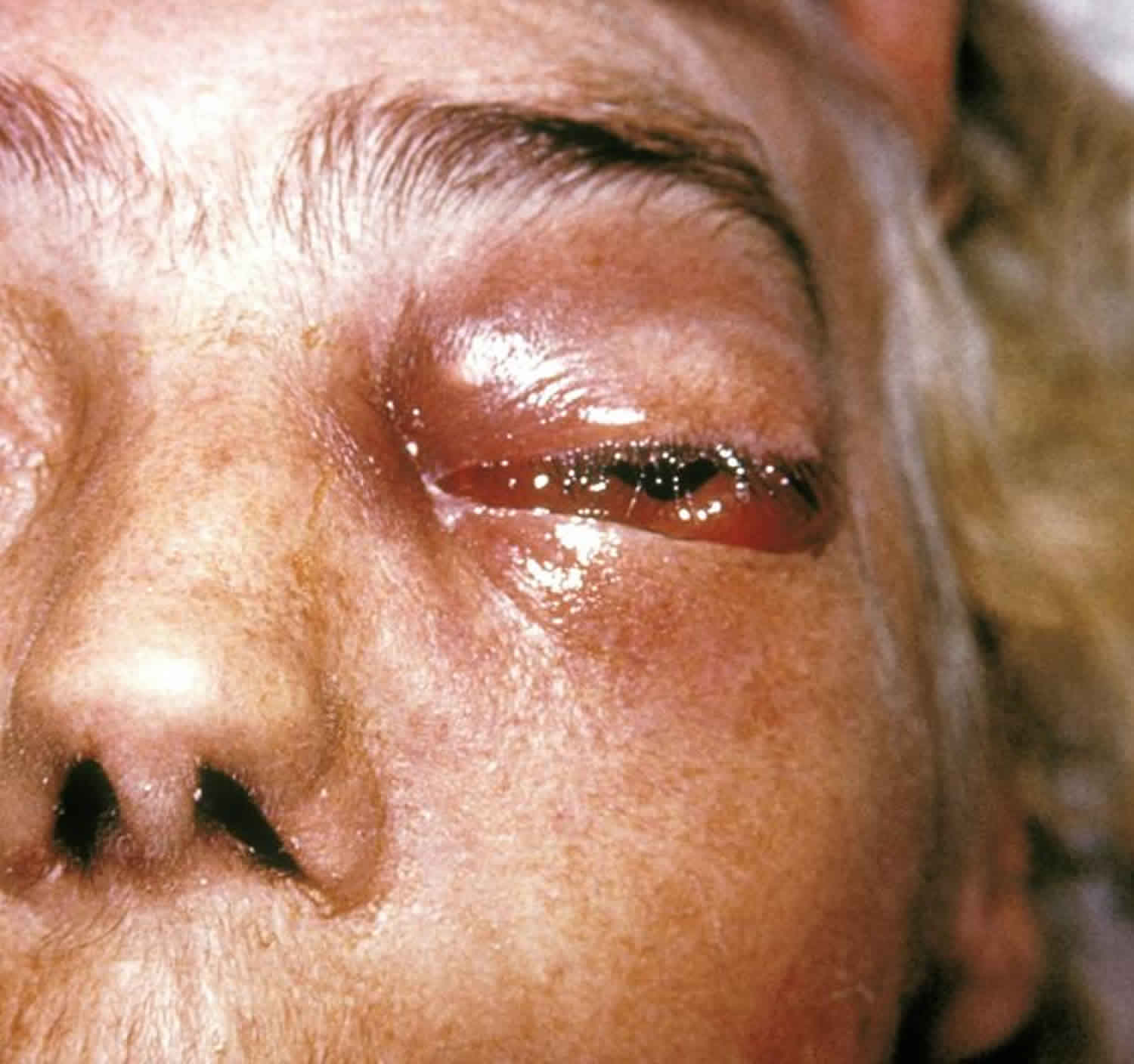
Rhinocerebral zygomycosis is the most common form of all invasive mucormycoses form with one third to half of all cases 52. It is seen primarily in uncontrolled diabetic patients. It occurs by inhalation or hematogenous or lymphatic dissemination. The classical form involves the sinuses, especially maxillary, but can easily spread to the neighboring tissue including nose, orbits, eyes, brain, cranial nerves, hard and soft palates, both mandibles and the rest of the face. It has great variety of clinical presentations from that of a simple acute sinusitis with purulent rhinorrhea 53, where the initial exam of the nasal mucosa may be normal, to a more dramatic presentation caused by progressive thrombosis and infarction. The exam might reveal violaceous discoloration, black eschar or frank tissue necrosis. Fever can be present or absent. Another well-recognized and severe form of rhino-cerebral zygomycosis is the presentation of periorbital cellulitis. It can be uni or bilateral. Initially it presents with tissue edema and erythema around the eye(s), later proptosis, ophtalmoplegia, and visual loss can ensue 54. The spread of the infection to the hard palate can cause perforation 55. Its extension towards the brain can cause utmost devastation; cases with brain abscesses 56, cerebral arteries aneurysms 57, hydrocephalus 58 and stroke 59 have been reported. Even the rare Garcin syndrome, where multiple cranial neuropathies occur, was documented in mucormycosis 60.
Pulmonary mucormycosis
Pulmonary mucormycosis is thought to be second most common form. It occurs by inhalation or hematogenous or lymphatic spread. It can present with mild to severe symptoms including fever, cough, sputum production, dyspnea, hypoxia, chest pain and hemoptysis 59. Mucormycosis can cause lobar consolidation 61, multiple disseminated lung nodules 62, fungal ball 63 or mycotic abcess formation 64. It can also present as a single tracheal 65 or multiple endobronchial lesions 66. Obviously, and because of its proximity, pulmonary zygomycosis can disseminate to the pleural space 67, chest wall 68 or the mediastinum where it can cause catastrophic rupture of large vessels 29.
Gastrointestinal zygomycosis
Gastrointestinal zygomycosis is relatively rare. It is thought to be caused by ingestion of zygospores especially in the malnourished and alcoholics 69 or it can be secondary to trauma 42. It can cause fever, abdominal pain and bloating, nausea and vomiting, hematemesis, melena or bowel perforation. It can be observed in the stomach where it can cause ulceration, bleeding or perforation 70. It is interesting to notice that cases of iatrogenic gastric mucormycosis were reported after use of naso-gastric tubes or even tongue depressors colonized by the fungus 71. Other possible gastrointestinal sites include terminal ileum 72 and large bowel 73. Mucor can infect other parts of the digestive system including liver 74, bile duct 75. It can be severe enough to involve many adjacent organs including pancreas 76.
Cutaneous mucormycosis
Intact skin forms a barrier against mucor penetration. Cutaneous zygomycosis takes hold when this barrier is disrupted. This disruption can be caused by skin maceration, burns 43 or trauma 77. It usually carries a better prognosis than other forms of mucormycosis until the fungal reaches deeper into muscle, bone or fascia where it causes severe necrosis; the mortality rate then becomes very high 78.
Disseminated zygomycosis
Disseminated zygomycosis has the worse prognosis. Its mortality rate approaches 100%. Since mucor is an angiotropic fungus, any prior form of this mycosis can cause severe fungemia in immune compromised individuals with subsequent hematogenous spread to many body organs including brain, heart, lungs, and kidneys among others. Moreover, disseminated mucormycosis was described with intravenous drug use 79 and diabetics utilizing self-monitoring material 80. Antemortem diagnosis can be very challenging. Blood cultures, in these severely ill patients, are usually negative. The diagnosis is suspected in the presence of disseminated organ infarction and necrosis 81. Cases of endocarditis 82 and myocarditis 83 were observed.
Miscellaneous mucormycosis
In miscellaneous mucormycosis, the infecting agents from the mucorales order can infect any part of the body. Indeed, documented cases of this fungal infection were reported in the ear 84 or limited to the parotid gland 85 or in the intravascular system where it can cause severe micro-aneurysms or migratory thrombi 86, adjacent to the spinal cord 87, inside joints like the knee 88 or affecting whole upper or lower limbs 89, within the urinary tract 90 and genital organs and pelvic floor 91. The consequences can be disastrous regardless of the affected site.
Zygomycosis diagnosis
Diagnosing invasive zygomycosis is not an easy task. Its clinical picture can be extremely variable. Further, because of its relative rarity, it is usually missed in its early stage when the chances of cure are still reasonable. Unfortunately, close to half of phycomycosis cases are diagnosed post-mortem 92. A high index of suspicion should always be kept when facing these variable presentations of mucormycosis especially when dealing with immune-compromised patients. When this infection is suspected, physicians have nowadays an extensive armamentarium at their disposition to try to pin down its elusive diagnosis. Initial blood work can reveal non-specific findings such as leukocytosis, hyperglycemia or acidosis. Frequently, stigmata of immunosuppression, including neutropenia, are encountered. Blood cultures are usually negative but, exceptionally, fungal growth in the blood can be observed 93. Until now, there is no specific serologic test for mucormycosis. Radiological investigations, such plain x-rays or computed tomography, can be completely normal or demonstrate variable abnormal findings depending on the infection size and location. That location will guide the clinician to use further diagnostic tools in order to further clarify the diagnosis. Examples of such tools include bronchoscopy with bronchoalveolar lavage in case of pulmonary disease, upper or lower endoscopy for gastric or bowel lesions, or video assisted device for abdominal or thoracic infection 94. However, obtaining tissue biopsy remains the gold standard for diagnosing invasive zygomycosis. Indeed, clinicians should not hesitate to obtain a good sample of the infected tissue as soon as possible to clinch the diagnosis. This task is relatively easy in cutaneous and rhinomaxillary mucormycosis; it becomes more challenging with deeper forms. Histopathology will reveal irregular broad non-septate hyphae and spores pathognomonic of mucor; with evidence of surrounding neutrophilic infiltration, necrosis and vasculature invasion.
Zygomycosis treatment
Most medical textbooks and literature emphasizes three important cornerstones in the treatment of invasive zygomycosis. They are: reversal of the underlying condition(s), medical therapy and surgical debridement.
- Reversal of underlying condition(s): any predisposing factor, such as hyperglycemia or acidosis or malnutrition or immunosuppression, must be corrected if possible. This easy initial intervention improves the chances of survival 95.
- Medical therapy: until recently, the natural course of mucormycosis was usually fatal. A breakthrough in the treatment of deep mycoses occurred in 1953 when Drs Charles Smith and William Winn discovered amphotericin B from a soil isolate brought from the Orinoco Basin in Venezuela 96. This discovery opened doors to the parenteral therapy for such mycoses including histoplasmosis, cryptococcosis, and mucormycosis. The first case of cure from this severe disease was reported by Harris in 1955 97. There are two types of antifungal treatment for invasive zygomycosis:
- Standard therapy: amphtericin B is a polyene macrolide. It continues to play a major role in the treatment of invasive zygomycosis. Both conventional and liposomal amphotericin B are effective against it; the liposomal form offers less infusion site side effects and milder nephrotoxicity, however, it generally costs more 98. The duration of therapy varies from weeks to months depending on the site and severity of the infection.
- Experimental therapy: newer antifungal medications are being currently developed. The orally administered posaconazole, from the family of azoles, recently showed promising results against the mucorales species 99. Iron chelation is a novel adjunctive therapy that has potential role in the treatment of mucormycosis 100. Future immunotherapy will probably hold some key answers in the management of zygomycosis.
- Surgical intervention: surgical debridement is another cornerstone in treating invasive zygomycosis. It is usually extensive and can be disfiguring. It has to be done in earnest in addition to other therapeutic interventions.
- Psychological support: patients infected with invasive mucomycosis face many difficult challenges. This infection can be prolonged and exhausting, it adds to the heavy burden(s) of their uncontrolled chronic condition, such as diabetes and its complications, immunosuppression from malignancy or AIDS, etc. These patients are clearly prone to psychological setbacks and major depression. Health care providers must pay special attention to these possibilities and provide necessary supportive and therapeutic treatment.
- Physical rehabilitation: if the infected person with invasive zygomycosis escapes death he could carry severe stigmata, from the infection or its treatment, such as disfigurement, partial/complete loss of an extremity or organ function. In many instances, these patients need prolonged course of physical or occupational rehabilitation depending on the degree of their disability.
The current standard of care for zygomycosis treatment includes aggressive surgical debridement and treatment with liposomal amphotericin B. If there is any suspicion for mucormycosis, amphotericin B should be initiated immediately. Correction of any underlying pathophysiology, such as diabetic ketoacidosis, should be done as well 101.
Hyperbaric oxygen therapy is commonly used for necrotizing soft tissue infections, chronic osteomyelitis, and compromised grafts and flaps 6. Hypoxia is a problem when it comes to nonhealing tissue. hyperbaric oxygen allows for saturation of oxygen within the blood and hemoglobin and a 10-fold increase of dissolved oxygen in plasma. Oxygen delivery to compromised tissue is increased, and normal tissue oxygenation for healing is restored. Hyperbaric oxygen therapy has also been shown to improve vascularity and stimulate new blood vessel growth. These factors are important in zygomycosis because of the angioinvasive nature of the infection, tissue becomes hypoxic and blood vessels become thrombosed. In addition, zygomycosis patients often undergo extensive debridement surgery. In cases of rhinocerebral murcomycosis, patients can be left disfigured. Addition of postsurgical hyperbaric oxygen therapy could help the formation of granulation tissue and bone healing 102.
Due to the rareness and severity of this rapidly progressive infection, it is extremely difficult to conduct randomized, controlled trials on human patients. Therefore, the majority of the research on using hyperbaric oxygen therapy for zygomycosis largely relies on case reports and retrospective studies. In a review of the literature done by Ferguson et al. 103, 12 patients with rhinocerebral mucormycosis were investigated. They were all treated with surgery and amphotericin B. Half of the patients were treated with adjunctive hyperbaric oxygen therapy. Of the patients who did not receive hyperbaric oxygen therapy, 4 out of the 6 patients died as a result of the infection. Of the 6 patients who received adjunctive hyperbaric oxygen therapy, only 2 died. However, this case review is too small to draw any statistical conclusions.
One of the most interesting case reports comes from Couch et al. in which 2 diabetic patients with rhinocerebral mucormycosis were treated with adjunctive hyperbaric oxygen therapy. A high fatality rate occurs when the cerebral extension of the infection occurs. Both of these patients were critically ill and had aggressive brain mucor abscesses and one patient even had total occlusion of the internal carotid artery. Despite surgical debridement and medical therapy, their infections progressed and hyperbaric oxygen therapy was added as salvage therapy. Both patients had marked clinical improvement upon addition of hyperbaric oxygen therapy. They each received treatments 6 days a week at 2.5 atmosphere absolute (ATA) for 90 minutes. One received 79 total treatments and the other received 85 treatments. Both patients remained disease free 21 months after discharge. De La Paz et al. and Melero et al. also published case reports on successfully treating bilateral rhinocerebral mucormycosis with adjunctive hyperbaric oxygen therapy. A more recent review of cases done in 2004 by John BV et al. explored the use of hyperbaric oxygen therapy in 28 cases. The hyperbaric oxygen treatment sessions 90 minutes long at a pressure of 2 to 3 ATA, ranging from 90 to 120 minutes. In general, hyperbaric oxygen therapy was not started until after surgical debridement. The survival rate was 86% with diabetic patients having a much higher survival rate of 94%. There is a higher rate of survival when the underlying condition is correctable such as diabetic ketoacidosis. Conversely, 2 out of the 3 patients with malignancies died as a result of the infection.
Considering the pathophysiology of invasive zygomycosis, its high morbidity and mortality, and the benefits of hyperbaric oxygen treatment, it is reasonable to consider hyperbaric oxygen therapy as adjunctive therapy in the treatment of this disease.
Mucormycosis prognosis
The prognosis of mucormycosis depends on several factors, including the infection site, rapidity of diagnosis, and type and severity of immunosuppression.
Even with prompt diagnosis and treatment, overall mortality rates average about 50%, although rhinocerebral and gastrointestinal forms of the infection carry a mortality rate of approximately 85%. Mortality rates are high because of the difficulty in establishing the diagnosis and the lack of adequate antifungal therapy. By the time a diagnosis of mucormycosis is confirmed, it has frequently spread via either local invasion with extensive tissue destruction or widely disseminated infection. Mortality is higher in patients who have hematological disorders or malignancies as their underlying conditions are more difficult to treat. The disseminated form has a mortality of over 90%. Patients who do survive are often left disfigured due to extensive debridement surgeries.
References- Zygomycosis. https://emedicine.medscape.com/article/232465-overview
- Kwon-Chung KJ. Taxonomy of fungi causing mucormycosis and entomophthoramycosis (zygomycosis) and nomenclature of the disease: molecular mycologic perspectives. Clin Infect Dis. 2012 Feb. 54 Suppl 1:S8-S15.
- Rammaert B, Lanternier F, Zahar JR, et al. Healthcare-associated mucormycosis. Clin Infect Dis. 2012 Feb. 54 Suppl 1:S44-54.
- Del Valle Zapico A, Rubio Suárez A, Mellado Encinas P, Morales Angulo C, Cabrera Pozuelo E. Mucormycosis of the sphenoid sinus in an otherwise healthy patient: Case report and literature review. J Laryngol Otol. 1996;110:471–3.
- Mucormycosis (Zygomycosis). https://emedicine.medscape.com/article/222551-overview
- Quandahl R, Cooper JS. Hyperbaric, Zygomycotic Infections. [Updated 2019 May 7]. In: StatPearls [Internet]. Treasure Island (FL): StatPearls Publishing; 2019 Jan-. Available from: https://www.ncbi.nlm.nih.gov/books/NBK493208
- Vaideeswar P, Shah R. Zygomycotic infective endocarditis in pregnancy. Cardiovasc. Pathol. 2017 May – Jun;28:28-30.
- Spellberg B, Edwards J Jr, Ibrahim A. Novel perspectives on mucormycosis: pathophysiology, presentation, and management. Clin Microbiol Rev. 2005;18:556–69.
- Liang KP, Tleyjeh IM, Wilson WR, Roberts GD, Temesgen Z. Rhino-orbitocerebral mucormycosis caused by Apophysomyces elegans. J Clin Microbiol. 2006;44:892–8.
- Chayakulkeeree M, Ghannoum MA, Perfect JR. Zygomycosis: the re-emerging fungal infection. Eur J Clin Microbiol Infect Dis. 2006;25:215–29.
- Roden MM, Zaoutis TE, Buchanan WL, Knudsen TA, Sarkisova TA, Schaufele RL, Epidemiology and outcome of zygomycosis: a review of 929 reported cases. Clin Infect Dis. 2005;41:634–53.
- Chamilos G, Lewis RE, Kontoyiannis DP. Delaying amphotericin B–based frontline therapy significantly increases mortality among patients with hematologic malignancy who have zygomycosis. Clin Infect Dis. 2008;47:503–9.
- Jung SH, Kim SW, Park CS, Song CE, Cho JH, Lee JH, et al. Rhinocerebral Mucormycosis: Consideration of prognostic factors and treatment modality. Auris Nasus Larynx. 2009;36:274–9.
- Pagano L, Offidani M, Fianchi L, Nosari A, Candoni A, Piccardi M, et al. Infection Program Mucormycosis in hematologic patients. Haematologica. 2004;89:207–14.
- Roden MM, Zaoutis TE, Buchanan WL, Knudsen TA, Sarkisova TA, Schaufele RL, et al. Epidemiology and outcome of zygomycosis: A review of 929 reported cases. Clin Infect Dis. 2005;41:634–53.
- Song YM, Shin SY. Bilateral ophthalmic artery occlusion in rhino-orbito-cerebral mucormycosis. Korean J Ophthalmol. 2008;22:66–9.
- Bhadani PP, Bhadani UK, Thapliyal N, Sen R. A rare presentation of invasive rhino-orbital mucormycosis in an immunocompetent young girl: A case report. Indian J Pathol Microbiol. 2007;50:785–6.
- Fukushima T, Sumazaki R, Shibasaki M, Saitoh H, Fujigaki Y, Kaneko M, et al. Successful treatment of invasive thoracopulmonary mucormycosis in a patient with acute lymphocytic leukemia. Cancer. 1995;76:895–9.
- Welk B, House AA, Ralph E, Tweedy E, Luke PP. Successful treatment of primary bilateral renal mucormycosis with bilateral nephrectomy. Urology. 2004;64:590.
- Marty FM, Cosimi LA, Baden LR. Breakthrough zygomycosis after voriconazole treatment in recipients of hematopoietic stem-cell transplants. N Engl J Med. 2004;350:950–2.
- Greenberg RN, Mullane K, van Burik JA, Raad I, Abzug MJ, Anstead G, Posaconazole as salvage therapy for zygomycosis. Antimicrob Agents Chemother. 2006;50:126–33.
- Spellberg B, Andes D, Perez M, Anglim A, Bonilla H, Mathisen GE, Safety and outcomes of open-label deferasirox iron chelation therapy for mucormycosis. Antimicrob Agents Chemother. 2009;53:3122–5.
- Jain D, Kumar Y, Vasishta RK, Rajesh L, Pattari SK, Chakrabarti A. Zygomycotic necrotizing fasciitis in immunocompetent patients: a series of 18 cases. Mod. Pathol. 2006 Sep;19(9):1221-6.
- Kontoyiannis DP, Lewis RE. Agents of mucormycosis and Entomophthoramycosis. Mandell GL, Bennett GE, Dolin R, eds. Mandell, Douglas and Bennett’s Principles and Practice of Infectious Diseases. 7th ed. Philadelphia, Pa: Churchill Livingstone; 2010. 3257-69.
- Waness A, Dawsari GA, Al Jahdali H. The rise of an opportunistic infection called “Invasive Zygomycosis”. J Glob Infect Dis. 2009;1(2):131–138. doi:10.4103/0974-777X.56256 https://www.ncbi.nlm.nih.gov/pmc/articles/PMC2840956
- Ikeda T, Tabuchi K, Shirota K, Une Y, Nomura Y. Mucormycosis in a cow. Jpn J Vet Sci. 1987;48:527–30.
- Gust N, Griffiths J. Platypus mucormycosis and its conservation implications. Australasian Mycologist. 2009;28:1–8
- Ribes JA, Vanover-Sams CL, Baker DJ. Zygomycetes in human disease. Clin Microbiol Rev. 2000;13(2):236–301. doi:10.1128/cmr.13.2.236-301.2000 https://www.ncbi.nlm.nih.gov/pmc/articles/PMC100153
- Kitabayashi A, Hirokawa M, Yamaguchi A, Takatsu H, Miura AB. Invasive pulmonary mucormycosis with rupture of the thoracic aorta. Am J Hematol. 1998;58:326–9.
- Mucormycosis (zygomycosis). https://www.uptodate.com/contents/mucormycosis-zygomycosis
- Chadli-Chaieb M, Bchir A, Fathallah-Mili A, Ach K, Maaroufi A, Garrouche A, et al. Mucormycosis in the diabetic patient. Presse Med. 2005;34:218–22.
- Espinoza CG, Halkias DG. Pulmonary mucormycosis as a complication of chronic salicylate poisoning. Am J Clin Pathol. 1983;80:508–11.
- Johnson KE, Leahy K, Owens C, Blankson JN, Merz WG, Goldstein BJ. An atypical case of fatal zygomycosis: Simultaneous cutaneous and laryngeal infection in a patient with a non-neutropenic solid prostatic tumor. Ear Nose Throat J. 2008;87:152–5.
- Gadadhar H, Hawkins S, Huffstutter JE, Panda M. Cutaneous mucormycosis complicating methotrexate, prednisone, and infliximab therapy. J Clin Rheumatol. 2007;13:361–2.
- Einollahi B, Lessan-Pezeshki M, Pourfarziani V, Nemati E, Nafar M, Pour-Reza-Gholi F, et al. Invasive fungal infections following renal transplantation: A review of 2410 recipients. Ann Transplant. 2008;13:55–8.
- Pavie J, Lafaurie M, Lacroix C, Marie Zagdanski A, Debrosse D, Socié G, et al. Successful treatment of pulmonary mucormycosis in an allogenic bone-marrow transplant recipient with combined medical and surgical therapy. Scand J Infect Dis. 2004;36:767–9.
- Fahimzad A, Chavoshzadeh Z, Abdollahpour H, Klein C, Rezaei N. Necrosis of nasal cartilage due to mucormycosis in a patient with severe congenital neutropenia due to HAX1 deficiency. J Investig Allergol Clin Immunol. 2008;18:469–72.
- Grant JM, St-Germain G, McDonald JC. Successful treatment of invasive Rhizopus infection in a child with thalassemia. Med Mycol. 2006;44:771–5.
- Lagorce Pagès C, Fabre A, Bruneel F, Zimmermann U, Hénin D. Disseminated mucormycosis in AIDS. Ann Pathol. 2000;20:343–5.
- Hopkins RJ, Rothman M, Fiore A, Goldblum SE. Cerebral mucormycosis associated with intravenous drug use: Three case reports and review. Clin Infect Dis. 1994;19:1133–7.
- Raizman NM, Parisien M, Grafe MW, Gordon RJ, Rosenwasser MP. Mucormycosis of the upper extremity in a patient with alcoholic encephalopathy. J Hand Surg Am. 2007;32:384–8.
- Berne JD, Villarreal DH, McGovern TM, Rowe SA, Moore FO, Norwood SH. A fatal case of posttraumatic gastric mucormycosis. J Trauma. 2009;66:933–5.
- Ledgard JP, van Hal S, Greenwood JE. Primary cutaneous zygomycosis in a burns patient: A review. J Burn Care Res. 2008;29:286–90.
- Metallidis S, Chrysanthidis T, Kazakos E, Saraf A, Nikolaidis P. A fatal case of pacemaker lead endocarditis caused by Mucor spp. Int J Infect Dis. 2008;12:e151–2.
- Chew HH, Abuzeid A, Singh D, Tai CC. Surgical wound mucormycosis necessitating hand amputation: A case report. J Orthop Surg (Hong Kong) 2008;16:267–9.
- Lin CY, Lee SC, Lin CC, Chan SC, Lee CT. Isolated fatal renal mucormycosis in a patient with chronic obstructive pulmonary disease and tuberculosis. Int J Clin Pract. 2003;57:916–8.
- Aziz S, Merrell RC, Edwards MF. Mucormycosis in patients with multi-organ failure. Arch Surg. 1984;119:1189–91.
- Saltoglu N, Tasova Y, Midikli D, Aksu HS, Sanli A, Dündar IH. Fever of unknown origin in Turkey: Evaluation of 87 cases during a nine-year-period of study. J Infect. 2004;48:81–5.
- Melnick JZ, Latimer J, Lee E, Henrich WL. Systemic mucormycosis complicating acute renal failure: Case report and review of the literature. Ren Fail. 1995;17:619–27.
- Alloway JA, Buchsbaum RM, Filipov PT, Reynolds BN, Day JA. Mucormycosis in a patient with sarcoidosis. Sarcoidosis. 1995;12:143–6.
- Choudhury M, Kahkashan E, Choudhury SR. Neonatal gastrointestinal mucormycosis: A case report. Trop Gastroenterol. 2007;28:81–2.
- Spellberg B, Edwards J, Ibrahim A. Novel perspectives on mucormycosis: Pathophysiology, presentation, and management. Clin Microbiol Rev. 2005;18:556–69.
- Szalai G, Fellegi V, Szabó Z, Vitéz LC. Mucormycosis mimicks sinusitis in a diabetic adult. Ann N Y Acad Sci. 2006;1084:520–30.
- Rutar T, Cockerham KP. Periorbital zygomycosis (mucormycosis) treated with posaconazole. Am J Ophthalmol. 2006;142:187–8
- Barrak HA. Hard palate perforation due to mucormycosis: Report of four cases. J Laryngol Otol. 2007;121:1099–102.
- Mohindra S, Mohindra S, Gupta R, Bakshi J, Gupta SK. Rhinocerebral mucormycosis: The disease spectrum in 27 patients. Mycoses. 2007;50:290–6.
- Kasliwal MK, Reddy VS, Sinha S, Sharma BS, Das P, Suri V. Bilateral anterior cerebral artery aneurysm due to mucormycosis. J Clin Neurosci. 2009;16:156–9.
- Sweeney PJ, Hahn JF, McHenry MC, Mitsumoto H. Mucormycosis presenting as positional nystagmus and hydrocephalus: Case report. J Neurosurg. 1980;52:270–2.
- Liu MF, Chen FF, Hsiue TR, Liu CC. Disseminated zygomycosis simulating cerebrovascular disease and pulmonary alveolar haemorrhage in a patient with systemic lupus erythematosus. Clin Rheumatol. 2000;19:311–4.
- Mutsukura K, Tsuboi Y, Imamura A, Fujiki F, Yamada T. Garcin syndrome in a patient with rhinocerebral mucormycosis. No To Shinkei. 2004;56:231–5.
- Holley A, Mayes D, Browning R. A 40-year-old man with neutropenic fever and lobar consolidation. Chest. 2008;133:816–9.
- Sharma A, Gupta V, Singh RS, Kakkar N, Singh S, Bambery P. Angioinvasive pulmonary mucormycosis presenting as multiple bilateral pulmonary nodules in a patient without obvious predisposing factors. Singapore Med J. 2008;49:e269–71.
- Lahiri TK, Agarwal D, Reddy GE, Bajoria A. Pulmonary mucoraceous fungal ball. Indian J Chest Dis Allied Sci. 2001;43:107–10.
- McAdams HP, Rosado de Christenson M, Strollo DC, Patz EF. Pulmonary mucormycosis: Radiologic findings in 32 cases. AJR Am J Roentgenol. 1997;168:1541–8.
- Sales-Badía JG, Hervás VZ, Galbis-Caravajal JM. Tracheal mucormycosis. Arch Bronconeumol. 2009;45:260–1.
- Fermanis GG, Matar KS, Steele R. Endobronchial zygomycosis. Aust N Z J Surg. 1991;61:391–3.
- Green WR, Bouchette D. Pleural mucormycosis (zygomycosis) Arch Pathol Lab Med. 1986;110:441–2.
- Asai K, Suzuki K, Takahashi T, Ito Y, Kazui T, Kita Y. Pulmonary resection with chest wall removal and reconstruction for invasive pulmonary mucormycosis during antileukemia chemotherapy. Jpn J Thorac Cardiovasc Surg. 2003;51:163–6.
- Shahapure AG, Patankar RV, Bhatkhande R. Gastric mucormycosis. Indian J Gastroenterol. 2002;21:231–2.
- Chung CS, Wang WL, Liu KL, Lin JT, Wang HP. Green ulcer in the stomach: Unusual mucormycosis infection. Gastrointest Endosc. 2008;68:566–7. discussion 567.
- Maraví-Poma E, Rodríguez-Tudela JL, de Jalón JG, Manrique-Larralde A, Torroba L, Urtasun J, et al. Outbreak of gastric mucormycosis associated with the use of wooden tongue depressors in critically ill patients. Intensive Care Med. 2004;30:724–8.
- Han JY, Cheon JH, Kim DH, Chon HJ, Kim SK, Kim TI, et al. Ileal mucormycosis diagnosed by colonoscopy in a patient with acute myeloid leukemia. Korean J Gastroenterol. 2008;52:179–82.
- Sakorafas GH, Tsolakides G, Grigoriades K, Bakoyiannis CN, Peros G. Colonic mucormycosis: An exceptionally rare cause of massive lower gastrointestinal bleeding. Dig Liver Dis. 2006;38:616–7.
- Mekeel KL, Hemming AW, Reed AI, Matsumoto T, Fujita S, Schain DC, et al. Hepatic mucormycosis in a renal transplant recipient. Transplantation. 2005;79:1636.
- Kantharia CV, Prabhu RY, Deshmukh H, Supe AN. Mucormycosis of the bile duct: A case report. Trop Gastroenterol. 2007;28:126.
- Bittencourt AL, Ayala MA, Ramos EA. A new form of abdominal zygomycosis different from mucormycosis: Report of two cases and review of the literature. Am J Trop Med Hyg. 1979;28:564–9.
- Simbli M, Hakim F, Koudieh M, Tleyjeh IM. Nosocomial post-traumatic cutaneous mucormycosis: A systematic review. Scand J Infect Dis. 2008;40:577–82.
- Devi SC, Kanungo R, Barreto E, Thomas AG, Shashikala N, Srinivasan S, et al. Favorable outcome of amphotericin B treatment of zygomycotic necrotizing fascitis caused by Apophysomyces elegans. Int J Dermatol. 2008;47:407–9.
- Leen CL, Brettle RP. Fungal infections in drug users. J Antimicrob Chemother. 1991;28:83–96.
- Hampson FG, Ridgway EJ, Feeley K, Reilly JT. A fatal case of disseminated zygomycosis associated with the use of blood glucose self-monitoring equipment. J Infect. 2005;51:e269–72.
- de Mol P, Meis JF. Disseminated Rhizopus microsporus infection in a patient on oral corticosteroid treatment: A case report. Neth J Med. 2009;67:25–8.
- Solano T, Atkins B, Tambosis E, Mann S, Gottlieb T. Disseminated mucormycosis due to Saksenaea vasiformis in an immunocompetent adult. Clin Infect Dis. 2000;30:942–3.
- Basti A, Taylor S, Tschopp M, Sztajzel J. Fatal fulminant myocarditis caused by disseminated mucormycosis. Heart. 2004;90:e60.
- Oktay MF, Askar I, Kilinç N, Tuzcu A, Topçu I. Auricular mucormycosis: A case report. Kulak Burun Bogaz Ihtis Derg. 2007;17:228–30.
- Chandu A, MacIsaac RJ, MacGregor DP, Campbell MC, Wilson MJ, Bach LA. A case of mucormycosis limited to the parotid gland. Head Neck. 2005;27:1108–11.
- Sochaj M, Claridge M, Green NJ, Fox AD. Intravascular mucormycosis as a cause of arm ischemia in an immunocompromised patient. J Vasc Surg. 2009;50:193–4.
- Suzuki G, Kurosawa M, Takanashi Y, Itaya T, Kunieda Y, Maeda S, et al. Transverse lesion of the spinal cord due to mucormycosis in an AML patient. Rinsho Ketsueki. 1996;37:694–700.
- Muscolo DL, Carbo L, Aponte-Tinao LA, Ayerza MA, Makino A. Massive bone loss from fungal infection after anterior cruciate ligament arthroscopic reconstruction. Clin Orthop Relat Res. 2009;467:2420–5.
- Moran SL, Strickland J, Shin AY. Upper-extremity mucormycosis infections in immunocompetent patients. J Hand Surg Am. 2006;31:1201–5.
- Pérez de la Espejo MP, Barrero Candau R, Chinchón Espino D, Campoy Martínez P. Bladder mucormycosis: Report of one case. Arch Esp Urol. 2004;57:67–9.
- Cohen-Ludmann C, Kerob D, Feuilhade M, Chaine B, Guermazi A, Janier M, et al. Zygomycosis of the penis due to rhizopus oryzae successfully treated with surgical debridement and a combination of high-dose liposomal and topical amphotericin B. Arch Dermatol. 2006;142:1657–8.
- Tietz HJ, Brehmer D, Janisch W, Martin H. Incidence of endomycoses in the autopsy material of the Berlin Charite´ Hospital. Mycoses. 1998;41:81–5.
- Disbay M, Adisen E, Kustimur S, Sari N, Cengiz B, Yalcin B, et al. Fungemia and cutaneous zygomycosis due to mucor circinelloides in an intensive care unit patient: Case report and review of literature. Jpn J Infect Dis. 2009;62:146–8.
- Ikedou Y, Emori M, Nagata N, Harada S, Horiuchi M, Kitahara Y, et al. A case of pulmonary mucormycosis diagnosed through video-assisted thoracoscopic surgery: Report of a case. Nihon Kokyuki Gakkai Zasshi. 2003;41:310–4.
- Perlroth J, Choi B, Spellberg B. Nosocomial fungal infections: Epidemiology, diagnosis, and treatment. Med Mycol. 2007;45:321–46.
- Dutcher JD. The discovery and development of amphotericin B. Dis Chest. 1968;54:296–8.
- Hilal AA, Taj-Aldeen SJ, Mirghani AH. Rhinoorbital mucormycosis secondary to Rhizopus oryzae: A case report and literature review. Ear Nose Throat J. 2004;83:556,558–60,562.
- Cagnoni P. Liposomal amphotericin B versus conventional amphotericin b in the empirical treatment of persistently febrile neutropenic patients. J Antimicrob Chemother. 2002;49:81–6.
- Rogers TR. Treatment of zygomycosis: Current and new options. J Antimicrob Chemother. 2008;61:i35–40.
- Ibrahim AS, Edwards JE, Fu Y, Spellberg B. Deferiprone iron chelation as a novel therapy for experimental mucormycosis. J Antimicrob Chemother. 2006;58:1070–3.
- Jenks JD, Mehta SR, Hoenigl M. Broad spectrum triazoles for invasive mould infections in adults: Which drug and when? Med. Mycol. 2019 Apr 01;57(Supplement_2):S168-S178.
- Hoenigl M, Gangneux JP, Segal E, Alanio A, Chakrabarti A, Chen SC, Govender N, Hagen F, Klimko N, Meis JF, Pasqualotto AC, Seidel D, Walsh TJ, Lagrou K, Lass-Flörl C, Cornely OA., European Confederation of Medical Mycology (ECMM). Global guidelines and initiatives from the European Confederation of Medical Mycology to improve patient care and research worldwide: New leadership is about working together. Mycoses. 2018 Nov;61(11):885-894.
- Ferguson B J, Mitchell T G, Moon R, Camporesi E M, Farmer J. Adjuvant hyperbaric oxygen treatment of rhinocerebral mucormycosis. Rev Infect Dis. 1988;10:551–559.


