close

Cancer

Cancer

Adrenal carcinoma

Adrenal cortical carcinoma

Adrenal cortical carcinoma

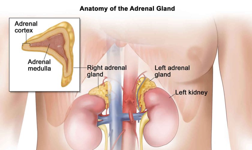
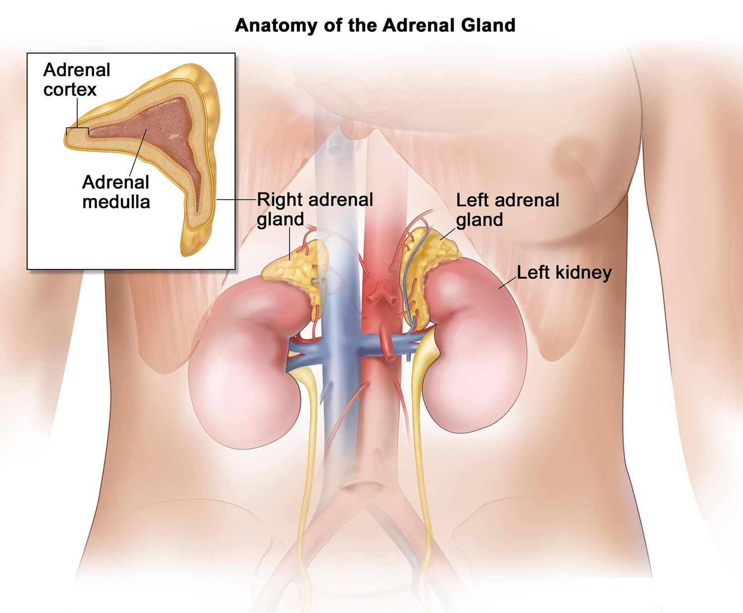
Adrenocortical carcinoma also called cancer of the adrenal cortex or adrenal cortical carcinoma, is a very rare cancer in which cancer cells form in the outer layer of your adrenal gland 1, 2. The outer layer of your adrenal gland is called the adrenal cortex (Figures 1 and 2). Cancer that forms in the adrenal medulla is called pheochromocytoma and is not discussed here in this article. You have 2 adrenal glands and one adrenal gland is located on top of each kidney. Your adrenal glands are responsible for releasing different classes of hormones. The adrenal cortex makes important hormones that:

  • Balance the water and salt in your body.
  • Help keep your blood pressure normal.
  • Help control your body’s use of protein, fat, and carbohydrates.
  • Cause your body to have masculine or feminine characteristics.

Adrenal cortical carcinoma is most common in children younger than 5 years old (median age 3–4 years) and adults in their 40s and 50s 3, 4, 5. Childhood adrenal cortical carcinoma typically present during the first 5 years of life (median age 3–4 years), although there is a second, smaller peak during adolescence 6, 7, 8. Both men and women can also develop adrenocortical carcinoma. Female sex is consistently predominant in most studies, with a female to male ratio of 1.6:1.0 5, 7, 8.

Adrenal cortical carcinomas are rare tumors with an annual incidence of 0.7–2.0 cases per million 9, 10. In children, 25 new adrenal cortical carcinoma cases are expected to occur annually in the United States 11. In southern Brazil, adrenal cortical carcinoma is approximately 10 to 15 times higher than that observed in the United States 12, 13, 14, 15.

Adrenocortical carcinoma may develop by chance alone (sporadic), but at least 50% of adrenal cortical carcinoma are thought to be linked to a cancer syndrome that is passed down through families (inherited) 16. The most common syndromes that are associated with adrenocortical carcinoma are Li-Fraumeni syndrome (TP53 gene germline and somatic mutation), Lynch syndrome (MSH2, MLH1, MSH6, PMS2, EPCAM genes), multiple endocrine neoplasia type 1 (MEN1 gene), Beckwith–Wiedemann syndrome (11p151 gene, IGF-2 overexpression), familial adenomatous polyposis (FAP gene, β catenin somatic mutations), neurofibromatosis type 1 (NF1 gene), Gardner’s syndrome and Carney complex (protein kinase A regulatory subunit 1A [PRKARIA] gene) 17, 18, 19, 20, 21, 22, 23, 24, 25. Although extremely rare, adrenocortical carcinomas have also been reported in patients with congenital adrenal hyperplasia (CAH) and myelolipoma 26, 27, 28. It is likely these are collision tumors rather than having been etiologically related 29.

There are a number of genes that have mutations that can cause sporadic adrenocortical carcinoma, including tumor protein 53 (TP53, p53), insulin-like growth factor 2 (IGF2) and β-catenin (CTNNB1 or ZNRF3) 30, 31, 32. Tumor protein 53 (TP53, p53) is a protein product of tumor suppressor gene located on chromosome 17 (17p13.1) 33. According to genomic analyses, germline mutations in TP53 were observed in 50–80% of children with sporadic adrenocortical carcinoma, while somatic TP53 mutation was observed in 20% to 30% of sporadic adrenocortical carcinoma patients where it correlates with poor outcome 34. In immunohistochemical studies diffuse TP53 staining correlates positively with increased Ki-67 expression 35.

There have been reports of both autosomal dominant inheritance and autosomal recessive inheritance.

Symptoms of adrenocortical carcinoma may include pain in your abdomen or back, a lump in your abdomen or a feeling of fullness in your abdomen.

Adrenocortical carcinoma can be classified into functioning and nonfunctioning tumors by clinical and biochemical assessment. Approximately 60% of adrenocortical carcinomas produce hormones 36. Nonfunctional adrenocortical carcinomas are rare (<10%) and tend to occur in older children 5.

Adrenocortical carcinomas in children are almost universally functional, they cause endocrine disturbances, and a diagnosis is usually made 5 to 8 months after the first signs and symptoms emerge 5, 6. Nonfunctional adrenocortical carcinomas are rare (<10%) and tend to occur in older children 5.

A functioning adrenal cortical carcinoma makes too much of one of the following hormones, as well as other hormones 37:

  • Cortisol. Too much cortisol may cause problem known as Cushing syndrome:
    • Weight gain in the face, neck, and trunk of the body and thin arms and legs.
    • Growth of fine hair on the face, upper back, or arms.
    • A round, red, full face (moon face).
    • Fat deposits behind the neck and shoulders (fatty hump or buffalo hump)
    • A deepening of the voice and swelling of the sex organs or breasts in both males and females.
    • Purple stretch marks on your abdomen
    • Muscle weakness and loss of muscle mass in your legs.
    • Weakened bones (osteoporosis), which can lead to fractures
    • High blood sugar levels, often leading to diabetes
    • High blood pressure
    • Easy bruising
    • Depression and/or moodiness
    • Menstrual irregularities in women.
  • Aldosterone. Too much aldosterone may cause:
    • High blood pressure.
    • Low blood potassium levels
    • Muscle weakness or cramps.
    • Frequent urination.
    • Feeling thirsty.
  • Testosterone. Men who make too much testosterone do not usually have signs or symptoms. However, too much testosterone in women may cause:
    • Growth of fine hair on the face, upper back, or arms.
    • Acne.
    • Balding.
    • A deepening of the voice.
    • No menstrual periods.
  • Estrogen (female-type hormone).
    • Too much estrogen in women may cause:
      • Irregular menstrual periods in women who have not gone through menopause.
      • Vaginal bleeding in women who have gone through menopause.
      • Weight gain.
    • Too much estrogen in men may cause:
      • Growth of breast tissue.
      • Lower sex drive.
      • Impotence.

These signs and symptoms may be caused by adrenocortical carcinoma or by other conditions. Check with your doctor if you have any of these problems.

Doctors diagnose adrenocortical carcinoma using imaging studies and tests that examine your blood and urine. An adrenocortical carcinoma is usually diagnosed based on urine tests for abnormal levels of cortisol, the hormone released by the adrenal glands. Blood tests can also be conducted to measure levels of potassium and sodium in your blood. A CT scan or MRI may be used to search for a visible tumor in the adrenal cortex.

Adrenal cortical carcinoma can spread to your liver, bone, lung, or other areas. Adrenocortical carcinoma can recur (come back) after it has been treated. The cancer may come back in the adrenal cortex or in other parts of the body.

The main treatment for adrenal cortical carcinoma is surgery to remove the tumor. You may get medicines (i.e., ketoconazole and metyrapone) to reduce production of cortisol, which causes many of your symptoms.

The prognosis (outlook) of adrenal cortical carcinomas varies: resectable tumors have a good prognosis, while metastatic and unresectable tumors have a poor prognosis 38. Overall the prognosis of adrenal cortical carcinoma is poor, with a 5-year survival rate of less than 40% 10, 39. Up to 70% of patients with the localized adrenal cortical carcinoma based on the American Joint Committee on Cancer (AJCC) Tumor, Node, and Metastases (TNM) staging system experienced recurrence within 3 years after surgery 39, while the survival duration of patients with metastatic adrenal cortical carcinomas or adrenal cortical carcinoma that has spread ranges from several months to 10 years or more 40, 41, 42.

Can adrenal cortical carcinoma be found early?

It is hard to find adrenal cancers early, and they are often quite large by the time they are diagnosed.

Adrenal cancers are often found earlier in children than in adults because cancers in children are more likely to secrete hormones that lead to signs and symptoms. For example, children may develop signs of puberty at an early age due to sex hormones made by adrenal cancer cells.

Adrenocortical carcinoma are sometimes found early by accident in adults, such as when a CT (computed tomography) scan of the abdomen is done for some other health concern.

Adrenal glands

The adrenal glands are two two triangle-shaped glands that measure about 1.5 inches in height and 3 inches in length. They are located on top of each kidney like a cap and is embedded in the mass of fatty tissue that encloses the kidney (Figure 1 and 2). Their name directly relates to their location (ad—near or at; renes—kidneys) 43.

The adrenal glands, located on the top of each kidney, are responsible for releasing different classes of hormones.

The outer part of the adrenal gland, called the adrenal cortex, produces the hormones cortisol, aldosterone, and hormones that can be changed into testosterone 43. The inner part of the gland, called the adrenal medulla, produces the hormones adrenaline and noradrenaline. These hormones are also called epinephrine and norepinephrine 43.

These hormones—cortisol, aldosterone, adrenaline, and noradrenaline—control many important functions in the body, including:

  • Maintaining metabolic processes, such as managing blood sugar levels and regulating inflammation.
  • Regulating the balance of salt and water.
  • Controlling the “fight or flight” response to stress.
  • Maintaining pregnancy.
  • Initiating and controlling sexual maturation during childhood and puberty.

The adrenal glands are also an important source of sex steroids, such as estrogen and testosterone.

When the adrenal glands produce more or less hormones than normal, you can become sick. This might happen at birth or later in life 44.

The adrenal glands can be affected by many diseases, such as autoimmune disorders, infections, tumors, and bleeding 44.

Conditions related to adrenal gland problems include:

  • Addison disease (also called adrenal insufficiency)
  • Congenital adrenal hyperplasia
  • Cushing syndrome
  • Diabetes – caused by another medical problem
  • Glucocorticoid medicines
  • Excessive or unwanted hair in women (hirsutism)
  • Hump behind shoulders (dorsocervical fat pad)
  • Hypoglycemia
  • Primary aldosteronism (Conn syndrome)
  • Waterhouse-Friderichsen syndrome

Location of adrenal glands

Figure 1. Location of the adrenal glands on top of each kidneys

adrenal glands location

adrenal-gland-location

Structure of the adrenal glands

Each adrenal gland is about the size of the top part of the thumb. Each adrenal gland is very vascular and consists of two parts:

  • The outer part is the adrenal cortex.
  • The central portion is the adrenal medulla.

These regions are not sharply divided, but they are functionally distinct structures that secrete different hormones.

The adrenal cortex and the adrenal medulla have very different functions. One of the main distinctions between them is that the hormones released by the adrenal cortex are necessary for life; those secreted by the adrenal medulla are not.

The adrenal medulla consists of irregularly shaped cells organized in groups around blood vessels. These cells are intimately connected with the sympathetic division of the autonomic nervous system. Adrenal medullary cells are actually modified postganglionic neurons. Preganglionic autonomic nerve fibers control their secretions. The adrenal medulla produces epinephrine and norepinephrine. These hormones are also called adrenaline and noradrenaline.

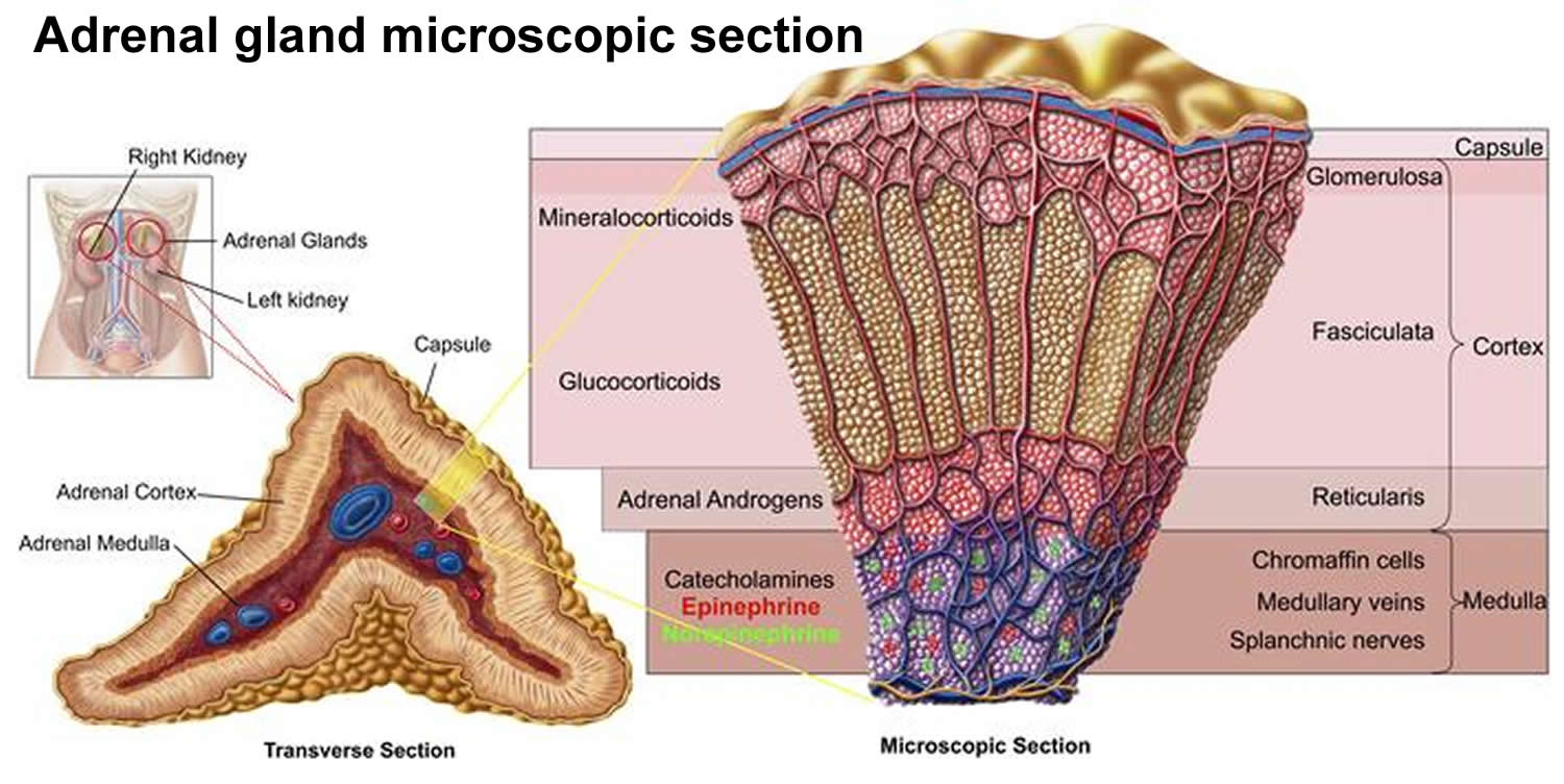
The adrenal cortex, which makes up the bulk of the adrenal gland, is composed of closely packed masses of epithelial cells, organized in layers. These layers form the outer (glomerulosa), middle (fasciculata), and inner (reticularis) zones of the cortex (Figure 3). As in the adrenal medulla, the cells of the adrenal cortex are well supplied with blood vessels. The adrenal cortex produces steroid hormones such as cortisol, aldosterone, and hormones that can be changed into testosterone.

Figure 2. Adrenal gland microscopic section

adrenal gland microscopic section

Adrenal Cortex Hormones

The adrenal cortex produces two main groups of corticosteroid hormones—glucocorticoids and mineralcorticoids. The release of glucocorticoids is triggered by the hypothalamus and pituitary gland. Mineralcorticoids are mediated by signals triggered by the kidney.

The principle mineralcorticoid is aldosterone, which maintains the right balance of salt and water while helping control blood pressure. Aldosterone is a hormone produced by the adrenal glands and helps to control the amount of fluid in the body by affecting how much salt and water the kidney retains or excretes. The adrenal glands produce aldosterone in response to another hormone called renin. Renin is produced by specialised cells in the kidney that detect when the body lacks salt; renin released by the kidney signals the adrenal glands to release aldosterone. The kidney detects an increase in aldosterone in the bloodstream and responds by retaining extra salt rather than excreting it in the urine. As the body regains the salt it needs, the level of renin in the bloodstream drops and therefore the amount of aldosterone in the blood also falls, meaning more water is excreted in the urine. This is an example of a feedback system.

When the hypothalamus produces corticotrophin-releasing hormone (CRH), it stimulates the pituitary gland to release adrenal corticotrophic hormone (ACTH). These hormones, in turn, alert the adrenal glands to produce corticosteroid hormones.

Glucocorticoids released by the adrenal cortex include:

  • 1) Cortisol: Also known as hydrocortisone, it regulates how the body converts fats, proteins, and carbohydrates to energy. It also helps regulate blood pressure and cardiovascular function.
  • 2) Corticosterone: This hormone works with hydrocortisone to regulate immune response and suppress inflammatory reactions.

There is a third class of hormone released by the adrenal cortex, known as sex steroids or adrenal sex hormones. The adrenal cortex releases small amounts of male and female sex hormones. These hormones are male types (adrenal androgens), but some are converted to female hormones (estrogens) in the skin, liver, and adipose tissue. The amounts of adrenal sex hormones are very small compared to the supply of sex hormones from the gonads, but they may contribute to
early development of reproductive organs. However, their impact is usually overshadowed by the greater amounts of hormones (such as estrogen and testosterone) released by the ovaries or testes. Cells in the inner zone of the adrenal cortex produce sex hormones.

The more important actions of cortisol include:

  1. Inhibition of protein synthesis in tissues, increasing the blood concentration of amino acids.
  2. Promotion of fatty acid release from adipose tissue, increasing the utilization of fatty acids and decreasing the use of glucose as energy sources.
  3. Stimulation of liver cells to synthesize glucose from noncarbohydrates, such as circulating amino acids and glycerol, increasing the blood glucose concentration.

These actions of cortisol help keep blood glucose concentration within the normal range between meals. This control is important, because a few hours without food can exhaust the supply of liver glycogen, a major source of glucose.

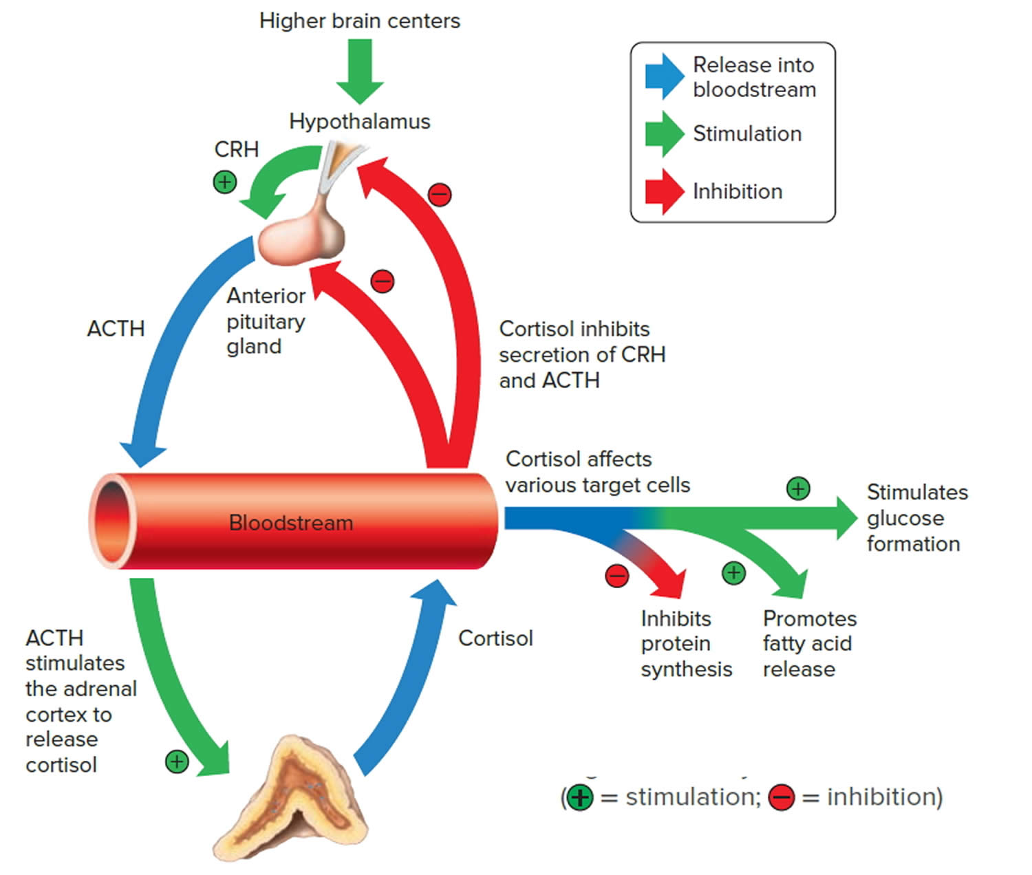
Negative feedback controls cortisol release (see Figure 4). This is much like control of thyroid hormones, involving the hypothalamus, anterior pituitary gland, and adrenal cortex. The hypothalamus secretes corticotropin-releasing hormone (CRH) into the pituitary gland portal veins, which carry CRH to the anterior pituitary, stimulating it to secrete adrenal corticotrophic hormone (ACTH). In turn, ACTH stimulates the adrenal cortex to release cortisol. Cortisol inhibits the release of CRH and ACTH, and as concentrations of these fall, cortisol production drops.

The set point of the feedback mechanism controlling cortisol secretion may change to meet the demands of changing conditions. For example, under stress—as from injury, disease, or emotional upset—information concerning the stressful condition reaches the brain. In response, brain centers signal the hypothalamus to release more corticotropin-releasing hormone (CRH), elevating the blood cortisol concentration until the stress subsides.

Figure 3. Negative feedback regulates cortisol secretion

regulation of cortisol secretion

Adrenal Medulla Hormones

Unlike the adrenal cortex, the adrenal medulla does not perform any vital functions. That is, you don’t need it to live 45. But that hardly means the adrenal medulla is useless. The hormones of the adrenal medulla are released after the sympathetic nervous system is stimulated, which occurs when you’re stressed. As such, the adrenal medulla helps you deal with physical and emotional stress.

You may be familiar with the fight-or-flight response—a process initiated by the sympathetic nervous system when your body encounters a threatening (stressful) situation. The hormones of the adrenal medulla contribute to this response.

Hormones secreted by the adrenal medulla are:

  • Epinephrine: Most people know epinephrine by its other name—adrenaline. This hormone rapidly responds to stress by increasing your heart rate and rushing blood to the muscles and brain. It also spikes your blood sugar level by helping convert glycogen to glucose in the liver. (Glycogen is the liver’s storage form of glucose.)
  • Norepinephrine: Also known as noradrenaline, this hormone works with epinephrine in responding to stress. However, it can cause vasoconstriction (the narrowing of blood vessels). This results in high blood pressure.

These adrenal medullary hormones have similar molecular structures and physiological functions. In fact, adrenaline (epinephrine), which makes up 80 percent
of the adrenal medullary secretion, is synthesized from noradrenaline (norepinephrine).

The effects of the adrenal medullary hormones resemble those of sympathetic neurons stimulating their effectors. The hormonal effects, however, last up to ten times longer than nervous stimulation because hormones are broken down more slowly than are neurotransmitters. Adrenaline (epinephrine) and noradrenaline (norepinephrine) increase heart rate, the force of cardiac muscle contraction, and blood glucose level. They also dilate airways, which makes breathing easier, elevate blood pressure, and decrease digestive activity.

Impulses arriving on sympathetic preganglionic nerve fibers stimulate the adrenal medulla to release its hormones at the same time that sympathetic impulses are stimulating other effectors. These sympathetic impulses originate in the hypothalamus in response to stress. In this way, adrenal medullary secretions function with the sympathetic division of the autonomic nervous system in preparing the body for energy-expending action, also called “fight-or-flight responses.”

Adrenal cortical carcinoma causes

The cause of most adrenal cortical carcinomas is currently unknown. However, scientists have found few risk factors that make a person more likely to develop adrenal cancer. Even if a patient does have one or more risk factors for adrenal cancer, it is impossible to know for sure how much that risk factor contributed to causing the cancer. But having a risk factor, or even several, does not mean that you will get the disease. Many people with risk factors never develop adrenal cancer, while others with this disease may have few or no known risk factors. Risk factors such as being overweight, smoking, living a sedentary lifestyle, and being exposed to cancer-causing substances in the environment can affect a person’s risk of many types of cancer. Although none of these factors has been found to definitely influence a person’s risk of developing adrenal cancer, smoking has been suggested as a risk factor by some researchers.

The majority of adrenal cortex cancers are not inherited (sporadic), but some (up to 15%) are caused by a genetic defect. This is more common in adrenal cancers in children 46.

Li-Fraumeni syndrome

Li-Fraumeni syndrome is a rare condition that is most often caused by a defect in the TP53 tumor suppressor gene. People with Li-Fraumeni syndrome have a high risk of several types of cancer, including include breast cancer, bone cancer, brain cancer, and adrenal cortex cancer. Li-Fraumeni syndrome causes a small portion of adrenal cancer in adults (about 1 of every 20), but it’s often the cause of adrenal cancer in children. In fact, about 8 of every 10 cases of adrenal cancer in children are caused by Li-Fraumeni syndrome. Many other adrenal cancers have also been found to have TP53 gene changes that were acquired after birth (not inherited).

Beckwith-Wiedemann syndrome

People with Beckwith-Wiedemann syndrome have large tongues, are large themselves, and have an increased risk for developing cancers of the kidney, liver and adrenal cortex.

Multiple endocrine neoplasia (MEN1)

People with MEN1 have a very high risk of developing tumors of 3 glands: the pituitary, parathyroid, and pancreas. About one-third to one-half of people with this condition also develop adrenal adenomas (benign tumors) or enlarged adrenal glands. These usually do not cause any symptoms. This syndrome is caused by defects in a gene called MEN1. People who have a family history of MEN1 or pituitary, parathyroid, pancreas, or adrenal cancers should ask their doctor if they might benefit from genetic counseling.

Familial adenomatous polyposis (FAP)

People with familial adenomatous polyposis (FAP) develop hundreds of polyps in the large intestine. These polyps will lead to colon cancer if the colon is not removed. Familial adenomatous polyposis (FAP) also increases the risk of other cancers, and may increase the risk for adrenal cancer. Still, most adrenal tumors in patients with familial adenomatous polyposis are benign adenomas. This syndrome is caused by defects in a gene called APC.

Lynch syndrome or hereditary nonpolyposis colorectal cancer (HNPCC)

Lynch syndrome also known as hereditary nonpolyposis colorectal cancer (HNPCC) is an inherited genetic disorder that increases the risk of colorectal cancer, stomach cancer, and some other cancers, including adrenal cortex cancer. In most cases, this disorder is caused by a defect in either the MLH1 or MSH2 gene, but other genes can cause Lynch syndrome, including MLH3, MSH6, TGFBR2, PMS1, and PMS2.

Adrenal cortical carcinoma prevention

Since there are no known preventable risk factors for this cancer, it is not possible to prevent adrenal cortical carcinoma at this time. Tobacco use has been suggested as a risk factor for adrenal cancer by some researchers, so not smoking might help reduce your risk.

Adrenal cortical carcinoma symptoms

In about half of people with adrenal cancer, symptoms are caused by the hormones made by the tumor. In the other half, symptoms occur because the tumor has grown so large that it presses on nearby organs.

As an adrenal cancer grows, it presses on nearby organs and tissues. This may cause pain near the tumor, a feeling of fullness in the abdomen, pain in your abdomen or back, or trouble eating because of a feeling of filling up easily.

Adrenocortical carcinoma symptoms may also include high blood pressure (hypertension), weight gain, frequent urination and possibly deepening of your voice. These symptoms are due to adrenocortical carcinoma causing excess secretion of hormones from the adrenal glands.

Symptoms of increased cortisol or other adrenal gland hormones may include:

  • Fatty, rounded hump high on the back just below the neck (buffalo hump)
  • Flushed, rounded face with pudgy cheeks (moon face)
  • Obesity
  • Stunted growth (short stature)
  • Virilization — the appearance of male characteristics, including increased body hair (especially on the face), pubic hair, acne, deepening of the voice, and enlarged clitoris (females). Virilization is caused by an excess of androgen (male-type hormone) secretion is seen, alone or in combination with hypercortisolism, in more than 80% of patients 12, 47.

Symptoms of increased aldosterone are the same as symptoms of low potassium, and include:

  • Muscle cramps
  • Weakness
  • Pain in your abdomen

A functioning adrenal cortical carcinoma makes too much of one of the following hormones, as well as other hormones 37:

  • Cortisol. Too much cortisol may cause problem known as Cushing syndrome:
    • Weight gain in the face, neck, and trunk of the body and thin arms and legs.
    • Growth of fine hair on the face, upper back, or arms.
    • A round, red, full face (moon face).
    • Fat deposits behind the neck and shoulders (fatty hump or buffalo hump)
    • A deepening of the voice and swelling of the sex organs or breasts in both males and females.
    • Purple stretch marks on your abdomen
    • Muscle weakness and loss of muscle mass in your legs.
    • Weakened bones (osteoporosis), which can lead to fractures
    • High blood sugar levels, often leading to diabetes
    • High blood pressure
    • Easy bruising
    • Depression and/or moodiness
    • Menstrual irregularities in women.
  • Aldosterone. Too much aldosterone may cause:
    • High blood pressure.
    • Low blood potassium levels
    • Muscle weakness or cramps.
    • Frequent urination.
    • Feeling thirsty.
  • Testosterone. Men who make too much testosterone do not usually have signs or symptoms. However, too much testosterone in women may cause:
    • Growth of fine hair on the face, upper back, or arms.
    • Acne.
    • Balding.
    • A deepening of the voice.
    • No menstrual periods.
  • Estrogen (female-type hormone).
    • Too much estrogen in women may cause:
      • Irregular menstrual periods in women who have not gone through menopause.
      • Vaginal bleeding in women who have gone through menopause.
      • Weight gain.
    • Too much estrogen in men may cause:
      • Growth of breast tissue.
      • Lower sex drive.
      • Impotence.

Adrenocortical carcinoma can be classified into functioning and nonfunctioning tumors by clinical and biochemical assessment. Approximately 60% of adrenocortical carcinomas produce hormones 36. Nonfunctional adrenocortical carcinomas are rare (<10%) and tend to occur in older children 5.

These signs and symptoms may be caused by adrenocortical carcinoma or by other conditions. Check with your doctor if you have any of these problems.

Adrenal cortical carcinoma diagnosis

To diagnose adrenal cortical carcinoma your doctor will take your medical history, perform a physical exam and ask about your symptoms.

Medical history

  • Your doctor will want to know if anyone in your family has had adrenal cancer or any other type of cancer.
  • Your doctor might also ask about your menstrual or sexual function and about any other symptoms you may be having.

Physical examination

A physical exam will give other information about possible signs of adrenal cancer or other health problems.

  • Your doctor will thoroughly examine your abdomen for evidence of a tumor (or mass).
  • Your blood and urine will likely be tested to look for high levels of the hormones made by some adrenal tumors.
  • If an adrenal tumor is suspected, imaging tests will be done to look for it. These tests can also help see if it has spread.

If a mass is seen on an imaging test and it is likely to be an adrenal cancer, doctors will recommend surgery to remove the cancer. Generally, doctors do not recommend a biopsy (removing a sample of the tumor to look at under the microscope to see if it is cancer) before surgery to remove the tumor. This is because doing a biopsy can increase the risk that an adrenal cancer will spread outside of the adrenal gland.

Blood and urine tests

Blood and urine tests to measure levels of adrenal hormones are important in deciding whether a patient with signs and symptoms of adrenal cancer has the disease. For urine tests, you may be asked to collect all of your urine for 24 hours. Blood and urine tests are as important as imaging tests in diagnosing adrenal cancer. Doctors might choose which tests to do based on the patient’s symptoms. But often doctors will check hormone levels even when symptoms of high hormone levels are not present. This is because symptoms of abnormal hormone levels can be very subtle, and blood tests might be able to detect changes in hormone levels even before symptoms occur.

Blood and urine tests will be done to check hormone levels:

  • Adrenocorticotropic hormone (ACTH) level will be low.
  • Aldosterone level will be high. The level of aldosterone will be measured and will be high if the tumor is making aldosterone. High aldosterone can also lead to low blood levels of potassium and renin (a hormone made by the kidneys) .
  • Cortisol level will be high. The levels of cortisol are measured in the blood and in the urine. If an adrenal tumor is making cortisol, these levels will be abnormally high. These tests may be done after giving the patient a dose of dexamethasone. Dexamethasone is a drug that acts like cortisol. If given to someone who does not have an adrenal tumor, it will lower levels of cortisol and similar hormones. In someone with an adrenal cortex tumor, these hormone levels will remain high after they receive dexamethasone. Blood levels of another hormone called adrenocorticotropic hormone (ACTH) will also be measured to help distinguish adrenal tumors from other diseases that can cause high cortisol levels.
  • Blood chemistry study. A procedure in which a blood sample is checked to measure the amounts of certain substances, such as potassium or sodium, released into the blood by organs and tissues in the body.
  • Potassium level will be low.
  • Male or female hormones may be abnormally high. Patients with androgen-producing tumors will have high levels of dehydroepiandrosterone sulfate (DHEAS) or testosterone. Patients with estrogen-producing tumors will have high levels of estrogen in their blood.
  • Twenty-four-hour urine test. A test in which urine is collected for 24 hours to measure the amounts of cortisol or 17-ketosteroids. A higher than normal amount of these in the urine may be a sign of disease in the adrenal cortex.
  • Low-dose dexamethasone suppression test. A test in which one or more small doses of dexamethasone are given. The level of cortisol is checked from a sample of blood or from urine that is collected for three days. This test is done to check if the adrenal gland is making too much cortisol.
  • High-dose dexamethasone suppression test. A test in which one or more high doses of dexamethasone are given. The level of cortisol is checked from a sample of blood or from urine that is collected for three days. This test is done to check if the adrenal gland is making too much cortisol or if the pituitary gland is telling the adrenal glands to make too much cortisol.

Imaging studies

Imaging tests may include:

  • Chest x-ray. A chest x-ray can show if the cancer has spread to the lungs. It may also be useful to determine if there are any serious lung or heart diseases.
  • Ultrasound. Ultrasound tests use sound waves to make pictures of parts of the body. A device called a transducer makes the sound waves, which are reflected off of tissues and organs in your body. The pattern of sound wave echoes is detected by the transducer and analyzed by a computer to create an image of these tissues and organs. This test can show if there is a tumor in the adrenal gland. It can also show tumors in the liver if the cancer has spread there. In general, ultrasound is not used to look for adrenal tumors unless a CT scan can’t be done for some reason.
  • CT scan (CAT scan). A procedure that makes a series of detailed pictures of areas inside your body, taken from different angles. The pictures are made by a computer linked to an x-ray machine. A dye may be injected into your vein or swallowed to help the organs or tissues show up more clearly. This procedure is also called computed tomography, computerized tomography, or computerized axial tomography. CT scans show the adrenal glands fairly clearly and often can confirm the location of the cancer. CT scans can also show lymph nodes and distant organs where metastatic cancer might be present. The CT scan can help determine if surgery is a good treatment option.
  • MRI (magnetic resonance imaging). A procedure that uses a magnet, radio waves, and a computer to make a series of detailed pictures of areas inside the body. This procedure is also called nuclear magnetic resonance imaging (NMRI). An MRI of the abdomen is done to diagnose adrenocortical carcinoma. MRI may sometimes provide more information than CT scans because it can better distinguish adrenal cancers from benign tumors. MRI scans are particularly helpful in examining the brain and spinal cord. In people with suspected adrenal tumors, an MRI of the brain may be done to examine the pituitary gland. Tumors of the pituitary gland, which lies underneath the front of the brain, can cause symptoms and signs similar to adrenal tumor.
  • PET scan (positron emission tomography scan). A procedure to find malignant tumor cells in the body. A small amount of radioactive glucose (sugar) is injected into your vein. The PET scanner rotates around your body and makes a picture of where glucose is being used in your body. Malignant tumor cells show up brighter in the picture because they are more active and take up more glucose than normal cells do. PET scans can be helpful in deciding if an adrenal tumor is likely to be benign or malignant (cancer), and if it may have spread. Some machines do both a PET and CT scan at the same time (PET/CT scan). This lets the doctor see areas that “light up” on the PET scan in more detail.
  • Adrenal angiography. A procedure to look at the arteries and the flow of blood near the adrenal glands. A contrast dye is injected into the adrenal arteries. As the dye moves through the arteries, a series of x-rays are taken to see if any arteries are blocked.
  • Adrenal venography. A procedure to look at the adrenal veins and the flow of blood near the adrenal glands. A contrast dye is injected into an adrenal vein. As the contrast dye moves through the veins, a series of x-rays are taken to see if any veins are blocked. A catheter (very thin tube) may be inserted into the vein to take a blood sample, which is checked for abnormal hormone levels.
  • MIBG scan. A very small amount of radioactive material called MIBG is injected into a vein and travels through the bloodstream. Adrenal gland cells take up the radioactive material and are detected by a device that measures radiation. This scan is done to tell the difference between adrenocortical carcinoma and pheochromocytoma.

Laparoscopy

A laparoscope, a thin, flexible tube with a tiny video camera on the end, is inserted through a small surgical opening in the patient’s side to allow the surgeon to see where the cancer is growing. A laparoscope can be used to help spot distant spread as well as enlarged lymph nodes (which might contain cancer). Sometimes it is combined with ultrasound to give a better picture of the cancer. Laparoscopy may be done to help predict whether it will be possible to completely remove the cancer by surgery. In addition to viewing adrenal tumors through the laparoscope, surgeons can sometimes remove small benign adrenal tumors through this instrument.

Biopsy

The removal of cells or tissues so they can be viewed under a microscope by a pathologist to check for signs of cancer. The sample may be taken using a thin needle, called a fine-needle aspiration (FNA) biopsy or a wider needle, called a core biopsy.

Since adrenal adenomas (benign tumors) and cancers can look alike under the microscope, a biopsy may not be able to tell whether or not an adrenal tumor is cancerous 48. A needle biopsy of an adrenal cancer also can actually spread tumor cells. For these reasons, a biopsy is generally not done before surgery if an adrenal tumor’s size and certain features seen on imaging tests suggest it is most likely cancer 48. Blood tests of hormone levels and imaging tests are more useful than biopsies in diagnosing adrenal cancer 48.

If the cancer appears to have metastasized (spread) to another part of the body such as the liver, then a needle biopsy of the metastasis may be done. If a patient is known to have an adrenal tumor and a liver biopsy shows adrenal cells are present in the liver, then the tumor is cancer.

In general, a biopsy is only done in a patient with adrenal cancer when there are tumors outside the adrenals and the doctor needs to know if these tumors are from the adrenal cancer or are caused by some other cancer or disease. Tumors in the adrenal glands are sometimes biopsied when the patient is known to have a different type of cancer (like lung cancer), and knowing if it has spread to the adrenal glands would alter treatment.

Adrenal cortical carcinoma staging

After your adrenocortical carcinoma has been diagnosed, tests are done to find out if cancer cells have spread within the adrenal gland or to other parts of your body. This process is called staging. The stage of a cancer describes how much cancer is in the body. It helps determine how serious the cancer is and how best to treat it. Doctors also use a cancer’s stage when talking about survival statistics.

In adults with adrenal cortical carcinoma, both the American Joint Committee on Cancer (AJCC) Tumor, Node, and Metastases (TNM) staging system and the European Network for the Study of Adrenal Tumors (ENSAT) staging system is used 49.

Both the American Joint Committee on Cancer (AJCC) and the European Network for the Study of Adrenal Tumors (ENSAT) are based on the same TNM categories, which are based on 3 key pieces of information for adults with adrenal cortical carcinoma:

  • The extent (size) of the tumor (T): T describes the size of the main (primary) tumor and whether it has grown into nearby areas. How large is the cancer? Has it grown into nearby areas?
  • The spread to nearby lymph nodes (N): Has the cancer spread to nearby lymph nodes? If so, how many? Lymph nodes are small bean-sized collections of immune system cells, to which cancers often spread first.
  • The spread (metastasis) to distant sites (M): Has the cancer spread to distant organs such as your liver, bone, lung, or other areas?

The adrenal cancer stages range from stages I (1) through IV (4). As a general rule, the lower the number, the less the cancer has spread. A higher number, such as stage 4( IV), means a more advanced cancer.

The following American Joint Committee on Cancer (AJCC) and European Network for the Study of Adrenal Tumors (ENSAT) Tumor, Node, and Metastases (TNM) stages are used for adrenocortical carcinoma in adults:

  1. Stage 1. In stage 1, the tumor is 5 centimeters or smaller and is found in the adrenal gland only.
  2. Stage 2. In stage 2, the tumor is larger than 5 centimeters and is found in the adrenal gland only.
  3. Stage 3. In stage 3, the tumor is any size and has spread:
    • to nearby lymph nodes; or
    • to nearby tissues or organs (kidney, diaphragm, pancreas, spleen, or liver) or to large blood vessels (renal vein or vena cava) and may have spread to nearby lymph nodes.
  4. Stage 4. In stage 4, the tumor is any size, may have spread to nearby lymph nodes, and has spread to other parts of the body, such as the lung, bone, or peritoneum.

Table 1. the American Joint Committee on Cancer (AJCC) and the European Network for the Study of Adrenal Tumors (ENSAT) TNM staging system for adrenal cancer in adults

ENSAT stageAJCC StageStage groupingStage description
11T1
N0
M0
The tumor is 5 cm (about 2 inches) or less in size and it has not grown into tissues outside the adrenal gland (T1).
It has not spread to nearby lymph nodes (N0) or distant sites (M0).
22T2
N0
M0
The tumor is greater than 5 cm (2 inches) in size and it has not grown into tissues outside the adrenal gland (T2).
It has not spread to nearby lymph nodes (N0) or distant sites (M0).
33T1
N1
M0
The tumor is 5 cm (about 2 inches) or less in size and it has not grown into tissues outside the adrenal gland (T1).
The cancer has spread to nearby lymph nodes (N1) but not to distant sites (M0).
OR
T2
N1
M0
The tumor is greater than 5 cm (2 inches) in size and it has not grown into tissues outside the adrenal gland (T2).
The cancer has spread to nearby lymph nodes (N1) but not to distant sites (M0).
OR
T3
Any N
M0
The tumor is growing in the fat that surrounds the adrenal gland. The tumor can be any size (T3).
It might or might not have spread to nearby lymph nodes (Any N0).
It has not spread to distant sites (M0).
OR
T4
Any N
M0
The tumor is growing into nearby organs, such as the kidney, pancreas, spleen, and liver or large blood vessels (renal vein or vena cava). The tumor can be any size (T4).
It may or may not have spread to nearby lymph nodes (Any N).
It has not spread to distant organs (M0).
44Any T
Any N
M1
The cancer has spread to distant sites like the liver or lungs (M1). It can be any size (Any T) and may or may not have spread to nearby tissues (Any T) or lymph nodes (Any N).

Footnotes: The following additional categories are not listed on the table above:

  • TX: Main tumor cannot be assessed due to lack of information
  • T0: No evidence of a primary tumor
  • NX: Regional lymph nodes cannot be assessed due to lack of information
[Source 50 ]

In children with adrenal cortical carcinoma the Children’s Oncology Group (COG) based on clinical data from the International Pediatric Adrenocortical Tumor Registry is used for staging purposes 51. The Children’s Oncology Group for adrenal cortical carcinoma staging are divided into four groups 52:

  1. Stage 1. Completely resected tumors <100 g and 200 cm³ with normal postoperative hormone levels. Stage 1 disease appears to be associated with a better prognosis 53
  2. Stage 2. Completely resected tumors ≥100 g or ≥200 cm³ with normal postoperative hormone levels,
  3. Stage 3. Residual disease or inoperable tumors, and
  4. Stage 4. Distant metastatic disease.

Adrenal cancer staging can be complex. If you have any questions about your stage, please ask your doctor to explain it to you in a way you understand.

Overall, adverse prognostic factors for adrenocortical carcinoma include the following 54:

  • Large tumor size. Tumor weight heavier than 200 g and tumor volume greater than 200 mL have been associated with a worse outcome 55, 56. Patients with small tumors have an excellent outcome when treated with surgery alone, regardless of histological features 57, 53, 8
  • Metastatic disease 55, 56, 57, 53, 8
  • Age. Age older than 4 or 5 years 55, 56, 53, 8, 5
  • Microscopic tumor necrosis 53
  • Para-aortic lymph node involvement 53
  • Incomplete resection or spillage during surgery 55, 56, 8
  • Low HLA class 2 antigen expression. A low expression of the HLA class 2 antigens HLA-DRA, HLA-DPA1, and HLA-DPB1 has been associated with older age, larger tumor size, presence of metastatic disease, and worse outcome 58. In children, increased expression of MHC class 2 genes, especially HLA-DPA1, is associated with a better prognosis 59.
  • Higher Ki-67 labeling index. A retrospective analysis of patients with adrenocortical carcinoma reported that higher Ki-67 labeling index correlated with worse overall survival and disease-free survival 60

Adrenal cortical carcinoma treatment

The main treatment for adrenal cortical carcinoma is surgery to remove the tumor. For advanced adrenal cortical carcinoma may also be treated with chemotherapy mitotane and cisplatin-based regimens, usually incorporating doxorubicin and etoposide 12, 61, 7, 15, 62. You may also get medicines (i.e., ketoconazole and metyrapone) to reduce production of cortisol, which causes many of your symptoms.

The use of radiation therapy in pediatric patients with adrenocortical carcinomas has not been consistently investigated. Adrenocortical carcinomas are generally considered to be radioresistant. Furthermore, because many children with adrenocortical carcinomas carry germline TP53 mutations that predispose them to cancer, radiation may increase the incidence of secondary tumors. One study reported that three of five long-term survivors of pediatric adrenocortical carcinomas died of secondary sarcomas that arose within the radiation field 12, 63.

Treatment options for childhood adrenocortical carcinomas include the following 54:

  1. Surgery. An aggressive surgical approach toward the primary tumor and all metastatic sites is recommended when feasible 64, 65. Because of tumor friability, rupture of the capsule with resultant tumor spillage is frequent (approximately 20% of initial resections and 43% of resections after recurrence) 5. When the diagnosis of adrenocortical carcinoma is suspected, laparotomy and a curative procedure are recommended rather than fine-needle aspiration to avoid the risk of tumor rupture 65, 66.. Laparoscopic resection is associated with a high risk of rupture and peritoneal carcinomatosis; thus, open adrenalectomy remains the standard of care 67.
  2. Mitotane and cisplatin-based chemotherapy regimens. In adults, mitotane is commonly used as a single agent in the adjuvant setting after complete resection 61. Little information is available about the use of mitotane in children, although response rates appear to be similar to those seen in adults 61, 68.
    • A retrospective analysis in Italy and Germany identified 177 adult patients with completely resected adrenocortical carcinoma. Recurrence-free survival was significantly prolonged by the use of adjuvant mitotane. Benefit was present with 1 to 3 g per day of mitotane and was associated with fewer toxic side effects than doses of 3 to 5 g per day 69
    • In a review of 11 children with advanced adrenocortical tumors treated with mitotane and a cisplatin-based chemotherapeutic regimen, measurable responses were seen in seven patients. The mitotane daily dose required for therapeutic levels was approximately 4 g/m2, and therapeutic levels were achieved after 4 to 6 months of therapy 61
    • In the GPOH-MET 97 trial, mitotane levels greater than 14 mg/L correlated with better survival 12, 7
    • The Children’s Oncology Group conducted a prospective, single-arm, risk-stratified, interventional study between 2006 and 2013 70. Stage 1 patients had small tumors limited to the adrenal gland and were treated with adrenalectomy alone. Stage 2 patients had tumors larger than 200 mL or heavier than 100 g and were treated with adrenalectomy and retroperitoneal lymph node dissection (retroperitoneal lymph node dissection). Stage 3 patients had incompletely resected primary tumors. Stage 4 patients had distant metastatic disease. Patients with stage 3 and stage 4 disease were treated with surgery of the primary tumor, retroperitoneal lymph node dissection, cisplatin, etoposide, doxorubicin, and mitotane.
      • The 5-year event-free survival rate estimates were 86.2% for stage 1 (24 patients), 53.3% for stage 2 (15 patients), 81% for stage 3 (24 patients), and 7.1% for stage 4 (14 patients).
      • On multivariable analysis, only stage and age were significantly associated with outcome.
      • The authors reported that the combination of mitotane and chemotherapy resulted in significant toxicity. One-third of patients with advanced disease could not complete the scheduled treatment.
    • The stated goal of the study was to determine if retroperitoneal lymph node dissection would improve outcome for stage 2 patients 71. The operative notes to assess the adequacy of the retroperitoneal lymph node dissection were available for 11 of 15 patients. The median number of lymph nodes resected was 4 (range, 1–30). In a multivariable analysis performed in a cohort of 283 adult patients with adrenocortical carcinoma, patients who underwent retroperitoneal lymph node dissection (defined as >5 nodes resected) had a significantly reduced recurrence risk and disease-related death rate than patients who did not undergo nodal dissection 71. The authors speculate that the retroperitoneal lymph node dissections performed in this study may not have been as thorough as the procedures carried out in the adult series, which could confound conclusions about the potential value of retroperitoneal lymph node dissection.
  3. Treatment options for relapsed childhood adrenocortical tumors include the checkpoint inhibitors. In a phase 1/2 trial of pediatric patients with advanced or relapsed solid tumors who were treated with pembrolizumab, two of four patients with adrenocortical carcinoma achieved partial responses 72.

Surgery

The main treatment for adrenal cancer is removal of the adrenal gland, an operation called an adrenalectomy. The surgeon will try to remove as much of the cancer as possible, including any areas of cancer spread. If nearby lymph nodes are enlarged, they also will need to be removed and checked for cancer spread.

One way to remove the adrenal gland is through an incision in the back, just below the ribs. This works well for small tumors, but it can be hard to see larger tumors well.

More often, the surgeon makes the incision through the front of the abdomen. This lets the surgeon see the tumor more clearly and makes it easier to see if it has spread. It also gives the surgeon room to remove a large cancer that has grown into tissues and organs near the adrenal gland. For example, if the cancer has grown into the kidney, all or part of the kidney must also be removed. If it has grown into the muscle and fat around the adrenal gland, these tissues will need to be removed as well.

Sometimes, the cancer can grow into the inferior vena cava, the large vein that carries blood from the lower body to the heart. If this is the case, it requires a very extensive operation to remove the tumor completely and preserve the vein. To remove the tumor from the vein, the surgeon may need to bypass the body’s circulation by putting the patient on a heart-lung bypass pump like that used in heart surgery. If the cancer has grown into the liver, the part of the liver containing the cancer might need to be removed, too.

It is also possible to remove some small adrenal tumors through a thin hollow, lighted tube (with a tiny video camera on the end) called a laparoscope. Instead of a large incision in the skin to remove the tumor, several small ones are made. The surgeon inserts the laparoscope through one of them to look inside the belly. Then, other instruments inserted through this tube or through other small incisions are used to remove the adrenal gland. The main advantage of this method is that because the incisions are smaller, patients recover from surgery more quickly.

Although laparoscopic surgery is used to treat adrenal adenomas (benign tumors), it often is not an option for treating larger adrenal cancers. This is because it’s important to remove the tumor in one piece whenever possible. To remove a large tumor with a laparoscope, the surgeon might have to break it up into small pieces first. Doing that raises the risk of the cancer spreading. Adrenal cancers that have grown into nearby tissues or lymph nodes can also be hard to remove completely using laparoscopy.

Chemotherapy

Chemotherapy (chemo) is the use of certain types of drugs to treat cancer. Typically, the drugs are given into a vein or by mouth (in pill form). These drugs enter the bloodstream and reach throughout the body, making this treatment useful for cancer that has spread (metastasized) to organs beyond the adrenal gland. Chemo does not work very well for adrenal cancer, so it is most often used for adrenal cancer that has become too widespread to be removed with surgery (although it is very unlikely to cure the cancer).

Mitotane

Mitotane is the drug most often used for people with adrenal cancer. It blocks hormone production by the adrenal gland and also destroys both adrenal cancer cells and healthy adrenal tissue. This drug can also suppress the usual adrenal steroid hormone production from your other, normal adrenal gland. This can lead to low levels of cortisol and other hormones, which can make you feel weak and sick. If this occurs, you’ll need to take steroid hormone pills to bring your hormone levels up to normal. Mitotane can also alter levels of other hormones, such as thyroid hormone or testosterone. If that occurs, you’d need drugs to replace these hormones as well.

Sometimes mitotane is given for a period of time after surgery has removed all the (visible) cancer. This is called adjuvant therapy and is meant to kill any cells that were left behind but were too small to see. Giving the drug this way may prevent or delay the cancer’s return. .

If the cancer has not been completely removed by surgery or has come back, mitotane will shrink the cancer in some patients. On average, the response lasts about a year, but it can be longer for some patients.

Mitotane is particularly helpful for people with adrenal cancers who have problems caused by excessive hormone production. Even when it doesn’t shrink the tumor, mitotane can reduce abnormal hormone production and relieve symptoms. Most patients with excess hormone production are helped by mitotane.

This drug can cause major side effects. The most common are nausea, vomiting, diarrhea, rashes, confusion, and sleepiness. Sometimes lower doses of the drug can still be effective and cause fewer side effects.

This drug is taken as a pill 3 to 4 times a day. Like other types of chemo, treatment with mitotane needs to be supervised closely by a doctor.

Other chemo drugs used for adrenal cancer

Drugs are sometimes combined with mitotane to treat advanced adrenal cancer. The drugs used most often are:

  • The combination of cisplatin, doxorubicin (Adriamycin), and etoposide (VP-16) plus mitotane
  • Streptozocin plus mitotane

Chemo drugs used less often, include:

  • Paclitaxel (Taxol)
  • 5-fluorouracil (5-FU)
  • Vincristine (Oncovin)

These drugs may be given in different combinations and are often given with mitotane.

Chemo drug side effects

Chemotherapy drugs kill cancer cells but also damage some normal cells, which can cause some side effects. Side effects from chemo depend on the type of drugs, their doses, and how long treatment lasts. Common side effects of chemo include:

  • Nausea and vomiting
  • Loss of appetite
  • Loss of hair
  • Hand and foot rashes
  • Mouth sores
  • Diarrhea
  • Increased risk of infection (due to a shortage of white blood cells)
  • Problems with bleeding or bruising after minor cuts or injuries (due to a shortage of blood platelets)
  • Anemia, fatigue, or shortness or breath (due to low red blood cell counts)

Along with the risks above, some chemo drugs can cause other side effects.

Ask your doctor what side effects you can expect based on the specific drugs you will get. Be sure to tell your doctor or nurse if you do have side effects, as there are often ways to help with them. For example, drugs can be given to help prevent or reduce nausea and vomiting.

Radiation therapy

Radiation therapy is not used often as the main initial treatment for adrenal cancer because the cancer cells are not easy to kill with x-rays. Radiation may be used after surgery to help keep the tumor from coming back. This is called adjuvant therapy. Radiation can also be used to treat areas of cancer spread, such as in the bones or brain.

Common side effects of radiation therapy include:

  • Nausea and vomiting
  • Diarrhea (if an area of the abdomen is treated)
  • Skin changes in the area being treated, which can range from redness to blistering and peeling
  • Hair loss in the area being treated
  • Fatigue
  • Low blood counts.

Drugs to block hormones made by adrenal cortical carcinoma

Drugs other than mitotane may be used to block hormones made by adrenal carcinoma or to lower the effects of the hormones. Treatment with some of these drugs may need to be supervised by an endocrinologist (hormone doctor) because they affect several hormone systems and might make it necessary to replace other hormones.

Ketoconazole and metyrapone can reduce adrenal steroid hormone production. This can help relieve symptoms caused by these hormones, but it doesn’t shrink the cancer.

Some drugs block the effects of the hormones made by the tumor. These include:

  • Spironolactone (Aldactone), which decreases effects of aldosterone
  • Mifepristone (Korlym), which decreases cortisol effects
  • Tamoxifen, toremifene (Fareston), and fulvestrant (Faslodex), can block the effects of estrogen. These drugs are more often used to treat breast cancer, but can be useful in some patients (often men) who have adrenal tumors that make estrogen.

Stage 1 adrenocortical carcinoma treatment

Treatment of stage 1 adrenocortical carcinoma may include the following:

  • Surgery (adrenalectomy). The entire adrenal gland will be removed. Nearby lymph nodes may also be removed if they are larger than normal.
  • A clinical trial of a new treatment.

Since a person has 2 adrenal glands, removal of the diseased one does not generally cause problems for the patient. If nearby lymph nodes are enlarged, they will be removed as well and checked to see if they contain cancer cells. Most surgeons do not remove these lymph nodes if they’re not enlarged.

In many cases, no further treatment is necessary. If the tumor was not removed completely, treatment with radiation and/or mitotane may be given after surgery to help keep the cancer from coming back.

These treatments may also be given if the tumor has a higher chance of coming back later because it was large or appears to be growing quickly (when looked at with a microscope). When treatment is given after surgery has removed all visible cancer, it is called adjuvant therapy. The goal of adjuvant therapy is to kill any cancer cells that may have been left behind but are too small to be seen. Killing these cells lowers the chance of the cancer coming back later.

Stage 2 adrenocortical carcinoma treatment

Treatment of stage 2 adrenocortical carcinoma may include the following:

  • Surgery (adrenalectomy). The entire adrenal gland will be removed. Nearby lymph nodes may also be removed if they are larger than normal.
  • A clinical trial of a new treatment.

Since a person has 2 adrenal glands, removal of the diseased one does not generally cause problems for the patient. If nearby lymph nodes are enlarged, they will be removed as well and checked to see if they contain cancer cells. Most surgeons do not remove these lymph nodes if they’re not enlarged.

In many cases, no further treatment is necessary. If the tumor was not removed completely, treatment with radiation and/or mitotane may be given after surgery to help keep the cancer from coming back.

These treatments may also be given if the tumor has a higher chance of coming back later because it was large or appears to be growing quickly (when looked at with a microscope). When treatment is given after surgery has removed all visible cancer, it is called adjuvant therapy. The goal of adjuvant therapy is to kill any cancer cells that may have been left behind but are too small to be seen. Killing these cells lowers the chance of the cancer coming back later.

Stage 3 adrenocortical carcinoma treatment

Treatment of stage 3 adrenocortical carcinoma may include the following:

  • Surgery (adrenalectomy). The entire adrenal gland will be removed. Nearby lymph nodes may also be removed if they are larger than normal.
  • A clinical trial of radiation therapy.
  • A clinical trial of a new treatment.

Surgery is the main treatment for stage 3 adrenal cancer. The goal of surgery is to remove all of the cancer. The adrenal gland with the tumor is always removed, and the surgeon might also need to remove some tissue around the adrenal gland, including part (or all) of the nearby kidney and part of the liver. The lymph nodes near the adrenal gland will also be removed. After surgery, adjuvant treatment with radiation and/or mitotane may be given to help keep the cancer from coming back.

Stage 4 adrenocortical carcinoma treatment

Treatment of stage 4 adrenocortical carcinoma may include the following as palliative therapy to relieve symptoms and improve the quality of life:

  • Chemotherapy or combination chemotherapy.
  • Radiation therapy to bones or other sites where cancer has spread.
  • Surgery to remove cancer that has spread to tissues near the adrenal cortex.
  • A clinical trial of chemotherapy, immunotherapy, or targeted therapy.

If it is possible to remove all of the cancer, then surgery may be done. When the cancer has spread to other parts of the body, it usually cannot be cured with surgery. Some doctors may still recommend surgery to remove as much of the tumor as possible. This type of surgery is called debulking. Removing most of the cancer may help reduce symptoms by lowering the production of hormones. Radiation therapy may also be used to treat any areas of cancer that are causing symptoms. For example, radiation can help when cancer that has spread to the bones is causing pain. Mitotane therapy is also an option. Treatment may begin right away, or it may be postponed until the cancer is causing symptoms. Other chemotherapy (chemo) drugs may also be used.

Recurrent adrenocortical carcinoma treatment

Cancer is called recurrent when it comes back after treatment. Recurrence can be local (in or near the same place it started) or distant (in other organs such as the lungs or bones). Local recurrence may be treated with surgery to remove the cancer. This is more likely to be done if all of the cancer can be removed. Distant recurrence is treated like stage IV disease. Debulking (removing as much of the cancer as possible) surgery may be done to relieve symptoms. People with recurrent adrenocortical carcinoma are often treated with mitotane and/or other chemo drugs. They may also receive radiation therapy. If the mitotane doesn’t work or cannot be tolerated, other drugs can be given to lower hormone production.

Treatment of recurrent adrenocortical carcinoma may include the following as palliative therapy to relieve symptoms and improve the quality of life:

  • Surgery.
  • Radiation therapy.
  • A clinical trial of chemotherapy or immunotherapy.

Most of the time, these treatments provide only temporary help because the tumor will eventually continue to grow. When this happens and these treatments are no longer helping, treatment aimed at providing as good a quality of life as possible may be the best choice. The best medicines to treat pain are morphine and other opioids. Many studies have shown that taking morphine as directed for pain does not mean a person will become addicted.

There are many other ways your doctor can help maintain your quality of life and control your symptoms. This means that you must tell your doctor how you are feeling and what symptoms you are having. Many patients don’t like to disappoint their doctors by telling them they are not feeling well. This does no one any good.

Follow-up care

Follow-up care will be very important after treatment for adrenal cancer. One reason for this is that the cancer can come back (recur), even after treatment for early-stage disease. Your doctor will want to see you frequently in the first months and years after treatment, but this might become less often as time goes on. This is a good time for you to talk to your cancer care team about any changes or problems you notice and any questions or concerns you have.

If you are still taking mitotane, your follow-up appointments may need to be more frequent to see if the mitotane levels in your blood are in a good range and if there are any side effects from this drug. Remember that mitotane will also suppress the usual adrenal steroid hormone production from your other, normal adrenal gland. As a result, you will need to take hormone replacement tablets to protect you against cortisol deficiency.

CT scans may be done periodically to see if the cancer has returned or is continuing to grow. Periodic tests of your blood and urine hormone levels will be done to evaluate the success of drugs in suppressing hormone production by the cancer.

Nutrition

Eating right can be hard for anyone, and may have gotten tougher during cancer treatment. The cancer, varying hormone levels, and your treatment can all affect how you eat and absorb nutrition. Nausea can be a problem during and after some treatments, and you may have lost your appetite and some weight.

If you have lost or are losing weight, or if you are having trouble eating, do the best you can. Eat what appeals to you. Eat what you can, when you can. You might find it helps to eat small portions every 2 to 3 hours until you feel better. Now is not the time to restrict your diet. Try to keep in mind that these problems usually improve over time. Your cancer team may refer you to a dietitian, an expert in nutrition who can give you ideas on how to fight some of the side effects of your treatment.

Adrenal cortical carcinoma life expectancy

The overall prognosis (outcome) of adrenal cortical carcinoma depends on how early the diagnosis is made and whether the tumor has spread (metastasized). Overall the prognosis of adrenal cortical carcinoma is poor, with a 5-year survival rate of less than 40% 10, 39. Up to 70% of patients with the localized adrenal cortical carcinoma based on TNM staging experienced recurrence within 3 years after surgery 39, while the survival duration of patients with metastatic adrenal cortical carcinomas or adrenal cortical carcinoma that has spread ranges from several months to 10 years or more 40.

Table 2. 5-year relative survival rates for adrenal cancer

Surveillance, Epidemiology, and End Results (SEER) stage5-year relative survival rate
Localized73%
Regional53%
Distant38%
All SEER stages combined50%

Footnotes: Survival rates can give you an idea of what percentage of people with the same type and stage of cancer are still alive a certain amount of time (usually 5 years) after they were diagnosed. They can’t tell you how long you will live, but they may help give you a better understanding of how likely it is that your treatment will be successful. The Surveillance, Epidemiology, and End Results (SEER) database tracks 5-year relative survival rates for adrenal cancer in the United States, based on how far the cancer has spread. The SEER database, however, does not group cancers by AJCC/ENSAT stages (stage 1, stage 2, stage 3, etc.). Instead, it groups cancers into localized, regional, and distant stages:

  • Localized: There is no sign that the cancer has spread outside of the adrenal gland.
  • Regional: The cancer has spread outside the adrenal gland to nearby structures or lymph nodes.
  • Distant: The cancer has spread to distant parts of the body, such as the liver or lungs.

A relative survival rate compares people with the same type and stage of cancer to people in the overall population. For example, if the 5-year relative survival rate for a specific stage of adrenal cancer is 73%, it means that people who have that cancer are, on average, about 73% as likely as people who don’t have that cancer to live for at least 5 years after being diagnosed.

  • These numbers apply only to the stage of the cancer when it is first diagnosed. They do not apply later on if the cancer grows, spreads, or comes back after treatment.
  • These numbers don’t take everything into account. Survival rates are grouped based on how far the cancer has spread. But other factors, such as your age and overall health, and how well the cancer responds to treatment, can also affect your outlook.
  • People now being diagnosed with adrenal cancer may have a better outlook than these numbers show. Treatments improve over time, and these numbers are based on people who were diagnosed and treated at least five years earlier.
[Source 73 ]

Most recent reviews report 5-year survival for adults with adrenal cortical carcinoma after curative surgery around 20–50% 74, 75, 76. In children, 5-year survival has been reported to range from 30% to 70% with substantial variation based on disease presentation 5, 6, 51. For example, children with completely resected adrenal cortical carcinomas have a 5-year survival around 80–90% while those with metastatic disease have a 5-year survival closer to 0–20% 77, 55, 78, 5, 6, 41.

References
  1. Mete O, Erickson LA, Juhlin CC, de Krijger RR, Sasano H, Volante M, Papotti MG. Overview of the 2022 WHO Classification of Adrenal Cortical Tumors. Endocr Pathol. 2022 Mar;33(1):155-196. doi: 10.1007/s12022-022-09710-8
  2. Hodgson A, Pakbaz S, Mete O. A Diagnostic Approach to Adrenocortical Tumors. Surg Pathol Clin. 2019 Dec;12(4):967-995. doi: 10.1016/j.path.2019.08.005
  3. Adrenocortical carcinoma. https://medlineplus.gov/ency/article/001663.htm
  4. Wooten MD, King DK. Adrenal cortical carcinoma. Epidemiology and treatment with mitotane and a review of the literature. Cancer. 1993 Dec 1;72(11):3145-55. doi: 10.1002/1097-0142(19931201)72:11<3145::aid-cncr2820721105>3.0.co;2-n
  5. Michalkiewicz E, Sandrini R, Figueiredo B, Miranda EC, Caran E, Oliveira-Filho AG, Marques R, Pianovski MA, Lacerda L, Cristofani LM, Jenkins J, Rodriguez-Galindo C, Ribeiro RC. Clinical and outcome characteristics of children with adrenocortical tumors: a report from the International Pediatric Adrenocortical Tumor Registry. J Clin Oncol. 2004 Mar 1;22(5):838-45. doi: 10.1200/JCO.2004.08.085
  6. Wieneke JA, Thompson LD, Heffess CS. Adrenal cortical neoplasms in the pediatric population: a clinicopathologic and immunophenotypic analysis of 83 patients. Am J Surg Pathol. 2003 Jul;27(7):867-81. doi: 10.1097/00000478-200307000-00001
  7. Redlich A, Boxberger N, Strugala D, Frühwald MC, Leuschner I, Kropf S, Bucsky P, Vorwerk P. Systemic treatment of adrenocortical carcinoma in children: data from the German GPOH-MET 97 trial. Klin Padiatr. 2012 Oct;224(6):366-71. doi: 10.1055/s-0032-1327579
  8. Gulack BC, Rialon KL, Englum BR, Kim J, Talbot LJ, Adibe OO, Rice HE, Tracy ET. Factors associated with survival in pediatric adrenocortical carcinoma: An analysis of the National Cancer Data Base (NCDB). J Pediatr Surg. 2016 Jan;51(1):172-7. doi: 10.1016/j.jpedsurg.2015.10.039
  9. Kebebew E., Reiff E., Duh Q.Y., Clark O.H., McMillan A. Extent of disease at presentation and outcome for adrenocortical carcinoma: Have we made progress? World J. Surg. 2006;30:872–878. doi: 10.1007/s00268-005-0329-x
  10. Kerkhofs T.M., Verhoeven R.H., Van der Zwan J.M., Dieleman J., Kerstens M.N., Links T.P., Van de Poll-Franse L.V., Haak H.R. Adrenocortical carcinoma: A population-based study on incidence and survival in the Netherlands since 1993. Eur. J. Cancer. 2013;49:2579–2586. doi: 10.1016/j.ejca.2013.02.034
  11. Berstein L, Gurney JG: Carcinomas and other malignant epithelial neoplasms. In: Ries LA, Smith MA, Gurney JG, et al., eds.: Cancer incidence and survival among children and adolescents: United States SEER Program 1975-1995. National Cancer Institute, SEER Program, 1999. NIH Pub.No. 99-4649, Chapter 11, pp 139-148.
  12. Rodriguez-Galindo C: Adrenocortical tumors in children. In: Schneider DT, Brecht IB, Olson TA: Rare Tumors in Children and Adolescents. Springer-Verlag, 2012, pp 436-44.
  13. Figueiredo BC, Sandrini R, Zambetti GP, Pereira RM, Cheng C, Liu W, Lacerda L, Pianovski MA, Michalkiewicz E, Jenkins J, Rodriguez-Galindo C, Mastellaro MJ, Vianna S, Watanabe F, Sandrini F, Arram SB, Boffetta P, Ribeiro RC. Penetrance of adrenocortical tumours associated with the germline TP53 R337H mutation. J Med Genet. 2006 Jan;43(1):91-6. doi: 10.1136/jmg.2004.030551
  14. Pianovski, M.A.D., Maluf, E.M.C.P., de Carvalho, D.S., Ribeiro, R.C., Rodriguez-Galindo, C., Boffetta, P., Zancanella, P. and Figueiredo, B.C. (2006), Mortality rate of adrenocortical tumors in children under 15 years of age in Curitiba, Brazil. Pediatr. Blood Cancer, 47: 56-60. https://doi.org/10.1002/pbc.20624
  15. Rodriguez-Galindo, C., Figueiredo, B.C., Zambetti, G.P. and Ribeiro, R.C. (2005), Biology, clinical characteristics, and management of adrenocortical tumors in children. Pediatr. Blood Cancer, 45: 265-273. https://doi.org/10.1002/pbc.20318
  16. Adrenocortical carcinoma. https://rarediseases.info.nih.gov/diseases/558/adrenocortical-carcinoma
  17. Vaidya A., Nehs M., Kilbridge K. Treatment of Adrenocortical Carcinoma. Surg. Pathol. Clin. 2019;12:997–1006. doi: 10.1016/j.path.2019.08.010
  18. Nakamura Y., Yamazaki Y., Felizola S.J., Ise K., Morimoto R., Satoh F., Arai Y., Sasano H. Adrenocortical carcinoma: Review of the pathologic features, production of adrenal steroids, and molecular pathogenesis. Endocrinol. Metab. Clin. N. Am. 2015;44:399–410. doi: 10.1016/j.ecl.2015.02.007
  19. Puglisi S., Perotti P., Pia A., Reimondo G., Terzolo M. Adrenocortical Carcinoma with Hypercortisolism. Endocrinol. Metab. Clin. N. Am. 2018;47:395–407. doi: 10.1016/j.ecl.2018.02.003
  20. Simonds W.F., Varghese S., Marx S.J., Nieman L.K. Cushing’s syndrome in multiple endocrine neoplasia type 1. Clin. Endocrinol. 2012;76:379–386. doi: 10.1111/j.1365-2265.2011.04220.x
  21. Domènech M., Grau E., Solanes A., Izquierdo A., Del Valle J., Carrato C., Pineda M., Dueñas N., Pujol M., Lázaro C., et al. Characteristics of Adrenocortical Carcinoma Associated With Lynch Syndrome. J. Clin. Endocrinol. Metab. 2021;106:318–325. doi: 10.1210/clinem/dgaa833
  22. Ferreira A.M., Brondani V.B., Helena V.P., Charchar H.L.S., Zerbini M.C.N., Leite L.A.S., Hoff A.O., Latronico A.C., Mendonca B.B., Diz M.D.P.E., et al. Clinical spectrum of Li-Fraumeni syndrome/Li-Fraumeni-like syndrome in Brazilian individuals with the TP53 p.R337H mutation. J. Steroid Biochem. Mol. Biol. 2019;190:250–255. doi: 10.1016/j.jsbmb.2019.04.011
  23. Menon R.K., Ferrau F., Kurzawinski T.R., Rumsby G., Freeman A., Amin Z., Korbonits M., Chung T.-T.L.L. Adrenal cancer in neurofibromatosis type 1: Case report and DNA analysis. Endocrinol. Diabetes Metab. Case Rep. 2014;2014:140074. doi: 10.1530/EDM-14-0074
  24. Anselmo J., Medeiros S., Carneiro V., Greene E., Levy I., Nesterova M., Lyssikatos C., Horvath A., Carney J.A., Stratakis C.A. A large family with Carney complex caused by the S147G PRKAR1A mutation shows a unique spectrum of disease including adrenocortical cancer. J. Clin. Endocrinol. Metab. 2012;97:351–359. doi: 10.1210/jc.2011-2244
  25. Traill Z., Tuson J., Woodham C. Adrenal carcinoma in a patient with Gardner’s syndrome: Imaging findings. Am. J. Roentgenol. 1995;165:1460–1461. doi: 10.2214/ajr.165.6.7484586
  26. Pakalniskis M.G., Ishigami K., Pakalniskis B.L., Fujita N. Adrenal collision tumour comprised of adrenocortical carcinoma and myelolipoma in a patient with congenital adrenal hyperplasia. J. Med. Imaging Radiat. Oncol. 2020;64:67–68. doi: 10.1111/1754-9485.12961
  27. Łebek-Szatańska A., Nowak K.M., Samsel R., Roszkowska-Purska K., Zgliczyński W., Papierska L. Adrenocortical carcinoma associated with giant bilateral myelolipomas in classic congenital adrenal hyperplasia. Pol. Arch. Intern. Med. 2019;129:549–550. doi: 10.20452/pamw.14788
  28. Varma T., Panchani R., Goyal A., Maskey R. A case of androgen-secreting adrenal carcinoma with non-classical congenital adrenal hyperplasia. Indian J. Endocrinol. Metab. 2013;17:S243–S245. doi: 10.4103/2230-8210.119585
  29. Lam AK. Adrenocortical Carcinoma: Updates of Clinical and Pathological Features after Renewed World Health Organisation Classification and Pathology Staging. Biomedicines. 2021 Feb 10;9(2):175. doi: 10.3390/biomedicines9020175
  30. Lodish M. Genetics of Adrenocortical Development and Tumors. Endocrinol. Metab. Clin. N. Am. 2017;46:419–433. doi: 10.1016/j.ecl.2017.01.007
  31. Creemers S.G., van Koetsveld P.M., van Kemenade F.J., Papathomas T.G., Franssen G.J.H., Dogan F., Eekhoff E.M., van der Valk P., de Herder W.W., Janssen J.A., et al. Methylation of IGF2 regulatory regions to diagnose adrenocortical carcinomas. Endocr. Relat. Cancer. 2016;23:727–737. doi: 10.1530/ERC-16-0266
  32. Szyszka P, Grossman AB, Diaz-Cano S, Sworczak K, Dworakowska D. Molecular pathways of human adrenocortical carcinoma – translating cell signalling knowledge into diagnostic and treatment options. Endokrynol Pol. 2016;67(4):427-50. doi: 10.5603/EP.a2016.0054
  33. Mizdrak M, Tičinović Kurir T, Božić J. The Role of Biomarkers in Adrenocortical Carcinoma: A Review of Current Evidence and Future Perspectives. Biomedicines. 2021 Feb 10;9(2):174. doi: 10.3390/biomedicines9020174). TP53 plays a role in regulation of the cell cycle, apoptosis, genomic stability and activation of DNA repair proteins. It is the most frequently altered gene in sporadic cancers ((Mizdrak M, Tičinović Kurir T, Božić J. The Role of Biomarkers in Adrenocortical Carcinoma: A Review of Current Evidence and Future Perspectives. Biomedicines. 2021 Feb 10;9(2):174. doi: 10.3390/biomedicines9020174
  34. Pereira S.S., Monteiro M.P., Bourdeau I., Lacroix A., Pignatelli D. Mechanisms of endocrinology: Cell cycle regulation in adrenocortical carcinoma. Eur. J. Endocrinol. 2018;179:R95–R110. doi: 10.1530/EJE-17-0976
  35. Wanis K.N., Kanthan R. Diagnostic and prognostic features in adrenocortical carcinoma: A single institution case series and review of the literature. World J. Surg. Oncol. 2015;13:117. doi: 10.1186/s12957-015-0527-4
  36. Allolio B, Fassnacht M: Clinical presentation and initial diagnosis. In: Hammer GD, Else T, eds.: Adrenocortical Carcinoma: Basic Science and Clinical Concepts. Springer, 2010, pp 31-47.
  37. Adrenocortical Carcinoma Treatment (PDQ®)–Patient Version. https://www.cancer.gov/types/adrenocortical/patient/adrenocortical-treatment-pdq
  38. Jang HN, Moon SJ, Jung KC, Kim SW, Kim H, Han D, Kim JH. Mass Spectrometry-Based Proteomic Discovery of Prognostic Biomarkers in Adrenal Cortical Carcinoma. Cancers (Basel). 2021 Aug 2;13(15):3890. doi: 10.3390/cancers13153890
  39. Jouinot A., Bertherat J. Adrenocortical carcinoma: Differentiating the good from the poor prognosis tumors. Eur. J. Endocrinol. 2018;178:R215–R230. doi: 10.1530/EJE-18-0027
  40. Else T., Kim A.C., Sabolch A., Raymond V.M., Kandathil A., Caoili E.M., Jolly S., Miller B.S., Giordano T.J., Hammer G.D. Adrenocortical carcinoma. Endocr. Rev. 2014;35:282–326. doi: 10.1210/er.2013-1029
  41. Fassnacht M, Allolio B: Epidemiology of adrenocortical carcinoma. In: Hammer GD, Else T, eds.: Adrenocortical Carcinoma: Basic Science and Clinical Concepts. Springer, 2010, pp 23-9.
  42. Bilimoria, K.Y., Shen, W.T., Elaraj, D., Bentrem, D.J., Winchester, D.J., Kebebew, E. and Sturgeon, C. (2008), Adrenocortical carcinoma in the United States. Cancer, 113: 3130-3136. https://doi.org/10.1002/cncr.23886
  43. US Department of Health and Human Services. National Institutes of Health. About Adrenal Gland Disorders. https://www.nichd.nih.gov/health/topics/adrenalgland/conditioninfo/Pages/default.aspx
  44. U.S. National Library of Medicine. Medline Plus. Adrenal glands. https://medlineplus.gov/ency/article/002219.htm
  45. Vertical Health Endocrine Web. An Overview of the Adrenal Glands. https://www.endocrineweb.com/endocrinology/overview-adrenal-glands
  46. Adrenal Cancer Risk Factors. https://www.cancer.org/cancer/adrenal-cancer/causes-risks-prevention/risk-factors.html
  47. Gönç EN, Özön ZA, Cakır MD, Alikaşifoğlu A, Kandemir N. Need for comprehensive hormonal workup in the management of adrenocortical tumors in children. J Clin Res Pediatr Endocrinol. 2014;6(2):68-73. doi: 10.4274/Jcrpe.1351
  48. Tests for Adrenal Cancer. https://www.cancer.org/cancer/adrenal-cancer/detection-diagnosis-staging/how-diagnosed.html
  49. Fassnacht, M., Johanssen, S., Quinkler, M., Bucsky, P., Willenberg, H.S., Beuschlein, F., Terzolo, M., Mueller, H.-H., Hahner, S., Allolio, B. and (2009), Limited prognostic value of the 2004 International Union Against Cancer staging classification for adrenocortical carcinoma. Cancer, 115: 243-250. https://doi.org/10.1002/cncr.24030
  50. Adrenal Cancer Stages. https://www.cancer.org/cancer/adrenal-cancer/detection-diagnosis-staging/staging.html
  51. Sandrini R, Ribeiro RC, DeLacerda L. Childhood adrenocortical tumors. J Clin Endocrinol Metab. 1997 Jul;82(7):2027-31. doi: 10.1210/jcem.82.7.4057
  52. Children’s Oncology Group. 2010. ARAR0332. Treatment of Adrenocortical Tumors with Surgery plus Lymph Node Dissection and Multiagent Chemotherapy: A Groupwide Phase III Study.
  53. Bulzico D, de Faria PA, de Paula MP, Bordallo MA, Pessoa CH, Corbo R, Ferman S, Vaisman M, Neto LV. Recurrence and mortality prognostic factors in childhood adrenocortical tumors: Analysis from the Brazilian National Institute of Cancer experience. Pediatr Hematol Oncol. 2016 May;33(4):248-58. doi: 10.3109/08880018.2016.1173148
  54. Childhood Adrenocortical Carcinoma Treatment (PDQ®)–Health Professional Version. https://www.cancer.gov/types/adrenocortical/hp/child-adrenocortical-treatment-pdq
  55. McAteer JP, Huaco JA, Gow KW. Predictors of survival in pediatric adrenocortical carcinoma: a Surveillance, Epidemiology, and End Results (SEER) program study. J Pediatr Surg. 2013 May;48(5):1025-31. doi: 10.1016/j.jpedsurg.2013.02.017
  56. Cecchetto, G, Ganarin, A, Bien, E, Vorwerk, P, Bisogno, G, Godzinsky, J, Dall’lgna, P, Reguerre, Y, Schneider, D, Laurence, B, Leblond, P, Ferrari, A, Brecht, I, DePaoli, A, Orbach, D. Outcome and prognostic factors in high-risk childhood adrenocortical carcinomas: A report from the European Cooperative Study Group on Pediatric Rare Tumors (EXPeRT). Pediatr Blood Cancer. 2017; 64:e26368. https://doi.org/10.1002/pbc.26368
  57. Klein JD, Turner CG, Gray FL, Yu DC, Kozakewich HP, Perez-Atayde AR, Voss SD, Zurakowski D, Shamberger RC, Weldon CB. Adrenal cortical tumors in children: factors associated with poor outcome. J Pediatr Surg. 2011 Jun;46(6):1201-7. doi: 10.1016/j.jpedsurg.2011.03.052
  58. Leite, F.A., Lira, R.C.P., Fedatto, P.F., Antonini, S.R.R., Martinelli, C.E., Jr, de Castro, M., Neder, L., Ramalho, L.N.Z., Tucci, S., Jr, Mastelaro, M.J., Seidinger, A.L., Cardinalli, I.A., Yunes, J.A., Brandalise, S.R., Tone, L.G. and Scrideli, C.A. (2014), Low expression of HLA-DRA, HLA-DPA1, and HLA-DPB1 is associated with poor prognosis in pediatric adrenocortical tumors (ACT). Pediatr Blood Cancer, 61: 1940-1948. https://doi.org/10.1002/pbc.25118
  59. Pinto EM, Rodriguez-Galindo C, Choi JK, Pounds S, Liu Z, Neale G, Finkelstein D, Hicks JM, Pappo AS, Figueiredo BC, Ribeiro RC, Zambetti GP. Prognostic Significance of Major Histocompatibility Complex Class II Expression in Pediatric Adrenocortical Tumors: A St. Jude and Children’s Oncology Group Study. Clin Cancer Res. 2016 Dec 15;22(24):6247-6255. doi: 10.1158/1078-0432.CCR-15-2738
  60. Martins-Filho SN, Almeida MQ, Soares I, Wakamatsu A, Alves VAF, Fragoso MCBV, Zerbini MCN. Clinical Impact of Pathological Features Including the Ki-67 Labeling Index on Diagnosis and Prognosis of Adult and Pediatric Adrenocortical Tumors. Endocr Pathol. 2021 Jun;32(2):288-300. doi: 10.1007/s12022-020-09654-x
  61. Zancanella P, Pianovski MA, Oliveira BH, Ferman S, Piovezan GC, Lichtvan LL, Voss SZ, Stinghen ST, Callefe LG, Parise GA, Santana MH, Figueiredo BC. Mitotane associated with cisplatin, etoposide, and doxorubicin in advanced childhood adrenocortical carcinoma: mitotane monitoring and tumor regression. J Pediatr Hematol Oncol. 2006 Aug;28(8):513-24. doi: 10.1097/01.mph.0000212965.52759.1c
  62. Hovi, L., Wikström, S., Vettenranta, K., Heikkilä, P. and Saarinen-Pihkala, U.M. (2003), Adrenocortical carcinoma in children: A role for etoposide and cisplatin adjuvant therapy? Preliminary report. Med. Pediatr. Oncol., 40: 324-326. https://doi.org/10.1002/mpo.10241
  63. Driver CP, Birch J, Gough DC, Bruce J. Adrenal cortical tumors in childhood. Pediatr Hematol Oncol. 1998 Nov-Dec;15(6):527-32. doi: 10.3109/08880019809018314
  64. Stewart JN, Flageole H, Kavan P. A surgical approach to adrenocortical tumors in children: the mainstay of treatment. J Pediatr Surg. 2004 May;39(5):759-63. doi: 10.1016/j.jpedsurg.2004.01.029
  65. Hubertus J, Boxberger N, Redlich A, von Schweinitz D, Vorwerk P. Surgical aspects in the treatment of adrenocortical carcinomas in children: data of the GPOH-MET 97 trial. Klin Padiatr. 2012 Apr;224(3):143-7. doi: 10.1055/s-0032-1304627
  66. Kardar AH. Rupture of adrenal carcinoma after biopsy. J Urol. 2001 Sep;166(3):984.
  67. Gonzalez RJ, Shapiro S, Sarlis N, Vassilopoulou-Sellin R, Perrier ND, Evans DB, Lee JE. Laparoscopic resection of adrenal cortical carcinoma: a cautionary note. Surgery. 2005 Dec;138(6):1078-85; discussion 1085-6. doi: 10.1016/j.surg.2005.09.012
  68. Ribeiro RC, Figueiredo B. Childhood adrenocortical tumours. Eur J Cancer. 2004 May;40(8):1117-26. doi: 10.1016/j.ejca.2004.01.031
  69. Terzolo M, Angeli A, Fassnacht M, Daffara F, Tauchmanova L, Conton PA, Rossetto R, Buci L, Sperone P, Grossrubatscher E, Reimondo G, Bollito E, Papotti M, Saeger W, Hahner S, Koschker AC, Arvat E, Ambrosi B, Loli P, Lombardi G, Mannelli M, Bruzzi P, Mantero F, Allolio B, Dogliotti L, Berruti A. Adjuvant mitotane treatment for adrenocortical carcinoma. N Engl J Med. 2007 Jun 7;356(23):2372-80. doi: 10.1056/NEJMoa063360
  70. Rodriguez-Galindo C, Krailo MD, Pinto EM, Pashankar F, Weldon CB, Huang L, Caran EM, Hicks J, McCarville MB, Malkin D, Wasserman JD, de Oliveira Filho AG, LaQuaglia MP, Ward DA, Zambetti G, Mastellaro MJ, Pappo AS, Ribeiro RC. Treatment of Pediatric Adrenocortical Carcinoma With Surgery, Retroperitoneal Lymph Node Dissection, and Chemotherapy: The Children’s Oncology Group ARAR0332 Protocol. J Clin Oncol. 2021 Aug 1;39(22):2463-2473. doi: 10.1200/JCO.20.02871
  71. Reibetanz J, Jurowich C, Erdogan I, Nies C, Rayes N, Dralle H, Behrend M, Allolio B, Fassnacht M; German ACC study group. Impact of lymphadenectomy on the oncologic outcome of patients with adrenocortical carcinoma. Ann Surg. 2012 Feb;255(2):363-9. doi: 10.1097/SLA.0b013e3182367ac3
  72. Geoerger B, Kang HJ, Yalon-Oren M, Marshall LV, Vezina C, Pappo A, Laetsch TW, Petrilli AS, Ebinger M, Toporski J, Glade-Bender J, Nicholls W, Fox E, DuBois SG, Macy ME, Cohn SL, Pathiraja K, Diede SJ, Ebbinghaus S, Pinto N. Pembrolizumab in paediatric patients with advanced melanoma or a PD-L1-positive, advanced, relapsed, or refractory solid tumour or lymphoma (KEYNOTE-051): interim analysis of an open-label, single-arm, phase 1-2 trial. Lancet Oncol. 2020 Jan;21(1):121-133. doi: 10.1016/S1470-2045(19)30671-0
  73. Survival Rates for Adrenal Cancer. https://www.cancer.org/cancer/adrenal-cancer/detection-diagnosis-staging/survival-by-stage.html
  74. Meyer A, Niemann U, Behrend M. Experience with the surgical treatment of adrenal cortical carcinoma. Eur J Surg Oncol. 2004 May;30(4):444-9. doi: 10.1016/j.ejso.2004.01.021
  75. Erdogan I, Deutschbein T, Jurowich C, Kroiss M, Ronchi C, Quinkler M, Waldmann J, Willenberg HS, Beuschlein F, Fottner C, Klose S, Heidemeier A, Brix D, Fenske W, Hahner S, Reibetanz J, Allolio B, Fassnacht M; German Adrenocortical Carcinoma Study Group. The role of surgery in the management of recurrent adrenocortical carcinoma. J Clin Endocrinol Metab. 2013 Jan;98(1):181-91. doi: 10.1210/jc.2012-2559
  76. Schulick RD, Brennan MF. Long-term survival after complete resection and repeat resection in patients with adrenocortical carcinoma. Ann Surg Oncol. 1999 Dec;6(8):719-26. doi: 10.1007/s10434-999-0719-7
  77. Icard P, Goudet P, Charpenay C, Andreassian B, Carnaille B, Chapuis Y, Cougard P, Henry JF, Proye C. Adrenocortical carcinomas: surgical trends and results of a 253-patient series from the French Association of Endocrine Surgeons study group. World J Surg. 2001 Jul;25(7):891-7. doi: 10.1007/s00268-001-0047-y
  78. Hanna AM, Pham TH, Askegard-Giesmann JR, Grams JM, Iqbal CW, Stavlo P, Moir CR. Outcome of adrenocortical tumors in children. J Pediatr Surg. 2008 May;43(5):843-9. doi: 10.1016/j.jpedsurg.2007.12.022
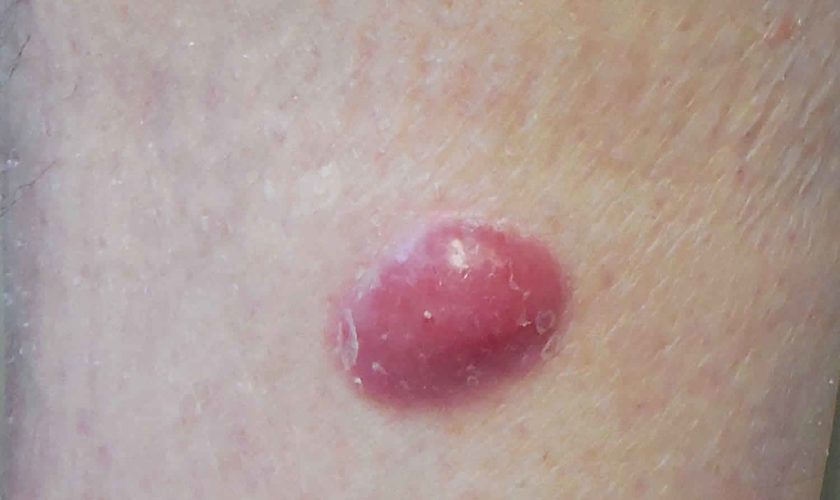
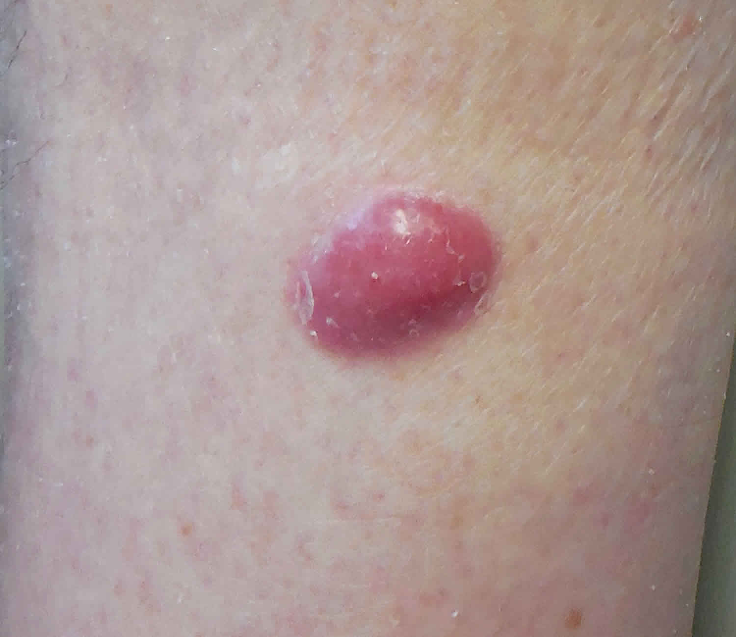
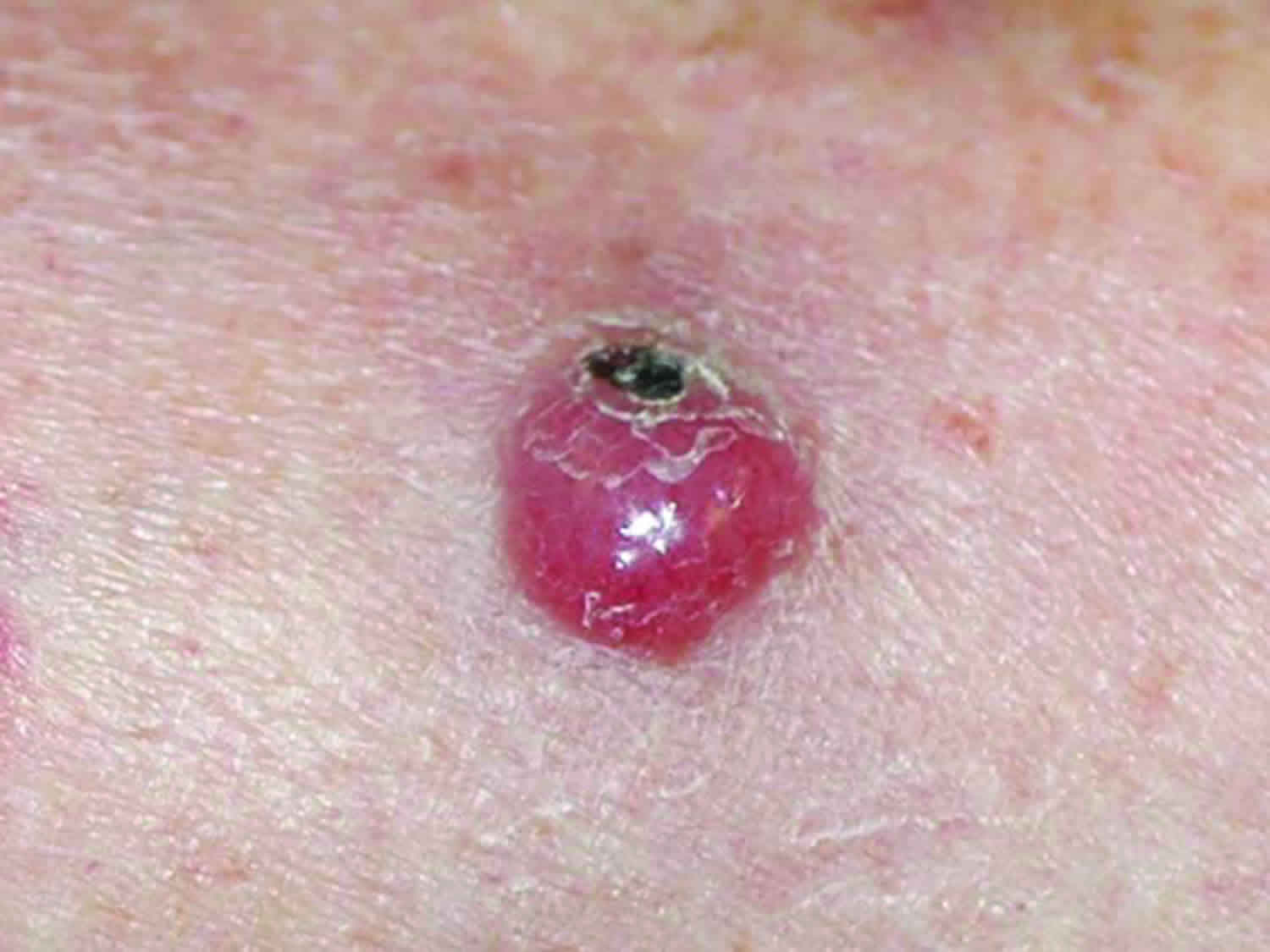
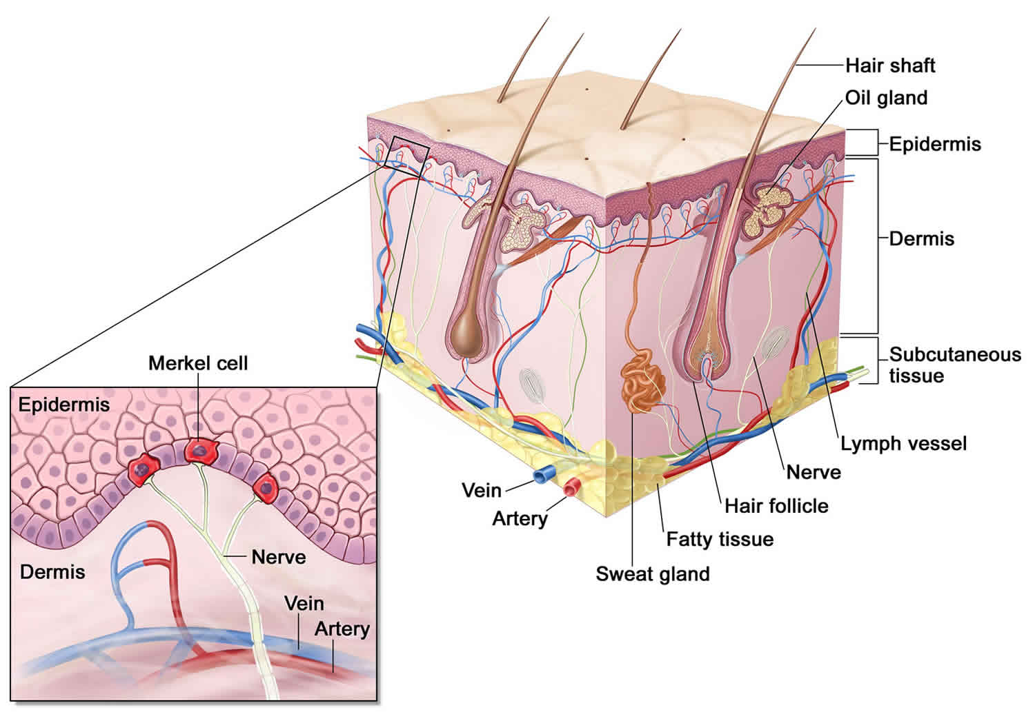
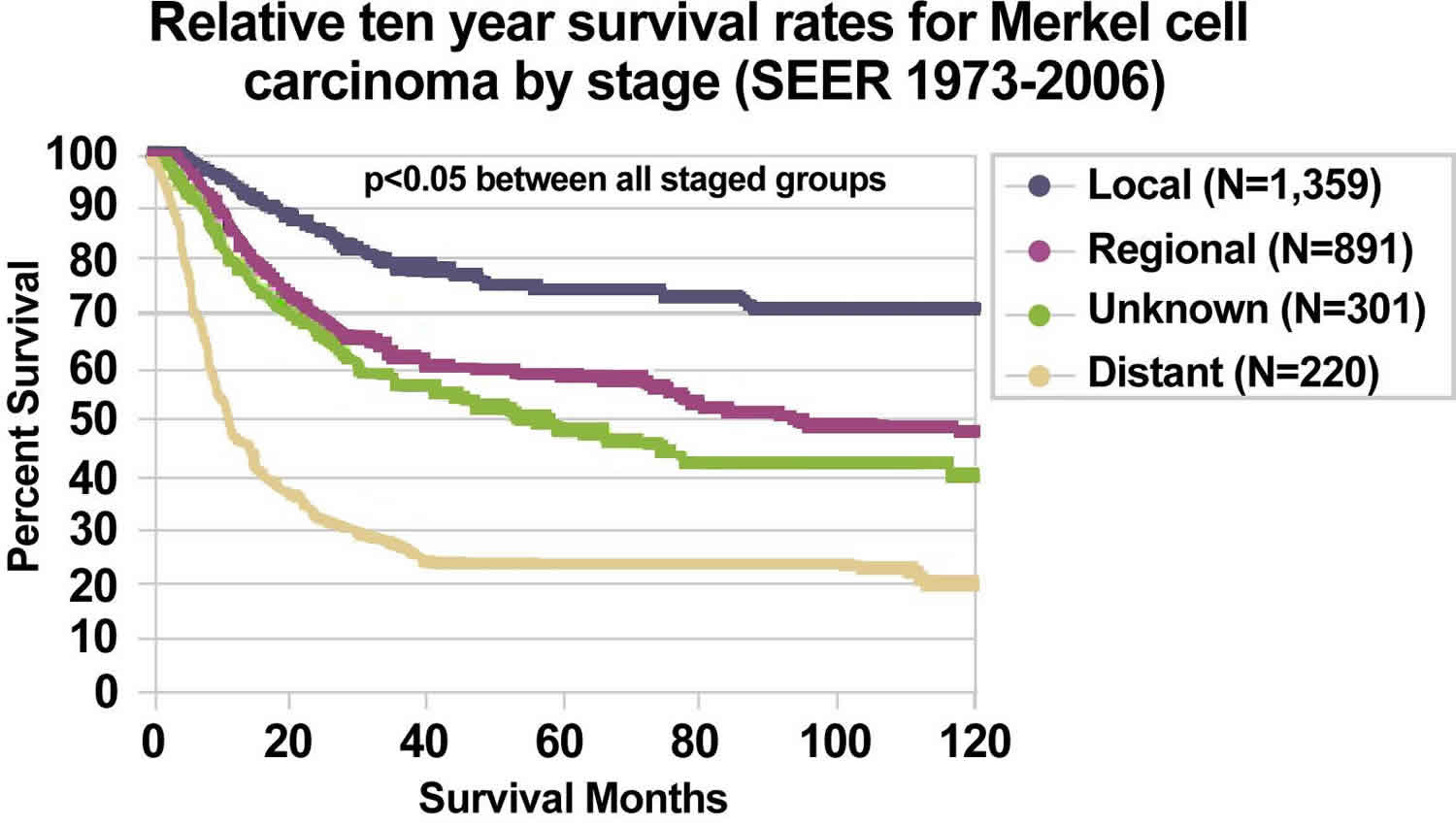
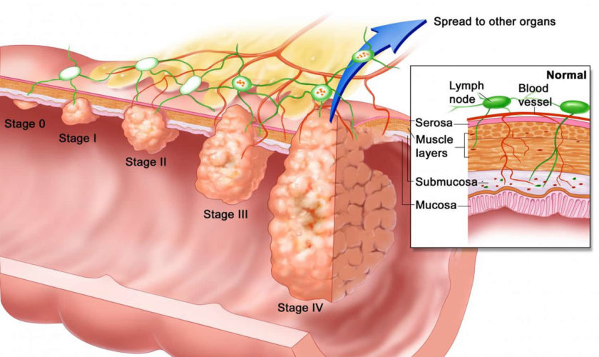
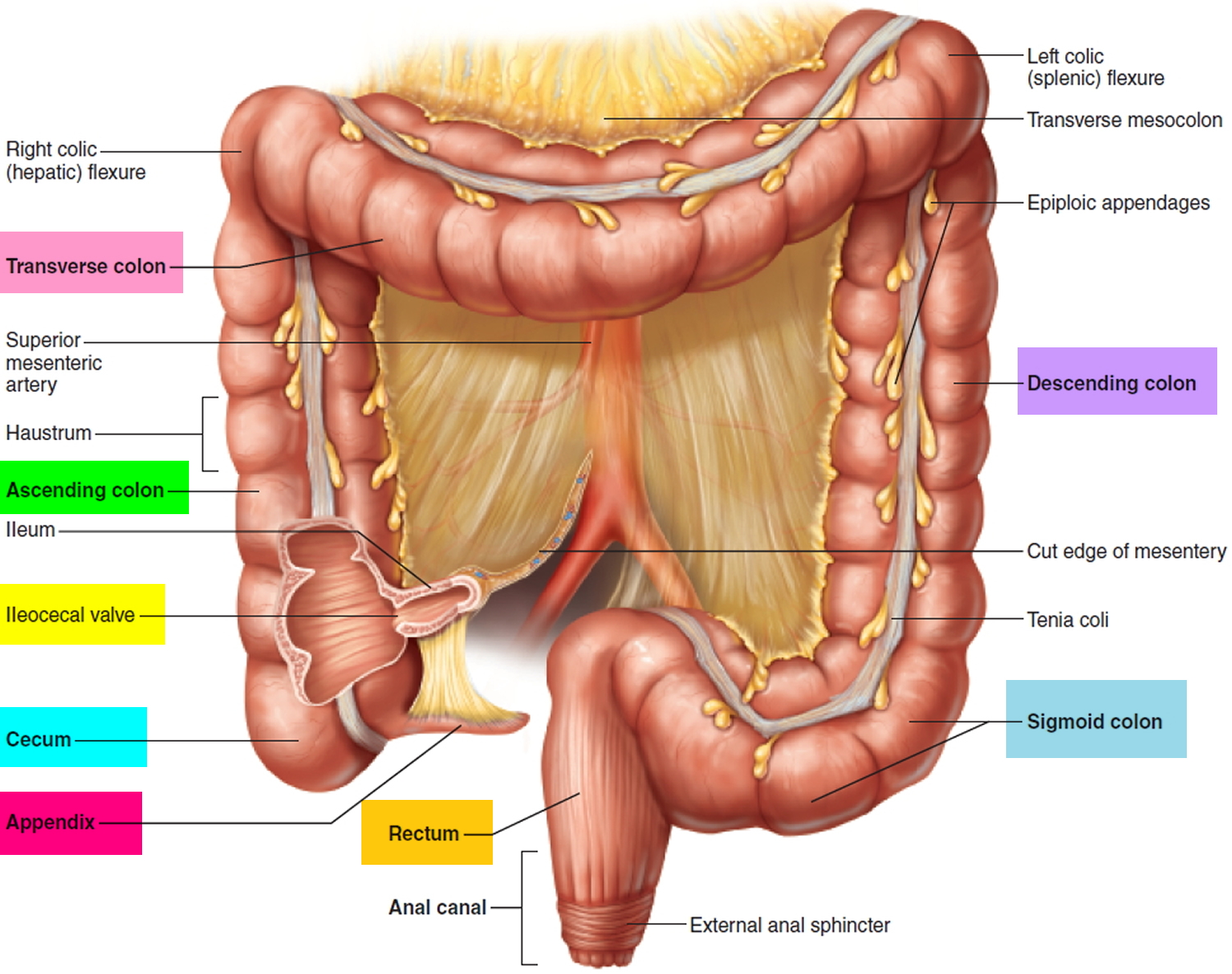
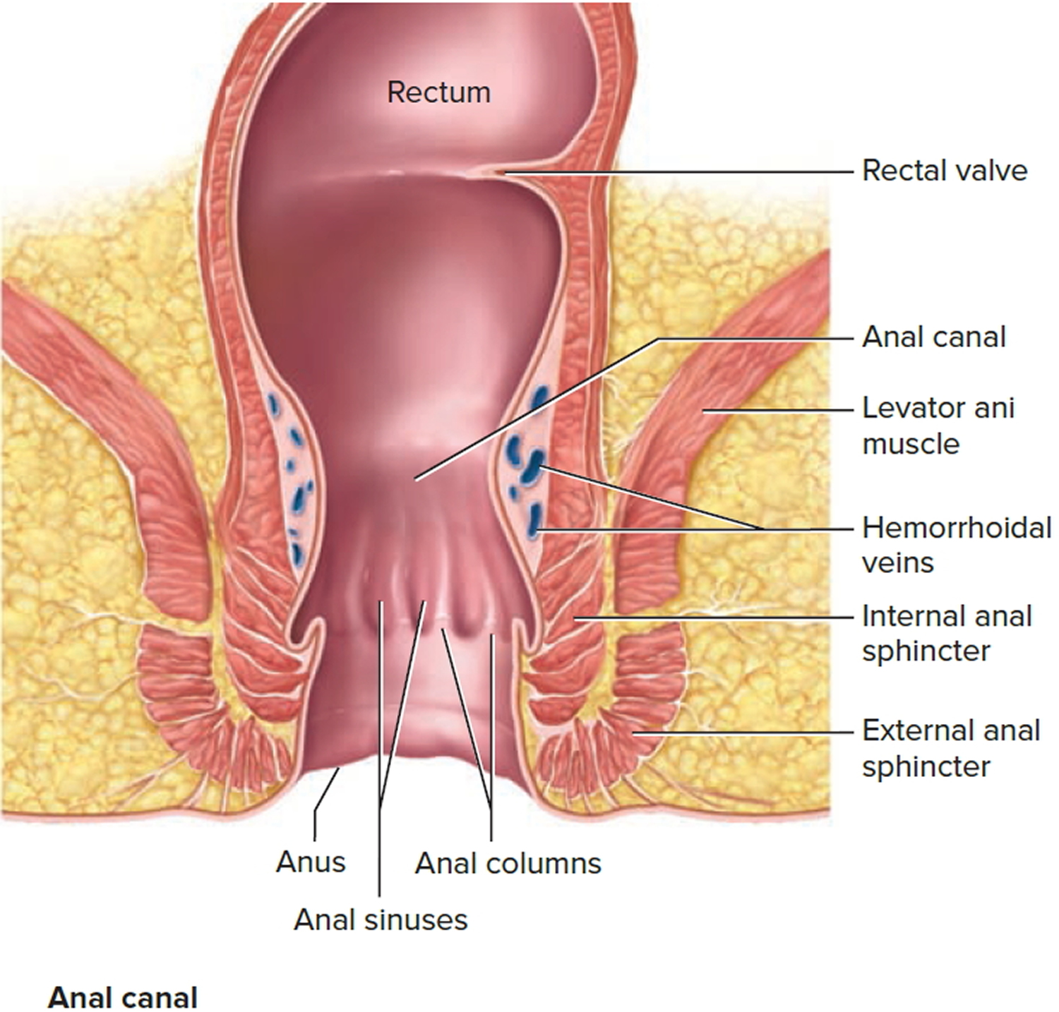
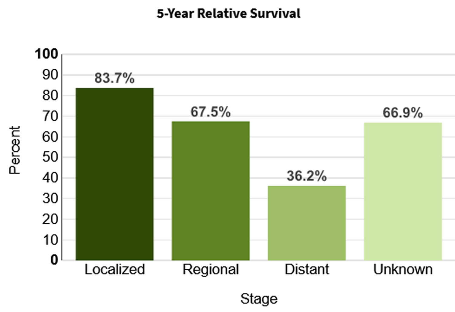
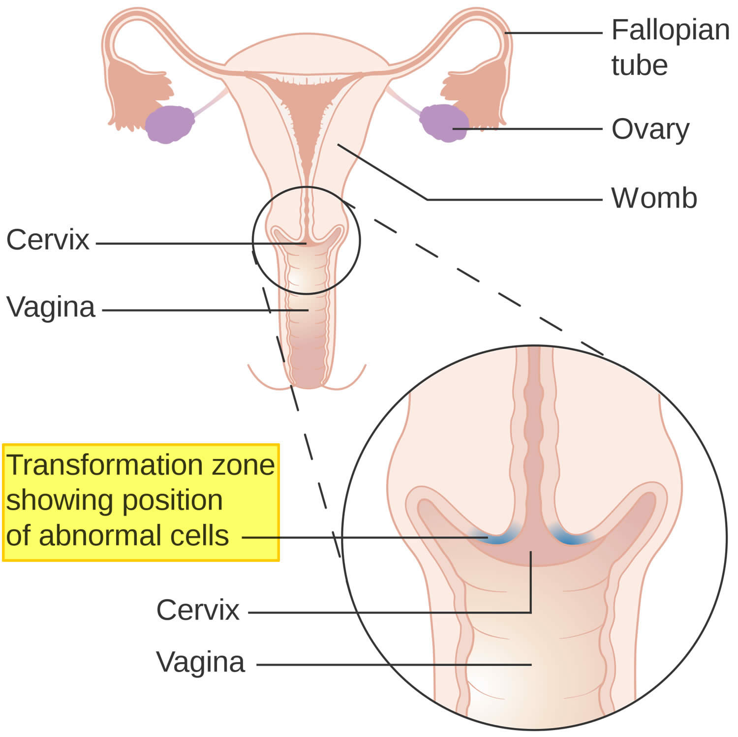
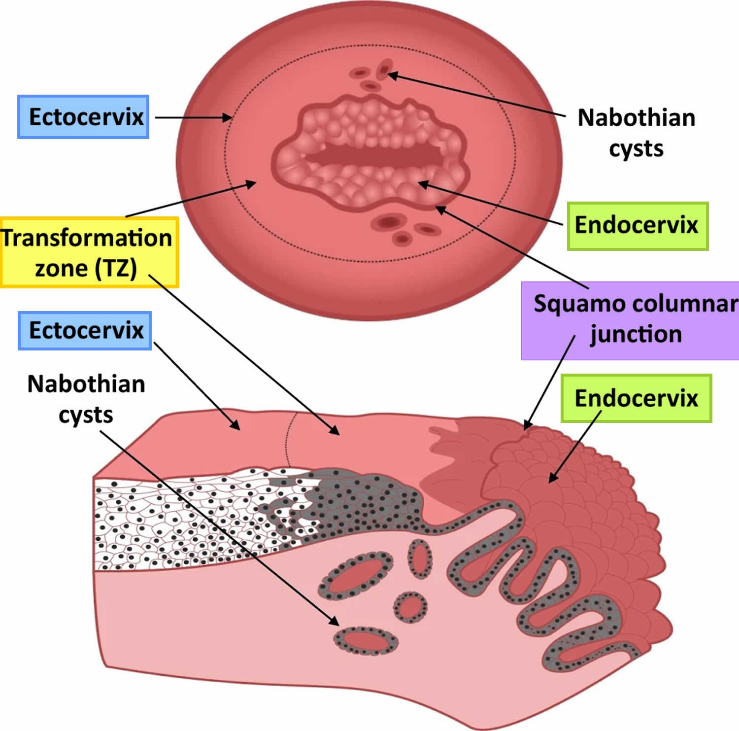
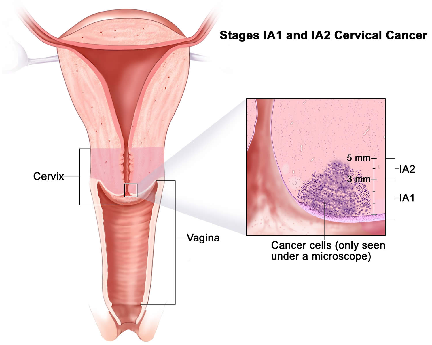
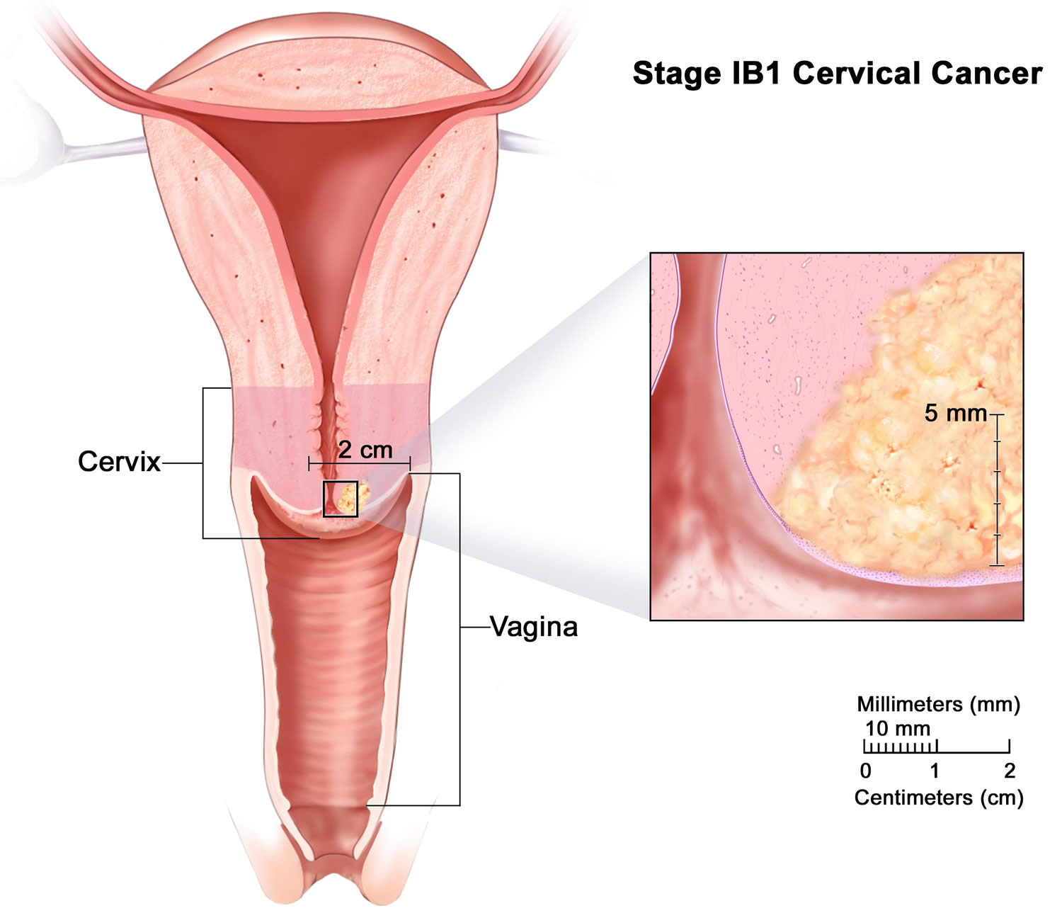
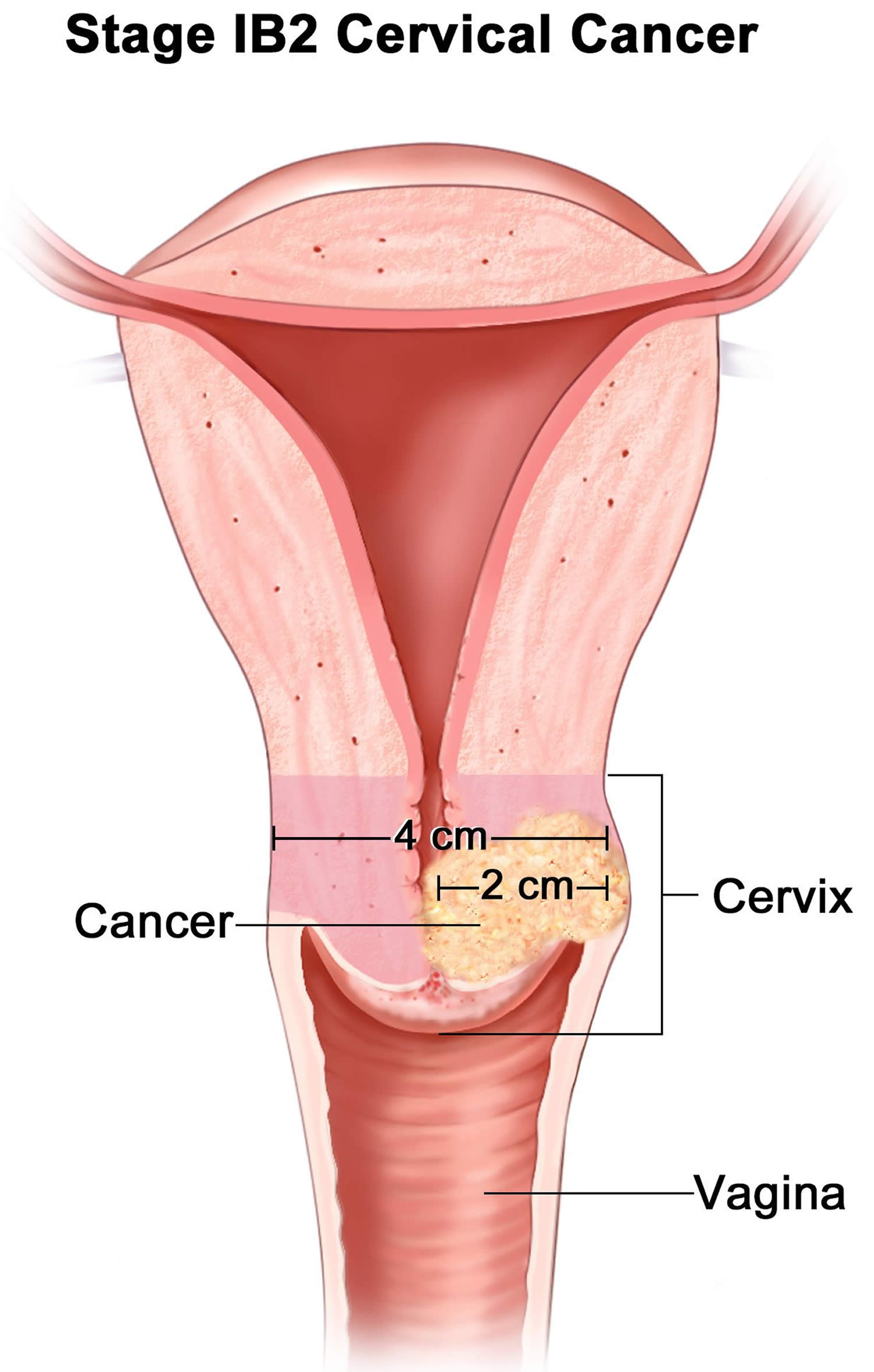
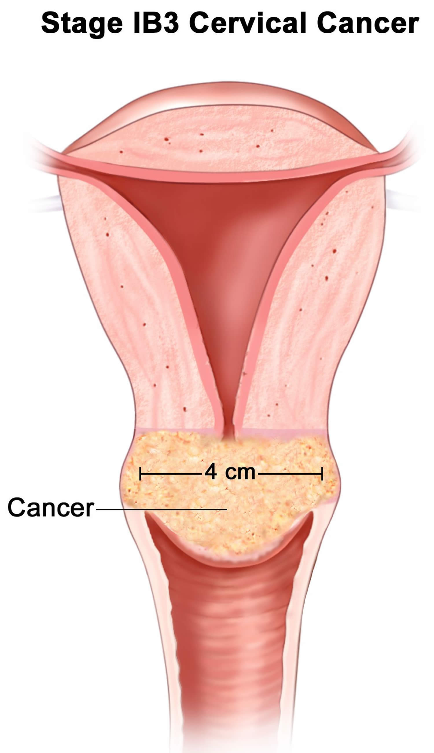
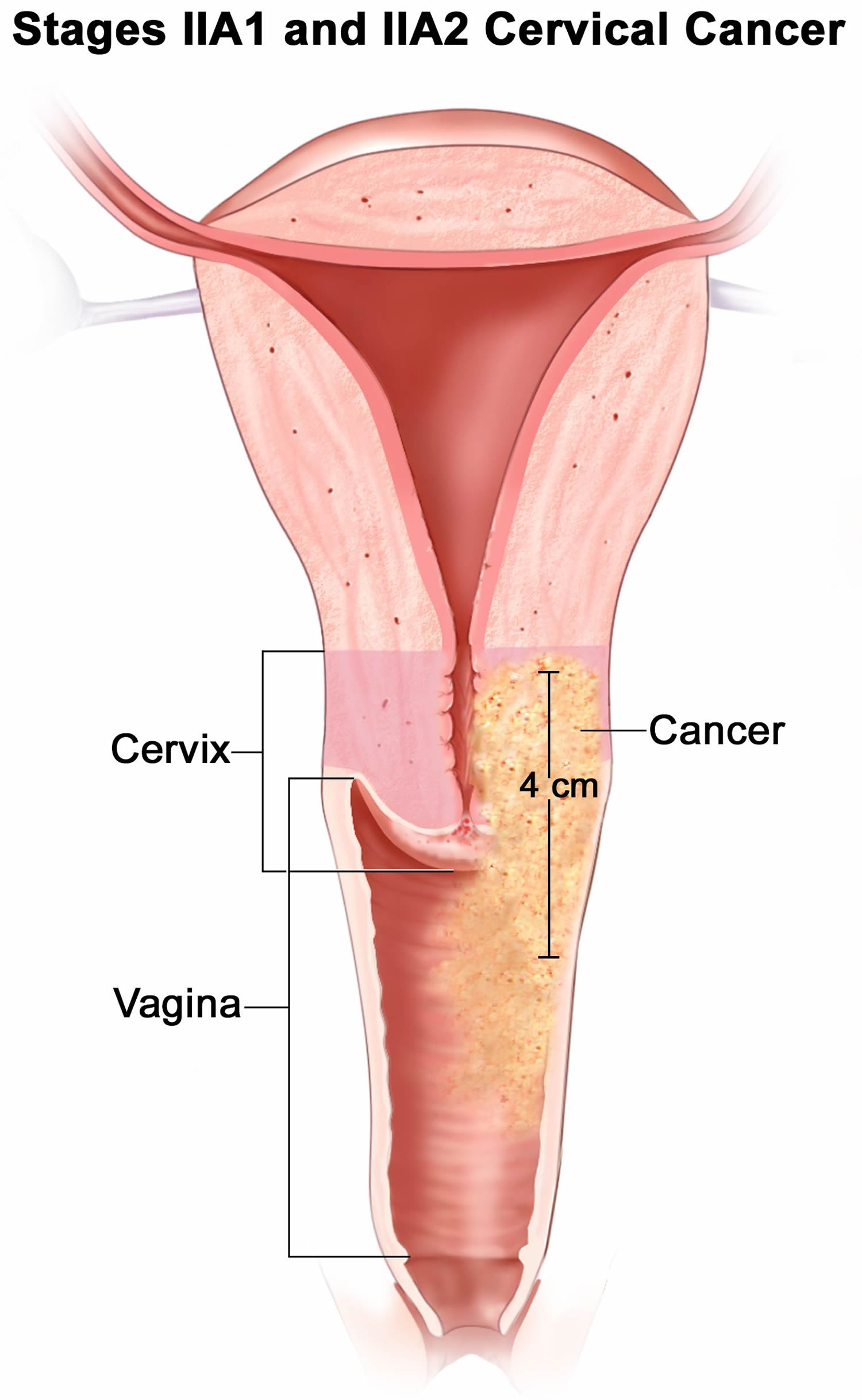
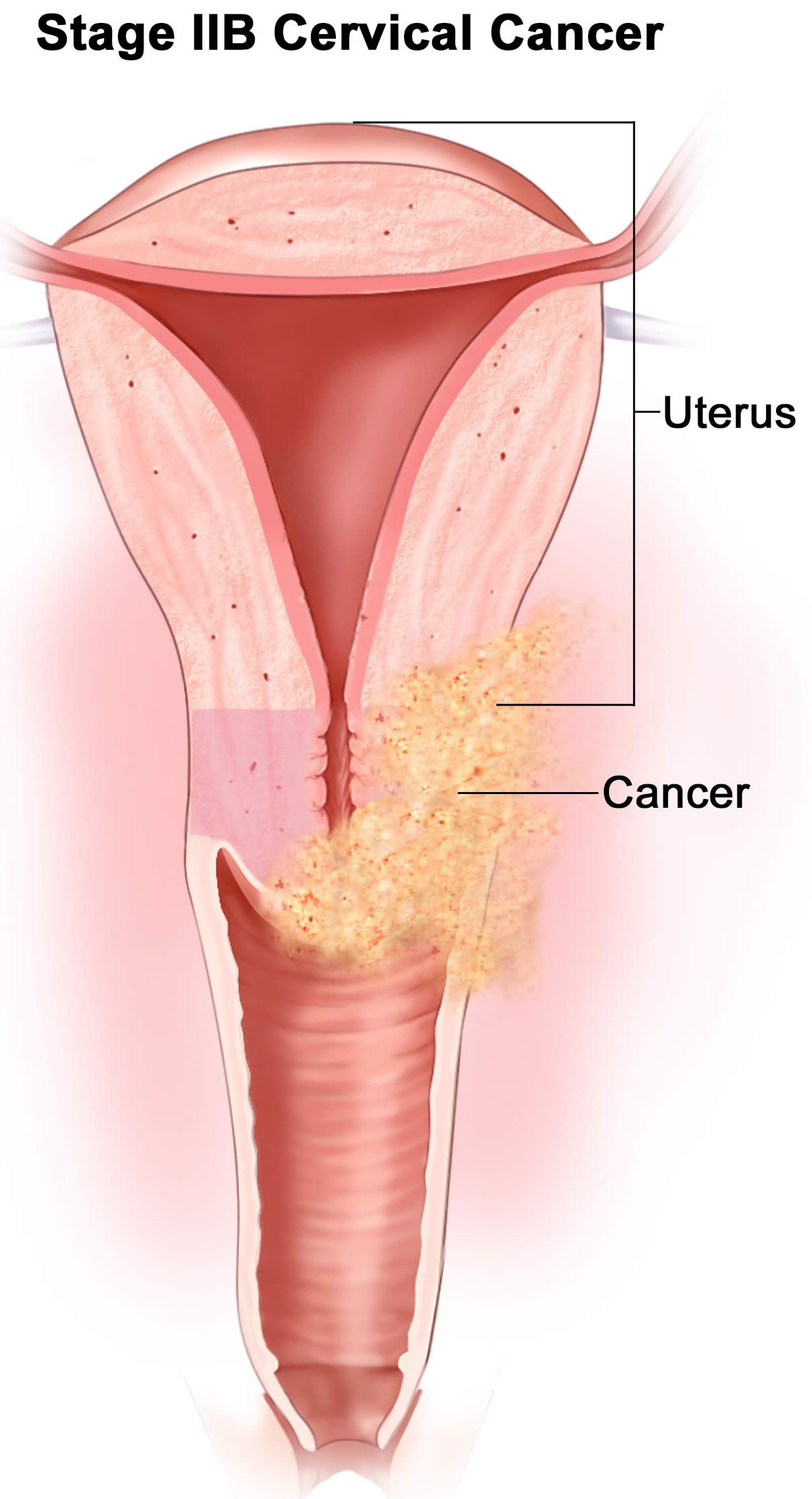
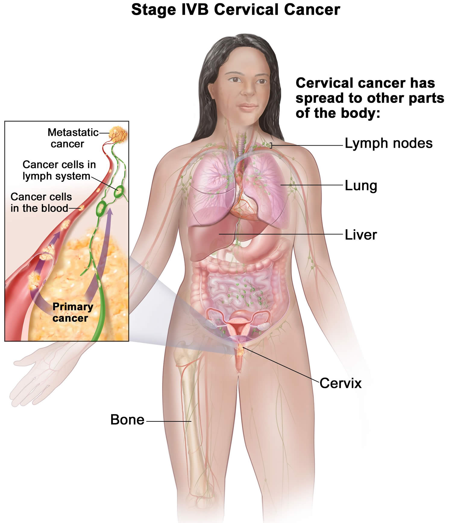
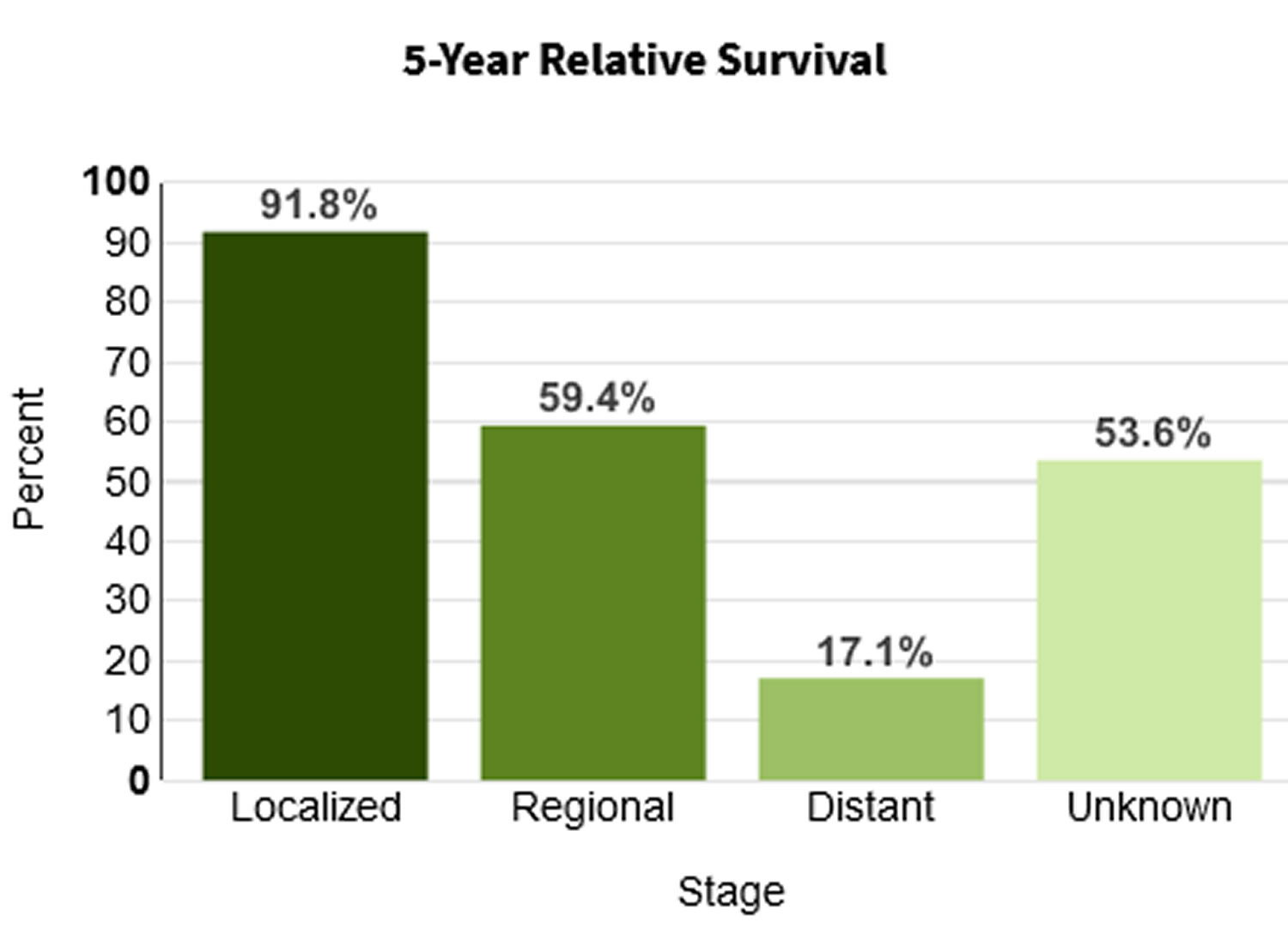
read more
Cancer

Merkel cell carcinoma

Merkel cell carcinoma

Merkel cell carcinoma

Merkel cell carcinoma also called Merkel cell cancer, neuroendocrine carcinoma of the skin or trabecular cancer of the skin, is a rare, highly aggressive neuroendocrine skin cancer with a high risk for returning (recurring) and spreading (metastasizing), often within two to three years after initial diagnosis 1, 2, 3, 4. Merkel cells are thought to be a type of skin neuroendocrine cell because they share some features with nerve cells and hormone-making cells. Merkel cells are found mainly at the base of the top layer of the skin (the epidermis) (see Figure 2). Merkel cells are very close to nerve endings in the skin. They help you sense light touch, which lets you do things like feel the fine details on an object’s surface. While rare, Merkel cell carcinomas are often aggressive and can advance rapidly which is why early detection and removal are especially important. Merkel cell carcinoma is 40 times more rare than melanoma, with an estimated one case per 130,000 people in the U.S. 5. About 2,000 cases of Merkel cell carcinoma are diagnosed in the United States each year 6. Experts expect that this will increase to 3,250 cases diagnosed annually by 2025, with similar increases expected in Australia and many European countries 7. Merkel cell carcinoma incidence increases progressively with age. There are few cases in patients younger than 50 years, and the median age at diagnosis is about 65 years 8. More than 4 out of 5 Americans diagnosed with Merkel cell carcinoma are older than age 70 6. And men are nearly 2-times more likely to have it compared with women, with women showing improved clinical outcomes 6, 9. Merkel cell carcinoma incidence is considerably greater in whites than blacks (more than 9 out of 10 cases of Merkel cell carcinoma in the United States are diagnosed in whites) and slightly greater in males than females 10, 11, 6.

Specific risk factors for developing Merkel cell carcinoma include ultraviolet (UV) exposure, advancing age, immunosuppression, and the Merkel cell polyomavirus (MCPyV) infection 12, 13, 14. Merkel cell carcinoma occurs most frequently in sun-exposed areas of skin, particularly the head and neck, followed by the extremities, and then the trunk 15, 16. Incidence has been reported to be greater in geographic regions with higher levels of ultraviolet B sunlight 17.

Merkel cell carcinoma is rare and dangerous but treatable, especially when found at an early stage 18. Be watchful for any new or changing lesions on your skin and look out for these warning signs.

If you’ve been treated for a previous Merkel cell carcinoma, pay close attention to the site and the surrounding region. Contact your medical team immediately if you see any suspicious changes.

Because Merkel cell carcinoma is such an uncommon form of skin cancer, it is best to seek treatment at an academic center with physicians who have specialized expertise in caring for people with this particular disease. A multi-disciplinary team experienced in the care of Merkel cell carcinoma is recommended. Dermatologists, surgeons, medical oncologists and radiation oncologists need to confer to determine the best plan for a given case.

While treatment options for Merkel cell carcinoma depend on the stage of the disease and the overall health of the patient, treatment includes surgical removal of the primary tumor along with:

  • Radiation therapy
  • Immunotherapy
  • Chemotherapy

Merkel cell carcinoma key points:

  • Increasing numbers of Merkel cell carcinomas have been reported by some centers in recent years.
  • Merkel cell carcinoma mainly affects older people, with most cases occurring after the age of 50.
  • Merkel cell carcinoma is slightly more common in men.
  • Merkel cell carcinoma occurs on parts of the body commonly exposed to sunlight, most often the head and neck.
  • Merkel cell carcinoma is also more common and more serious in those that are immune suppressed, such as patients with solid organ transplants, human immunodeficiency virus (HIV) infection, hematological malignancy or on drugs such as azathioprine.

Figure 1. Merkel cell carcinoma

Merkel cell carcinoma

Merkel cell carcinoma

Figure 2. Merkel Cell anatomy

Merkel Cell anatomy

Footnote: Merkel cells are located deep in the top layer of skin. Merkel cells are connected to nerves, signaling touch sensation as “touch receptors.” Merkel cell carcinoma was named after Merkel cells due to the similar microscopic features; however, recent research suggests that it is unlikely that Merkel cell carcinoma originates directly from normal Merkel cells with recent investigation pointing to their origin being early B-cells (lymphocytes) based on cellular morphology, the expression of early B-cell markers and clonal immunoglobulin chain rearrangement.

How dangerous is Merkel cell carcinoma?

While Merkel cell carcinoma is about three to five times more likely to be deadly than melanoma, with early detection, Merkel cell carcinoma can be treated successfully. Five-year survival rates are overall 30–50% 18. Survival rates are better for patients initially diagnosed with Stage 1 Merkel cell carcinoma and in younger patients than when it has already metastasized or patients are over 80 years of age.

If you think you might have Merkel cell carcinoma, see your doctor. Treatment becomes increasingly difficult once the disease has spread, but new options are now available. Thanks to advances in the field of immunotherapy, Merkel cell carcinoma survival rates are improving.

What you can do:

  • Examine your skin head-to-toe every month.
  • See your dermatologist every year for a professional skin exam. If you are newly diagnosed with Merkel cell carcinoma, seek a consultation with a multidisciplinary expert. Find a specialist here.
  • If you’ve had an Merkel cell carcinoma, follow up regularly with your dermatologist once treatment is complete. Follow the exam schedule your doctor recommends — it’s the best way to quickly detect a recurrence. Consider getting the AMERK biomarker blood test that can help detect recurrences early.
  • Safeguard your skin and avoid unprotected UV exposure. It is the single most effective way to reduce your risk of developing Merkel cell carcinoma.
  • Practice sun-safe habits, such as seeking shade and wearing protective clothing, a wide-brimmed hat and UV-blocking sunglasses. Use a broad-spectrum sunscreen every day. Sunscreen is a good defense against premature aging and skin cancer. It’s never too late to start wearing it.

Merkel cell carcinoma causes

Increased incidence of Merkel cell carcinoma has also been seen in people treated heavily with methoxsalen (psoralen) and ultraviolet A (PUVA) for psoriasis (3 of 1,380 patients, 0.2%), and those with chronic immune suppression, especially from chronic lymphocytic leukemia, human immunodeficiency virus, and previous solid organ transplant 19.

The immune system is believed to play a critical role in Merkel cell carcinoma pathogenesis with both Merkel cell polyoma virus (MCPyV) and ultraviolet (UV) radiation identified as risk factors for oncogenesis 2. In 2008, a novel polyomavirus called Merkel cell polyomavirus (MCPyV) was first reported in Merkel cell carcinoma tumor specimens 20, a finding subsequently confirmed in other laboratories 21. High levels of viral DNA and clonal integration of the Merkel cell polyomavirus in Merkel cell carcinoma tumors have also been reported 22 along with expression of certain viral antigens in Merkel cell carcinoma cells and the presence of antiviral antibodies. Merkel cell polyomavirus has been detected in about 80% Merkel cell carcinomas tested. However, not all cases of Merkel cell carcinoma appear to be associated with Merkel cell polyomavirus infection 23.

Merkel cell polyomavirus has been detected at very low levels in normal skin distant from the Merkel cell carcinoma primary, in a significant percentage of patients with non-Merkel cell carcinoma cutaneous disorders, in normal-appearing skin in healthy individuals, and in nonmelanoma skin cancers in immune-suppressed individuals 24. Various methods have been used to identify and quantify the presence of Merkel cell polyomavirus in Merkel cell carcinoma tumor specimens, other non-Merkel cell carcinoma tumors, blood, urine, and other tissues 25.

The significance of the new Merkel cell polyomavirus findings remains uncertain. The prognostic significance of viral load, antibody titer levels, and the role of underlying immunosuppression in hosts (from disease and medications) are under investigation.

Prevalence of Merkel cell polyomavirus appears to differ between Merkel cell carcinoma patients in the United States and Europe versus Australia. There may be two independent pathways for the development of Merkel cell carcinoma: one driven by the presence of Merkel cell polyomavirus, and the other driven primarily by sun damage, especially as noted in patient series from Australia 26.

Immunosuppression is an important factor for the development of Merkel cell carcinomas. The immune system is believed to play an important role in both the development of Merkel cell carcinoma and subsequent therapeutic response 2. Increased risk of Merkel cell carcinoma development has been shown in immunosuppressed patient populations including, but not limited to, patients with HIV/AIDS 27, patients undergoing solid organ transplant 28, 29 and patients with chronic lymphocytic leukemia 30 and non-Hodgkin lymphoma 31, 32. Furthermore, Merkel cell carcinoma in immunosuppressed patients has been observed to sometimes regress with improvement in immune function 33, 34, emphasizing the role immune surveillance in Merkel cell carcinoma development 35. Immunosuppression has been shown in multiple studies to be a negative independent prognostic factor for patients with Merkel cell carcinoma 36, 37, 38, 39, 40. With regard to differential impact on clinical outcomes of immunosuppression in Merkel cell carcinoma, Cook et al. 41 performed a retrospective analysis of 89 patients with non-metastatic Merkel cell carcinoma and found increased mortality risk for immunosuppressed patients with HIV/AIDS and organ transplant relative to patients with autoimmune disease (reference group). Yusuf et al. 36 found immune status to be an independent predictor of overall survival for patients with Merkel cell carcinoma and found immunosuppression to be associated with overall survival, with the lowest 3-year overall survival rates for immunosuppressed patients with solid organ transplants.

Merkel cell carcinoma was previously believed to arise from Merkel cells, which are pressure receptors in the skin. A recent investigation is pointing to their origin being early B-cells (lymphocytes) based on cellular morphology, the expression of early B-cell markers and clonal immunoglobulin chain rearrangement.

Although no unique marker for Merkel cell carcinoma has been identified, a variety of molecular and cytogenetic markers of Merkel cell carcinoma have been reported 42.

Risk factors for developing Merkel cell carcinoma

Anything that increases your likelihood of developing a disease like Merkel cell carcinoma (Merkel cell skin cancer) is called a risk factor.

These factors put you at increased risk for the disease.

  • History of unprotected exposure to ultraviolet (UV) radiation from the sun or indoor tanning. UV exposure creates a double threat for Merkel cell carcinoma. Not only does UV exposure cause damage that increases your skin cancer risk; it also suppresses your immune system, reducing its ability to repair damage and fight skin cancers and other diseases. No matter the source of UV radiation, there is a clear and dangerous correlation between exposure and Merkel cell carcinoma.
  • Weakened immune system, due to a medical condition or medications. If your immune system is persistently weakened or suppressed, you are about 15 times more likely to develop Merkel cell carcinoma than people with healthy immune systems. However, more than 90 percent of Merkel cell carcinoma cases arise in people with no known immune problems. Immunosuppression can be caused by:
    • Diseases such as HIV and certain cancers including chronic lymphocytic leukemia and lymphomas.
    • Immunosuppressant medications used for transplant recipients and for autoimmune diseases including lupus, psoriasis, Crohn’s disease and rheumatoid arthritis.
    • Immunosuppressed Merkel cell carcinoma patients often develop the disease at a far younger age; about half of Merkel cell carcinomas in these patients occur before age 50.
    • If your immune system is suppressed, talk to your doctors about your chances of developing Merkel cell carcinoma, and be sure to protect yourself against other risk factors, especially UV exposure.
  • History of skin cancer, If you’ve had melanoma, squamous cell carcinoma (SCC), basal cell carcinoma (BCC) or Bowen’s disease (an early form of SCC) — skin cancers frequently caused by UV exposure — you are more likely to develop Merkel cell carcinoma.
  • Age: Most people who develop Merkel cell carcinoma are over 50 years old.
  • Gender: Men are more likely to get the disease. This might be because they tend to get more sun exposure.
  • Fair skin: People with fair skin are at greater risk for Merkel cell carcinoma, but it can affect anyone.
  • Merkel cell polyomavirus. Evidence of the Merkel cell polyomavirus can be found in most, but not all, Merkel cell tumors. The virus, discovered in 2008, lives in the skin of most people, without signs and symptoms, and without ever developing into Merkel cell carcinoma. Since it’s a very common virus, and since Merkel cell carcinoma is an extremely rare disease, scientists are still not certain about exactly how or why the virus causes Merkel cell carcinoma in some people. What is clear is that other factors such as UV exposure and immunosuppression also play a role in initiating the growth of Merkel cell carcinoma tumors.

Merkel cell carcinoma prevention

While your risk of getting Merkel cell carcinoma (Merkel cell cancer) is low, and some risk factors for Merkel cell carcinoma, such as your age, sex, and skin color can’t be controlled, there are things you can do that might help lower your risk. These might also lower your risk of getting more common types of skin cancer, as well as some other types of cancer.

  • Limit your exposure to ultraviolet (UV) rays. The most important way to lower your risk of skin cancers (including Merkel cell carcinoma) is to limit your exposure to ultraviolet (UV) rays. The main types of UV rays that can affect your skin include UVA rays and UVB rays. UVB rays have more energy and are a more potent cause of at least some skin cancers, but both UVA and UVB rays can damage skin and cause skin cancer. There are no safe UV rays. Practice sun safety when you are outdoors. The US National Weather Service and the Environmental Protection Agency (EPA) have developed the UV Index, which gives you an idea of how strong the UV light is in your area on any given day, on a scale from 1 to 11+. A higher number means greater risk of exposure to UV rays and a higher chance of sunburn and skin damage that could ultimately lead to skin cancer. Further information about the UV Index, as well as your local UV Index forecast, can be found on the EPA’s website at https://www.epa.gov/sunsafety/uv-index-1
    • Simply staying in the shade is one of the best ways to limit your UV exposure. If you are going to be in the sun, “Slip! Slop! Slap! and Wrap” is a catchphrase that can help you remember some of the key steps you can take to protect yourself from UV rays:
      • Slip on a shirt. Many companies now make clothing that’s lightweight, comfortable, and protects against UV rays even when wet. It tends to be more tightly woven, and some have special coatings to help absorb UV rays. These sun-protective clothes may have a label listing the UV protection factor (UPF) value (the level of protection the garment provides from the sun’s UV rays, on a scale from 15 to 50+). The higher the UPF, the higher the protection from UV rays.
      • Slop on sunscreen (use a “broad spectrum SPF50+” sunscreen that filters out about 98% of the UV rays). Broad spectrum means it protect against both UVA and UVB rays. Sunscreens labeled with SPFs as high as 100+ are available. Only “broad spectrum” sunscreen products with an SPF of 15 or higher can state that they help protect against skin cancer and early skin aging if used as directed with other sun protection measures. However, no sunscreen protects you completely.
      • Slap on a hat. A hat with at least a 2- to 3-inch brim all around is ideal because it protects areas that are often exposed to intense sun, such as the ears, eyes, forehead, nose, and scalp. A dark, non-reflective underside to the brim can also help lower the amount of UV rays reaching the face from reflective surfaces such as water. A shade cap (which looks like a baseball cap with about 7 inches of fabric draping down the sides and back) also is good, and will provide more protection for the neck. If you don’t have a shade cap (or another good hat) available, you can make one by wearing a large handkerchief or bandana under a baseball cap. A baseball cap protects the front and top of the head but not the neck or the ears, where skin cancers commonly develop. Straw hats are not as protective as hats made of tightly woven fabric.
      • Wrap on sunglasses to protect the eyes and skin around them. The ideal sunglasses should block 99% to 100% of UVA and UVB rays. Before you buy, check the label to make sure they do. Labels that say “UV absorption up to 400 nm” or “Meets ANSI UV Requirements” mean the glasses block at least 99% of UV rays. Those labeled “cosmetic” block about 70% of UV rays. If there is no label, don’t assume the sunglasses provide any UV protection.
  • Don’t use tanning beds or sunlamps. Many people believe the UV rays of tanning beds are harmless. This is not true. Tanning lamps give off UV rays, which can cause long-term skin damage and can contribute to skin cancer. Most skin doctors and health organizations recommend not using tanning beds and sun lamps.
  • Protect children from the sun. Children need special care, since they tend to spend more time outdoors and can burn more easily. Parents and other caregivers should protect children from excess sun exposure by using the steps above. Children need to be taught about the dangers of too much sun exposure as they become more independent.
  • Keep your immune system strong. Having a weakened immune system greatly increases the risk of getting Merkel cell carcinoma, as well as other types of skin cancer. In some cases, such as organ transplant, you can’t control the things that must be done that weaken (suppress) your immune system. But something you can control is being infected with HIV. Infection with HIV, the virus that causes AIDS, weakens the immune system. Avoiding known risk factors for HIV infection, such as intravenous (IV) drug use and having unprotected sex with many partners, can also lower your risk of immune system problems. This, in turn, might help keep you from getting Merkel cell carcinoma and many other types of cancer.

Merkel cell carcinoma symptoms

Merkel cell carcinoma (MCC) tumors often, but not always, appear on sun-exposed areas of your body, especially your face, neck, arms, and legs, but it can occur anywhere on your body. The tumors are not nearly as distinctive as other more common skin cancers such as basal cell carcinoma and can appear as a pearly pimple-like lump, sometimes skin-colored, pink, red, purple or bluish-red, though they are rarely tender to the touch. Sometimes the skin on the top of the tumor breaks open and bleeds. The rapid speed at which they grow is what often causes patients and doctors to take notice. Because of its nonspecific clinical appearance, Merkel cell carcinoma is rarely suspected before a biopsy is performed 43.

Merkel cell cancers spread through the lymphatic system and multiple metastases can develop around the main tumor (local recurrence). Merkel cell carcinoma may also spread to lymph nodes in the neck, armpits and groin. This is more likely in thicker tumors. Most recurrences occur within the first two years after diagnosis.

Merkel cell carcinoma is rare, and it can look like many other, more common types of skin cancer or other skin problems when it first appears. It’s very important to have any new, growing, or changing lumps, bumps, or spots on your skin checked by a doctor as soon as possible so that the cause can be found and treated, if needed. The earlier any type of skin cancer is found, the easier it might be to treat.

Can Merkel cell carcinoma be found early?

While the American Cancer Society doesn’t have guidelines for the early detection of skin cancer, knowing your own skin is important in finding skin cancer early. Learn the patterns of moles, blemishes, freckles, and other marks on your skin so that you’ll notice any changes. Many doctors recommend checking your own skin once a month. Self-exams are best done in a well-lit room in front of a full-length mirror. Use a hand-held mirror for areas that are hard to see, such as the backs of your thighs.

Examine all of your skin, including your palms and soles, scalp, ears, nails, and your back. A friend or family member can also help you with these exams, especially for those hard-to-see places, like your scalp and back.

Be sure to show your doctor any skin changes that concern you and have them look at areas that may be hard for you to see. Any spots on your skin that are new or changing in size, shape, or color should be seen by a doctor right away. If you can’t see your doctor soon, you might want to take good close-up photos of the area so your doctor can see if it’s changing when you do get an appointment.

Any unusual sore, lump, blemish, marking, or change in the way an area of the skin looks or feels may be a sign of skin cancer or a warning that it might occur. The area might become red, swollen, scaly, crusty, or start oozing or bleeding. It may feel itchy, tender, or painful.

Merkel cell tumors usually look like firm, pink, red, or purple lumps or bumps on sun-exposed areas of the skin. They usually don’t hurt, but they can grow quickly and can sometimes open up as ulcers or sores.

Merkel cell carcinoma diagnosis

Merkel cell carcinoma should be considered in any tumor with “AEIOU” clinical features, which are present in about 90% of patients with Merkel cell skin cancer.

  • Asymptomatic or non-tender
  • Expanding rapidly
  • Immune suppressed
  • Older than 50
  • UV-exposed fair skin

Not all patients have every element in the “AEIOU” mnemonic; however, in this study 44, 89% of patients met three or more criteria, 52% met four or more criteria, and 7% met all five criteria.

The main test is a biopsy of the tumor. This shows characteristic Merkel cell carcinoma pathology. Immunohistochemistry can be helpful as cytokeratin-20 (CK20) is positive in up to 95% of tumors and thyroid transcription factor (TTF1) is usually negative.

A general examination, including evaluation of local lymph nodes, and staging investigations may be arranged to determine whether the tumor has spread to other sites.

Staging investigations may include:

  • Sentinel node biopsy. A sentinel lymph node biopsy can be used to find the lymph nodes that are likely to be the first place the Merkel cell carcinoma would go if it has spread. These lymph nodes are called sentinel nodes.
  • Lymph node ultrasound scan
  • Imaging using X-rays, computed tomography (CT), magnetic resonance imaging (MRI) and positron emission tomography (PET) scans. They can be used to see if Merkel cell carcinoma has spread to lymph nodes or to other organs in the body. Imaging tests can also be done to help see how well treatment is working or to look for possible signs of cancer coming back (recurring) after treatment.

If an imaging work-up is performed, it may include a computed tomography (CT) scan of the chest and abdomen to rule out primary small cell lung cancer as well as distant and regional metastases. Imaging studies designed to evaluate suspicious signs and symptoms may also be recommended. In one series, CT scans had an 80% false-negative rate for regional metastases 45. Head and neck presentations may require additional imaging. Magnetic resonance imaging has been used to evaluate Merkel cell carcinoma but has not been studied systematically 46. Fluorine F 18-fludeoxyglucose positron emission tomography results have been reported only in selected cases 47. Routine blood work as a baseline has been recommended but has not been studied systematically. There are no known circulating tumor markers specifically for Merkel cell carcinoma.

A specific Merkel cell carcinoma staging system is published by the American Joint Committee (AJCC) on Cancer.

Longterm survival is likely if the lymph nodes do not contain tumor cells.

The results of initial clinical staging of Merkel cell carcinoma vary widely in the literature, based on retrospective case series reported over decades. For invasive cancers, 48.6% were localized, 31.1% were regional, and 8.2% were distant 48.

Merkel cell carcinoma that presents in regional nodes without an identifiable primary lesion is found in a minority of patients, with the percent of these cases varying among the reported series. Tumors without an identifiable primary lesion have been attributed to either spontaneous regression of the primary or metastatic neuroendocrine carcinoma from a clinically occult site 49.

Challenges in diagnosis

Diagnosis and management of Merkel cell carcinoma presents distinct challenges, including:

  • Misdiagnosis: 56 percent of Merkel cell carcinomas are believed to be benign when initially examined by physicians, who may mistake the tumors for cysts or infected hair follicles.
  • Delayed medical attention: Medical care is often not sought early as these lesions do not appear concerning. About 5 percent of Merkel cell carcinomas occur in mucosal sites such as the mouth, nasal cavity and throat, where these painless lesions are hard to spot before the disease has advanced.
  • Undetectable: In one study, 14 percent of Merkel cell carcinomas were initially discovered in the lymph nodes already, without any tumor being identified.

Since Merkel cell carcinoma is a rare and aggressive cancer, delayed detection, diagnosis and treatment are especially concerning, because as the disease progresses, treatment becomes more difficult and the risk of recurrence increases.

Initial Clinical Evaluation

Because local-regional spread is common, newly diagnosed Merkel cell carcinoma patients require a careful clinical examination that includes looking for satellite lesions and regional nodal involvement and tests are done to find out if cancer cells have spread to other parts of the body.

Tailoring an imaging work-up to the clinical presentation and any relevant signs and symptoms should be considered. There has been no systematic study of the optimal imaging work-up for newly diagnosed patients, and it is not clear if all newly diagnosed patients, especially those with the smallest primaries, benefit from a detailed imaging work-up.

The process used to find out if cancer has spread to other parts of the body is called staging. The information gathered from the staging process determines the stage of the disease. It is important to know the stage in order to plan treatment.

The following tests and procedures may be used in the staging process:

  • CT scan (CAT scan): A procedure that makes a series of detailed pictures of areas inside the body, taken from different angles. The pictures are made by a computer linked to an x-ray machine. A dye may be injected into a vein or swallowed to help the organs or tissues show up more clearly. A CT scan of the chest and abdomen may be used to check for primary small cell lung cancer, or to find Merkel cell carcinoma that has spread. A CT scan of the head and neck may also be used to find Merkel cell carcinoma that has spread to the lymph nodes. This procedure is also called computed tomography, computerized tomography, or computerized axial tomography.
  • PET scan (positron emission tomography scan): A procedure to find malignant tumor cells in the body. A small amount of radioactive glucose (sugar) is injected into a vein. The PET scanner rotates around the body and makes a picture of where glucose is being used in the body. Malignant tumor cells show up brighter in the picture because they are more active and take up more glucose than normal cells do.
  • Lymph node biopsy: There are several types of lymph node biopsy used to stage Merkel cell carcinoma.
    • Sentinel lymph node biopsy: The removal of the sentinel lymph node during surgery. The sentinel lymph node is the first lymph node in a group of lymph nodes to receive lymphatic drainage from the primary tumor. It is the first lymph node the cancer is likely to spread to from the primary tumor. A radioactive substance and/or blue dye is injected near the tumor. The substance or dye flows through the lymph ducts to the lymph nodes. The first lymph node to receive the substance or dye is removed. A pathologist views the tissue under a microscope to look for cancer cells. If cancer cells are not found, it may not be necessary to remove more lymph nodes. Sometimes, a sentinel lymph node is found in more than one group of nodes.
    • Lymph node dissection: A surgical procedure in which the lymph nodes are removed and a sample of tissue is checked under a microscope for signs of cancer. For a regional lymph node dissection, some of the lymph nodes in the tumor area are removed. For a radical lymph node dissection, most or all of the lymph nodes in the tumor area are removed. This procedure is also called lymphadenectomy.
    • Core needle biopsy: A procedure to remove a sample of tissue using a wide needle. A pathologist views the tissue under a microscope to look for cancer cells.
    • Fine-needle aspiration biopsy: A procedure to remove a sample of tissue using a thin needle. A pathologist views the tissue under a microscope to look for cancer cells.
  • Immunohistochemistry: A laboratory test that uses antibodies to check for certain antigens (markers) in a sample of a patient’s tissue. The antibodies are usually linked to an enzyme or a fluorescent dye. After the antibodies bind to a specific antigen in the tissue sample, the enzyme or dye is activated, and the antigen can then be seen under a microscope. This type of test is used to help diagnose cancer and to help tell one type of cancer from another type of cancer.

Merkel cell carcinoma staging

After someone is diagnosed with Merkel cell cancer, your medical team will try to figure out if it has spread, and if so, how far. This process is called staging. Staging is a way to understand how much the cancer has grown and how far it has spread. It helps determine how serious the cancer is, how best to treat it and the risk of it coming back. Doctors also use a cancer’s stage when talking about survival statistics.

American Joint Committee on Cancer (AJCC) Stage Groupings and the TNM system is frequently used to stage Merkel cell carcinoma. It’s a classification based on three factors:

  • T represents the size of the original tumor, its growth rate and other factors.
  • N indicates whether the cancer has spread to the local lymph nodes and to what extent.
  • M stands for the spread or metastasis to distant lymph nodes and organs.

Numbers or letters after T, N, and M provide more details about each of these factors. Higher numbers mean the cancer is more advanced. Once the patient’s TNM categories have been established, the overall stage number is assigned. As a rule, the lower the stage number, the less the disease has progressed. For example, the earliest stage Merkel cell cancers are called stage 0 (or carcinoma in situ), and then range from stages I (1) through IV (4). Cancer staging can be complex, so ask your doctor to explain it to you in a way you understand.

The staging system in the Table 1 below uses the pathologic stage also called the surgical stage. This is the staging system most often used for Merkel cell cancer. It’s decided after testing the tissue taken out during an operation.

Sometimes, if surgery can’t be done right away or at all, the cancer will be given a clinical stage instead. This is based on the results of a physical exam, biopsy, and imaging tests. The clinical stage will be used to help plan treatment. In some cases, though, the cancer has spread further than the clinical stage estimates, and may not predict the patient’s outlook as well as a pathologic stage. Clinical staging for Merkel cell carcinoma follows a separate staging system that’s not covered in the table below. If your cancer has been clinically staged, ask your doctor for information about your specific stage.

The following stages are used for Merkel cell carcinoma:

Table 1. Merkel cell carcinoma stages

AJCC StageStage groupingStage description*
0Tis
N0
M0
The cancer is only in the epidermis, the outermost skin layer (Tis).
It has not spread to nearby lymph nodes (N0) or distant sites (M0).
This stage is also known as carcinoma in situ (Tis).
IT1
N0
M0
The cancer is no more than 2 centimeters (cm) across (about 4/5 inch).
It has not spread to nearby lymph nodes (N0) or to distant sites (M0).
IIAT2 or T3
N0
M0
The cancer is more than 2, but less than 5 cm (about 2 inches) across (T2) OR the tumor is more than 5 cm across (T3).
It has not spread to nearby lymph nodes (N0) or to distant sites (M0).
IIB
T4
N0
M0
The cancer has grown into nearby tissues such as muscles, bones, or cartilage (T4).
It has not spread to nearby lymph nodes (N0) or to distant sites (M0).
IIIA
T1, T2, T3, or T4
N1a(sn) or N1a
M0
The cancer can be any size or may have grown into nearby tissues (T1, T2, T3, or T4) AND the cancer has spread to nearby lymph nodes, but this was found during a lymph node biopsy or surgery and was not seen on exams or imaging tests (N1a[sn] or N1a).
It has not spread to distant sites (M0).
OR
T0
N1b
M0
There’s no sign of a primary cancer (T0) AND the cancer has spread to nearby lymph nodes, which was seen on exams or imaging tests and then confirmed by biopsy or surgery (N1b).
It has not spread to distant sites (M0).
IIIBT1, T2, T3, or T4
N1b, N2 or N3
M0
The cancer can be any size or may have grown into nearby tissues (T1, T2, T3, or T4) AND any of the following:
It has spread to nearby lymph nodes, which was seen on exams or imaging tests and then confirmed by biopsy or surgery (N1b).
It has spread toward a nearby lymph node area without reaching the lymph nodes (N2). This is called in transit metastasis.
It has spread toward a nearby lymph node area (called in transit metastasis) and has reached the lymph nodes (N3).
It has not spread to distant sites (M0).
IVT0, T1, T2, T3 or T4
Any N
M1
The cancer can be any size or may have grown into nearby tissues (T0, T1, T2, T3, or T4) AND might or might not have spread to nearby lymph nodes (Any N).
It has spread to distant lymph nodes or organs, such as the lungs or skin (M1).

Footnotes: * The following categories are not listed on the table above:

  • TX: Main tumor cannot be assessed due to lack of information.
  • T0: No evidence of a primary tumor.
  • NX: Regional lymph nodes cannot be assessed due to lack of information.
[Source 50 ]

Stage 0 (carcinoma in situ) Merkel cell carcinoma

In stage 0, abnormal Merkel cells are found in the top layer of skin. These abnormal cells may become cancer and spread into nearby normal tissue.

Stage 1 Merkel cell carcinoma

In stage 1, the tumor is 2 centimeters or smaller.

Stage 2 Merkel cell carcinoma

Stage 2 Merkel cell carcinoma is divided into stages 2A and 2B.

  • In stage 2A, the tumor is larger than 2 centimeters.
  • In stage 2B, the tumor has spread to nearby connective tissue, muscle, cartilage, or bone.

Stage 3 Merkel cell carcinoma

Stage 3 Merkel cell carcinoma is divided into stages 3A and 3B.

In stage 3A, either of the following is found:

  • the tumor may be any size and may have spread to nearby connective tissue, muscle, cartilage, or bone. A lymph node cannot be felt during a physical exam but cancer is found in the lymph node by sentinel lymph node biopsy or after the lymph node is removed and checked under a microscope for signs of cancer; or
  • a swollen lymph node is felt during a physical exam and/or seen on an imaging test. When the lymph node is removed and checked under a microscope for signs of cancer, cancer is found in the lymph node. The place where the cancer began is not known.

In stage 3B, the tumor may be any size and:

  • may have spread to nearby connective tissue, muscle, cartilage, or bone. A swollen lymph node is felt during a physical exam and/or seen on an imaging test. When the lymph node is removed and checked under a microscope for signs of cancer, cancer is found in the lymph node; or
  • cancer is in a lymph vessel between the primary tumor and lymph nodes that are near or far away. Cancer may have spread to lymph nodes.

Stage 4 Merkel cell carcinoma

In stage 4, the tumor has spread to skin that is not close to the primary tumor or to other parts of the body, such as the liver, lung, bone, or brain.

Recurrent Merkel Cell Carcinoma

Recurrent Merkel cell carcinoma is cancer that has recurred (come back) after it has been treated. The cancer may come back in the skin, lymph nodes, or other parts of the body. It is common for Merkel cell carcinoma to recur.

Merkel cell carcinoma treatment

Once Merkel cell carcinoma is diagnosed, multidisciplinary consultation is usual. As 5-year survival rates for Merkel cell carcinoma are only around 50%, early aggressive treatment is required, most often with a combination of surgery and radiation therapy.

Two competing philosophies underlie many of the controversies about the most appropriate method of treating Merkel cell carcinoma. In the first philosophy, Merkel cell carcinoma is treated like other nonmelanoma skin cancers, with an emphasis on treating local-regional disease with surgery and radiation as appropriate. In the second philosophy, Merkel cell carcinoma is treated according to its biologic features. This approach makes it analogous to small cell lung cancer, which is assumed to be a systemic disease, and leads to a more routine recommendation of systematic adjuvant chemotherapy 51.

Localized Merkel cell carcinoma

Surgical excision is the main treatment of primary Merkel cell carcinoma. Primary surgical resection with the intent of obtaining histologically negative margins may be achieved with wide local excision, narrow margin excision, or with Mohs micrographic surgery with sentinel lymph node biopsy (localized disease). The National Comprehensive Cancer Network (NCCN) and the European Association of Dermato-Oncology (EADO)–European Organisation for Research and Treatment of Cancer (EORTC) guidelines recommend a 1–2 cm excision margin down to the muscle fascia or the pericranium (the membrane that externally covers the skull), regardless of tumor size 52, 53, 54. However, there is limited clinical data with regard to the optimal resection margin for Merkel cell carcinoma. Furthermore, the European Association of Dermato-Oncology (EADO)–European Organisation for Research and Treatment of Cancer (EORTC) guidelines recommend complete histologic inspection of the margins of excised tissue using Mohs microscopically controlled surgery, but experience is limited in Merkel cell carcinoma 55, 56, 57. Of note, the safety margin is intended to remove microscopic satellite metastases rather than to ensure clear resection margins of the primary tumor 52, 53. Any reconstruction involving tissue displacement should be postponed until negative margins have been confirmed and sentinel lymph node biopsy is performed, if applicable. Surgical techniques for reconstruction of the skin defect should take further adjuvant radiotherapy into account.

With regard to optimal surgical resection technique for patients with early stage Merkel cell carcinoma, the available literature is limited to retrospective single institution or database analyses in the absence of level 1 evidence. Shaikh et al. 58 performed an analysis of the SEER database of patients with microscopically confirmed Merkel cell carcinoma (Merkel cell cancer) with 2093 patients (92.3%) treated with wide local excision and 174 patients (7.7%) treated with Mohs micrographic surgery. No significant differences in overall survival or Merkel cell carcinoma-specific survival were demonstrated in their cohort. Mohs micrographic surgery was more likely to be used for Merkel cell carcinoma of the head and neck. Singh et al. 59 performed an analysis of the National Cancer Database, including 1795 patients with localized (stage 1 or 2) Merkel cell carcinoma treated with wide local excision (n = 1685), and did not demonstrate a significant difference in overall survival between the two groups. In the absence of prospective data, either wide local excision or Mohs micrographic surgery appear to be efficacious for tumor ablation for well-selected tumors. Mohs micrographic surgery in particular may be beneficial for Merkel cell carcinoma of the head and neck where tissue preservation may be paramount.

Allen and colleagues performed a retrospective analysis of 251 patients with Merkel cell carcinoma treated at Memorial Sloan Kettering Cancer Center and did not find a surgical margin of ≥1 cm to be associated with decreased local recurrence in comparison to margins <1 cm 60. Perez et al. 61 performed a retrospective examination of 240 patients with Merkel cell carcinoma treated at the Moffitt Cancer Center and did not find local recurrence to be significantly different between patients with 1 cm resection margins (2.9%), 1.1 to 1.9 cm margins (2.8%), or ≥2 cm margins (5.2%). Of note, 69.2% of patients in their cohort received adjuvant radiation therapy. In contrast, Andruska and colleagues 62 performed a retrospective analysis of 79 patients with stage 1 or 2 Merkel cell carcinoma treated with wide local excision at Washington University in St. Louis and demonstrated higher disease-specific survival for patients at 1 year with ≥2 cm margins (87.8%) relative to patients with 1 to 1.9 cm margins (71.4%) and margins < 1 cm (57.7%). The majority of patients (68%) did not receive adjuvant radiotherapy in their cohort. Additional considerations include the ability to undergo primary wound closure, which may be associated with superior cosmesis and decreased postoperative care/costs relative to graft or flap closure, and may be difficult to perform with larger resection margins (56.5% of patients in the cohort presented by Perez et al. with 1 cm margins underwent primary closure compared to 34.1% of patients with margins ≥ 2 cm). When considering the available literature in total, 1 to 2 cm resection margins when clinically feasible appear appropriate with consideration for adjuvant radiotherapy in the setting of close (2 cm or less) resection margins along with additional clinical risk factors.

The primary site may be treated with radiotherapy postoperatively, especially for large lesions (> 2 cm). Radiation treatment leads to increased local and regional disease control and higher long-term survival rates.

The relevant lymph nodes may also be surgically removed or irradiated as a prophylactic measure.

In a review of 18 case series, 279 of 926 patients (30.1%) developed local recurrence during follow-up, excluding those presenting with distant metastatic disease at presentation. These recurrences have been typically attributed to inadequate surgical margins or possibly a lack of adjuvant radiation therapy 63.

Given the propensity of Merkel cell carcinoma to recur locally (sometimes with satellite lesions and/or in-transit metastases), wide local excision to reduce the risk of local recurrence has been recommended for patients with clinical stage 1 or stage 2 disease.

Recommendations about the optimal minimum width and depth of normal tissue margin to be excised around the primary tumor differ among the various retrospective case series, but this question has not been studied systematically 64. No definitive data suggest that extremely wide margins improve overall survival, although some reports suggest that wider margins appear to improve local control 63. Frozen-section evaluation of margins may be useful, especially when the tumor is in an anatomical site that is not amenable to wide margins.

Some authors have advocated the use of Mohs micrographic surgery as a tissue-sparing technique. The relapse rate has been reported to be similar to or better than that of wide excision, but comparatively few cases have been treated in this manner and none in randomized, controlled trials 65.

Radiation therapy

Because of the aggressive nature of Merkel cell carcinoma, its apparent radiosensitivity, and the high incidence of local and regional recurrences (including in-transit metastases after surgery alone to the primary tumor bed), some clinicians have recommended adjuvant radiation therapy to the primary site and nodal basin. The National Comprehensive Cancer Network suggests consideration of adjuvant radiation therapy targeting the primary tumor bed for patients with clinically node-negative localized Merkel cell carcinoma and no baseline risk factors (primary tumor > 2 cm, lymphovascular invasion, head and neck primary site, immunosuppression) in the setting of known clinical risk factors, including positive or close resection margins or lymphovascular invasion 52. Adjuvant radiation therapy consideration of the tumor bed is recommended for patients with one or more of the aforementioned baseline risk factors in the setting of a narrow resection margin (<1 cm). Nodal basin radiation in contiguity with radiation to the primary site has been considered, especially for patients with larger tumors, locally unresectable tumors, close or positive excision margins that cannot be improved by additional surgery, and those with positive regional lymph nodes, especially after sentinel lymph node dissection (stage 2) 66. Similar recommendations are given regarding primary tumor bed resection for clinically node-positive patients (without nonregional or distant disease). Immunosuppressed patients are at higher risk for recurrence and radiation therapy should be strongly considered for these patients 37, 67. Similarly, recommendations from the European Association of Dermato-Oncology (EADO)–European Organisation for Research and Treatment of Cancer (EORTC) support consideration of adjuvant radiation therapy for most patients with cN0 (nodes not clinically detectable but no pathologic examination) Merkel cell carcinoma 68.

With regard to the draining lymph node radiation therapy, observation is recommended for most patients with localized Merkel cell carcinoma and negative sentinel lymph node biopsy with radiation therapy consideration recommended for patients at higher risk for sentinel lymph node biopsy failure (prior surgery/resection, suboptimal sentinel lymph node biopsy such as failure to perform IHC, profound immunosuppression, or with head and neck primary tumors given potential multiple draining LN basins. Of note, elective nodal radiation therapy for head and neck tumors must be carefully weighed with the potential treatment effects of irradiating multiple nodal basins). The European Association of Dermato-Oncology (EADO)–European Organisation for Research and Treatment of Cancer (EORTC) in general does not recommend adjuvant radiation therapy of the draining nodal basin after therapeutic nodal dissection, but supports consideration of nodal radiation therapy at the discretion of a multidisciplinary tumor board, particularly in the case of involved lymph nodes with extracapsular extension 68.

With regard to time to radiation therapy initiation, the National Comprehensive Cancer Network recommends expeditious initiation of radiation therapy after appropriate postsurgical healing 52. Tsang et al. 69 found relatively high rates of disease progression after surgery but prior to radiation therapy initiation (five of 11 patients waiting for adjuvant radiation therapy). Two recent analyses of the National Cancer Database did not demonstrate time to radiation therapy initiation to be associated with overall survival for patients with localized Merkel cell carcinoma 70, 71, and may offer reassurance for patients requiring additional time for optimal postsurgical healing. Given the limitations of the National Cancer Database, including lack of cancer-specific outcomes, including locoregional control or disease progression prior to radiation therapy, attempts should be made to limit any unnecessary delays to radiation therapy initiation after wound healing.

Gillenwater et al. 72 performed a retrospective analysis of 66 patients with Merkel cell carcinoma of the head and neck and found the use of postoperative radiotherapy to be associated with improved local and regional control, though no difference in disease-specific survival was noted. Clark et al. 73 found adjuvant radiotherapy to be associated with improved locoregional control and disease-free survival in a retrospective analysis of 110 patients with Merkel cell carcinoma of the head and neck. Chen and colleagues 74 queried the National Cancer Database and found both postoperative radiation therapy and chemoradiation to be associated with improved overall survival for patients with Merkel cell carcinoma of the head and neck.

When recommended, the radiation dose given has been at least 50 Gy to the surgical bed with margins and to the draining regional lymphatics, delivered in 2 Gy fractions. For patients with unresected tumors or tumors with microscopic evidence of spread beyond resected margins, higher doses of 56 Gy to 65 Gy to the primary site have been recommended 75. Guidelines from the National Comprehensive Cancer Network recommend adjuvant radiation therapy doses (conventionally fractionated at 2 Gy/fraction) of 50–56 Gy in the setting of R0 resection, 56–60 Gy in the setting of R1 resection (microscopically positive margins), and 60 to 66 Gy in the setting of R2 resection (grossly positive margins) unamenable to further resection. radiation therapy doses of 60–66 Gy are recommended for patients unamenable to surgical resection 52. The National Comprehensive Cancer Network guidelines acknowledge limited evidence supporting dosing recommendations and mention that dose recommendations are provided based on clinical practice of National Comprehensive Cancer Network member institutions and evidence from other cutaneous malignancies. Similar radiation therapy doses are recommended by the European Association of Dermato-Oncology (EADO)–European Organisation for Research and Treatment of Cancer (EORTC) (50 Gy with a 10 Gy boost to tumor bed) 68. With regard to optimal adjuvant radiation therapy dose, Patel et al. 76 performed an analysis of the National Cancer Database of patients with Merkel cell carcinoma of the head and neck and found adjuvant radiation doses of 50 to 55 Gy to be associated with optimal survival. A subsequent analysis of the National Cancer Database of patients with stages 1–3 Merkel cell carcinoma suggested that conventionally fractionated (1.8 to 2 Gy per fraction) adjuvant radiation therapy doses of 50 to 57 Gy may be associated with optimal survival for these patients 77. Limitations inherent to analyses of the National Cancer Database, including lack of granularity regarding radiation target/portals, and cancer-specific endpoints, including Merkel cell carcinoma-specific death and local, regional, and distant control, are present and should be considered when evaluating such investigations. Furthermore, it is unclear if optimal adjuvant radiation therapy doses vary according to Merkel cell polyomavirus (MCPyV) status, which merits future investigation.

Both National Comprehensive Cancer Network and European Association of Dermato-Oncology (EADO)–European Organisation for Research and Treatment of Cancer (EORTC) guidelines recommend consideration of radiation therapy for palliation of symptomatic Merkel cell carcinoma unamenable to resection/radiation therapy as definitive treatment. Palliative dose fractionation schema include 30 Gy in 10 fractions, 20 Gy in 4 or 5 fractions, and 8 Gy in 1 fraction which can be considered for symptomatic primary, regional, and distant sites of disease. In particular, single fraction radiotherapy (8 Gy) has been demonstrated with excellent target control and favorable treatment effect profiles for patients with metastatic Merkel cell carcinoma 78, and intriguingly has been associated with durable local control and limited treatment effects in a retrospective analysis of 12 patients with localized (stage 1/2) Merkel cell carcinoma of the head and neck treated with surgical resection followed by single fraction radiation therapy 79. Such hypofractionated regimens merit further prospective study with larger patient cohorts, and currently may be reasonable to consider for patients with symptomatic metastases, or for adjuvant therapy for patients unable to receive conventionally fractionated radiation therapy.

With regard to optimal radiotherapy targeting of the resected primary tumor, the National Comprehensive Cancer Network recommends generous (~5 cm) margins around the resected tumor bed if clinically feasible 80. Such generous margins may be difficult to incorporate in Merkel cell carcinoma of the head and neck secondary to proximity of vital normal anatomy, and ultimate selection of radiotherapy margins must balance coverage of satellite/local in-transit disease and clinical risk factors for local recurrence (tumor size, lymph-vascular space invasion, immune status, etc.) with treatment morbidity. Elective targeting of the in-transit lymphatics or draining regional nodes is recommended in general only when the nodal bed is in close proximity to the primary tumor. If no sentinel lymph node biopsy or nodal dissection is performed, or if conditions exist which may increase the potential for false-negative sentinel lymph node biopsy (previous wide local excision, operator error, failure to perform appropriate immunohistochemistry on sentinel lymph nodes), elective radiotherapy of draining nodal beds can be considered. Elective nodal irradiation is also recommended to be considered in cases of unsuccessful sentinel lymph node biopsy for Merkel cell carcinoma of the head and neck, and for patients with Merkel cell carcinoma and profound immunosuppression who are at higher risk of presenting with nodal disease 36 and at higher risk for regional recurrence 81. In a randomized trial of patients with stage 1 Merkel cell carcinoma treated with wide local excision and adjuvant radiotherapy of the tumor bed plus or minus elective nodal irradiation (ENI, 50 Gy in 25 fractions) which prematurely closed after enrolling 83 patients, elective nodal irradiation was associated with significantly reduced risk of regional relapse, though no significant benefit in overall survial was demonstrated 82. In the setting of clinically evident adenopathy in the absence of sentinel lymph node biopsy for confirmation or therapeutic nodal dissection, radiotherapy targeting the involved nodal bed to radiotherapy doses is suggested for gross disease as noted above. Regional radiotherapy is also suggested for patients with positive sentinel lymph node biopsy or after lymph node dissection with multiple involved lymph nodes or extracapsular extension. Subsites of the head and neck may have complex nodal drainage, with potential multiple draining nodal basins. Malar cheek Merkel cell carcinoma tumors may drain to ipsilateral facial nodes, the ipsilateral submandibular (IB) basin, the ipsilateral parotid nodes, or preauricular nodes, amongst other draining basins. Merkel cell carcinoma of the ear may drain to the preauricular, postauricular, or upper jugulodigastric nodes (level II), amongst other basins. Primary tumors involving the pinna or posterior to the external auditory canal may additionally drain to the posterior cervical triangle nodes (level VA/VB) 83.

Recommendations from the European Association of Dermato-Oncology (EADO)–European Organisation for Research and Treatment of Cancer (EORTC) are similar with certain differences. sentinel lymph node biopsy is recommended whenever feasible for clinically node-negative disease, with recommendations for therapeutic nodal dissection for positive sentinel lymph node biopsy. In part due to the lack of overall survival benefit demonstrated in the aforementioned trial of elective nodal irradiation for patients with stage 1 Merkel cell carcinoma by Jouary et al. 82, and a lack of overall survival benefit demonstrated with adjuvant radiotherapy for patients with stage 3 (lymph node positive) Merkel cell carcinoma in an analysis of the National Cancer Database by Bhatia et al. 84, adjuvant radiotherapy of the draining lymph nodes is not recommended in general by the European multi-society guidelines 68. Given the paucity of level 1 evidence, and the differing therapeutic ratios with adjuvant radiotherapy pending anatomic subsite and other abovementioned clinical risk factors, both the National Comprehensive Cancer Network and European society guidelines recommend discussion of adjuvant therapy by an Merkel cell carcinoma-specific multidisciplinary tumor board for individualized recommendations. Such tumor boards may also be optimally situated with regard to definitive radiotherapy for patients unwilling or unable to undergo primary surgical resection.

As immunotherapy becomes increasingly integrated into the treatment of metastatic, locally advanced, and localized Merkel cell carcinoma, questions remain regarding the optimal integration of radiotherapy with regard to the target (should elective nodal irradiation using traditional dose/fractionation schema be omitted given the potential for abrogation of the host immune response due to treatment related lymphopenia and radiation-stimulated cytokine signaling/pathways which may have a net immunosuppressive effect), as well as fractionation (is there a benefit to hypofractionated radiotherapy regimens which preclinically have been associated with differing effects regarding the tumor microenvironment, including immune response in other tumor subtypes 85, 86, 87, 88, 89 and are being studied in ongoing prospective clinical trials for patients with Merkel cell carcinoma). Questions also remain regarding optimal fractionation/targets for patients with profound immunosuppression who may have suboptimal clinical outcomes with conventionally fractionated radiotherapy relative to immunocompetent patients 81.

Disease involving regional lymph nodes

If cancer has spread to involve the lymph nodes, these may be surgically removed or treated with radiotherapy. In some cases, systemic chemotherapy may also be administered.

The role of elective lymph node dissection in the absence of clinically positive lymph nodes has not been studied in formal clinical trials. In small case series, elective lymph node dissection has been recommended for larger primary tumors, tumors with more than ten mitoses per high-power field, lymphatic or vascular invasion, and the small-cell histologic subtypes 90.

Sentinel lymph node biopsy has been suggested as a preferred initial alternative to complete elective lymph node dissection for the proper staging of Merkel cell carcinoma. Sentinel lymph node biopsy has less morbidity than complete nodal dissection. Furthermore, for Merkel cell carcinoma sites with indeterminate lymphatic drainage, such as those on the back, sentinel lymph node biopsy techniques can be used to identify the pertinent lymph node bed(s). If performed, sentinel lymph node biopsy is done at the time of the wide resection when the local lymphatic channels are still intact.

Several reports have found the use of sentinel lymph node biopsy techniques in Merkel cell carcinoma to be reliable and reproducible 91. However, the significance of sentinel lymph node positivity remains unclear.

  • One meta-analysis of ten case series found that sentinel lymph node positivity strongly predicted a high short-term risk of recurrence and that subsequent therapeutic lymph node dissection was effective in preventing short-term regional nodal recurrence 92.
  • Another meta-analysis of 12 retrospective case series (only partially overlapping the collection of case series in the previous meta-analysis) found that 45:
    • Sentinel lymph node biopsy detected Merkel cell carcinoma spread in one-third of patients whose tumors would have otherwise been clinically and radiologically understaged.
    • The recurrence rate was three times higher in patients with a positive sentinel lymph node biopsy than in those with a negative sentinel lymph node biopsy.
  • Between 2006 and 2010, a large, retrospective, single-institutional series of 95 patients (with a total of 97 primary tumors) identified a sentinel lymph node in 93 instances, and nodal tumor was seen in 42 patients. Immunohistochemical techniques were used to assess node positivity. Various models of tumor and patient characteristics were studied to predict node positivity. There was no subgroup of patients predicted to have lower than 15% to 20% likelihood of sentinel lymph node positivity, suggesting that sentinel lymph node biopsy may be considered for all curative patients with clinically negative lymph nodes and no distant metastases 93.
  • From 1996 to 2010, another retrospective, single-institutional study of 153 patients with localized Merkel cell carcinoma who underwent sentinel lymph node biopsy analyzed factors associated with sentinel lymph node positivity. The best predictors of sentinel lymph node biopsy positivity were tumor size and lymphovascular invasion 94.

In the absence of adequately powered, prospective, randomized clinical trials, the following questions remain:

  • Should every positive sentinel lymph node biopsy be followed routinely by completion nodal surgery and/or radiation therapy?
  • Are outcomes demonstrably improved by routinely adding radiation if lymph node surgery reveals tumor in multiple nodes and/or extracapsular extension and/or lymphovascular invasion?
  • Should patients with Merkel cell carcinomas smaller than 1 cm routinely undergo sentinel lymph node dissection?
  • Should patients with negative or omitted nodal work-up routinely undergo local or local-regional radiation therapy?
  • Should immunohistochemical staining techniques be used to identify micrometastases in lymph nodes, and is micrometastatic disease in nodes clinically relevant?

At present, the primary role of lymph node surgery is for staging and guiding additional treatment.

Based on a small number of retrospective studies, therapeutic dissection of the regional lymph nodes after a positive sentinel lymph nodeD appears to minimize but not totally eliminate the risk of subsequent regional lymph node recurrence and in-transit metastases 95. There are no data from prospective randomized trials demonstrating that definitive regional nodal treatment with surgery improves survival.

Distant metastatic Merkel cell carcinoma

Any distant metastatic Merkel cell carcinoma is very serious and has a very poor prognosis. If time permits it’s often a good idea to get a second opinion from a team of experts. Treatment of the metastatic disease is aimed at improving quality of life. Treatment options might include surgery, radiation therapy, chemotherapy, immunotherapy, or some combination of these. The benefits of each treatment need to be weighed against the side effects they might cause. Be sure you understand the goal of each treatment and its possible downsides before starting treatment.

A positive response has been reported in up to 50% of patients with advanced Merkel cell carcinoma when treated with the immune checkpoint inhibitors avelumab (Bavencio), pembrolizumab (Keytruda), retifanlimab (Zynyz), and nivolumab (Opdivo). Pembrolizumab, nivolumab, retifanlimab and avelumab are anti-PD-1 and PD-L1 antibodies that function to restore active T cell response against the tumor. Response to PD-1 blockade can occur in virus-positive and virus-negative subtypes 96. These drugs have been shown to shrink or slow the growth of some advanced Merkel cell carcinoma tumors, sometimes even after other treatments have been tried 97, 98, 99, 100. Clinical trials are on-going. Note that new Merkel cell carcinoma has been reported during checkpoint inhibitor treatment of other forms of cancer.

In March 2017, the US the Food and Drug Administration granted accelerated approval to another PD-1/PD-L1 agent, avelumab (Bavencio®) to treat metastatic Merkel cell carcinoma. Approval was based on a trial of avelumab that showed a response in one-third of patients.

Experimental treatment with other agents is being explored.

Consensus treatment guidelines are published by the National Comprehensive Cancer Network. Guidelines from the National Comprehensive Cancer Network currently recommend consideration of immunotherapy for patients with metastatic Merkel cell carcinoma (pembrolizumab, nivolumab, or avelumab), or recurrent locally advanced Merkel cell carcinoma unamenable to definitive resection or radiation therapy (pembrolizumab) who do not have contraindications for receiving immunotherapy such as vital solid organ transplantation requiring immunosuppression or severe auto-immune conditions. Neoadjuvant or adjuvant immunotherapy off protocol is not currently recommended.

Chemotherapy

A variety of chemotherapy regimens have been used for patients with Merkel cell carcinoma in the settings of adjuvant, advanced, and recurrent therapy 101. Even though no phase 3 clinical trials have been conducted to demonstrate that adjuvant chemotherapy produces improvements in overall survival, some clinicians recommend its use in most cases because of the following:

  • A biologic analogy is made between Merkel cell carcinoma and the histologically similar small cell carcinoma of the lung, which is considered a systemic disease.
  • The risk of metastases and progression with Merkel cell carcinoma is high.
  • Good initial clinical response rates have been noted with some chemotherapy regimens.

When possible, patients encouraging to participate in clinical trials should be considered.

From 1997 to 2001, the Trans-Tasman Radiation Oncology Group performed a phase 2 evaluation of 53 Merkel cell carcinoma patients with high-risk, local-regional disease. High risk was defined as recurrence after initial therapy, involved lymph nodes, primary tumor larger than 1 cm, gross residual disease after surgery, or occult primary with positive lymph nodes. Therapy included local-regional radiation (50 Gy in 25 fractions), synchronous carboplatin (area under the curve = 4.5), and IV etoposide (89 mg/m² on days 1–3 in weeks 1, 4, 7, and 10). Surgery was not standardized for either the primary tumor or the lymph nodes, and 12 patients had close margins, positive margins, or gross residual disease. Twenty-eight patients had undissected nodal beds, and the remainder had a variety of nodal surgeries. With a median follow-up of 48 months, 3-year overall survival was 76%, local-regional control was 75%, and distant control was 76%. Radiation reactions in the skin and febrile neutropenia were significant clinical acute toxicities. Because of the heterogeneity of the population and the nonstandardized surgery, it is difficult to infer a clear treatment benefit from the chemotherapy 102.

In a subsequent report, the same investigators evaluated a subset of these protocol patients (n = 40, after excluding patients with unknown primaries) and compared them with 61 historical controls who received no chemotherapy, were treated at the same institutions, were diagnosed before 1997, and had no routine imaging staging studies. Radiation was given to 50 patients. There was no significant survival benefit seen for chemotherapy patients 103.

In a subsequent, pilot clinical trial of 18 patients from 2004 to 2006, the same investigators attempted to reduce the skin and hematological toxicity seen in Study 96-07. The drug schedule was changed to carboplatin (area under the curve = 2) administered weekly during radiation beginning day 1 for a maximum of five doses, followed by three cycles of carboplatin (area under the curve 4.5, and IV etoposide 80 mg/m² on days 1–3 beginning 3 weeks after radiation and repeated every 3 weeks for three cycles). The radiation was similar to the earlier trial 102. Early results suggest less toxicity, but other clinical outcomes have not yet been reported 104.

Use of chemotherapy has also been reported in selected patients with locally advanced and metastatic disease. In one retrospective study of 107 patients, 57% of patients with metastatic disease and 69% with locally advanced disease responded to initial chemotherapy. Median overall survival was 9 months for patients with metastatic disease and 24 months for patients with locally advanced disease. At 3 years, overall survival was projected to be 17% for those with metastatic disease and 35% for those with locally advanced disease. Toxicity was significant, however, and without clear benefit, particularly in older patients 105.

Treatment options for recurrent Merkel Cell carcinoma

If Merkel cell carcinoma comes back after treatment, further treatment depends on where it comes back and what types of treatment were used before.

Treatment of recurrent Merkel cell carcinoma may include the following:

  • Wide local excision to remove a larger area of tissue than was removed in earlier surgery. A lymph node dissection may also be done.
  • Radiation therapy after surgery.
  • Chemotherapy.
  • Radiation therapy and/or surgery as palliative treatment to relieve symptoms and improve quality of life.

If Merkel cell cancer comes back on the skin where it first started, surgery (with wider margins) can often be done to try to remove it. This might be followed by radiation therapy to the area if it hasn’t been given before. If the nearby lymph nodes haven’t been treated, they might be removed and/or treated with radiation. Some doctors might consider giving chemotherapy as well, but it’s not clear how helpful this might be.

If Merkel cell cancer comes back in the nearby lymph nodes and they have not been treated before, they might be removed and/or treated with radiation. Some doctors might consider giving chemotherapy too, but, again, it’s not clear how helpful this is.

Cancers that come back in distant parts of the body can be hard to treat. Surgery and/or radiation therapy might be used, but the goal is usually to ease symptoms rather than try to cure the cancer. Chemotherapy can often shrink or slow the growth of the cancer for a time and can help relieve symptoms. But chemotherapy can also cause side effects that need to be taken into account. Treatment with an immunotherapy drug might be another option. These drugs have been shown to be helpful against some advanced Merkel cell carcinomas.

The benefits of each treatment need to be weighed against the side effects they might cause. Be sure you understand the goal of each treatment and its possible downsides before starting treatment.

Follow-up

The most appropriate follow-up techniques and frequency for patients treated for Merkel cell carcinoma have not been prospectively studied. Because of the propensity for local and regional recurrence, clinicians should perform at least a thorough physical examination of the site of initial disease and the regional lymph nodes. Imaging studies may be ordered to evaluate signs and symptoms of concern, or they may be performed to identify distant metastases early; but, there are no data suggesting that early detection and treatment of new distant metastases results in improved survival.

Guidelines from the National Comprehensive Cancer Network recommend physical examination, including complete skin and nodal evaluation every 3 to 6 months for the first 3 years, and every 6–12 months thereafter 80. Imaging is recommended for patients deemed to be at increased risk of recurrence. Clinical examination, including consideration of ultrasound examination of at-risk nodal basins, is recommended every 4 months for the first 3 years, then every 6 months for 5 years 68. Imaging with either diagnostic CT or PET/CT is suggested yearly for the first five years. As mentioned in the section above regarding serology, serial measurement of T-antigen oncoprotein titers should be strongly considered for patients with baseline oncoprotein levels 106. Patients with negative baseline oncoprotein titers may also warrant consideration of increased imaging and physical examination frequency.

In one series of 237 patients presenting with local or regional disease, the median time-to-recurrence was 9 months (range, 2–70 months). Ninety-one percent of recurrences occurred within 2 years of diagnosis 107. It has been suggested that the intensity of follow-up can be gradually diminished after 2 to 3 years as the majority of recurrences are likely to have already occurred 107.

Merkel cell carcinoma prognosis

In a review of patients from 18 case series, 279 of 926 patients (30.1%) developed local recurrence during follow-up, excluding those presenting with distant metastatic disease. These events have been typically attributed to inadequate surgical margins and/or a lack of adjuvant radiation therapy. In addition, 545 of 982 patients (55.5%) had lymph node metastases at diagnosis or during follow-up 108.

In the same review of 18 case series, the most common sites of distant metastases were distant lymph nodes (60.1%), distant skin (30.3%), lung (23.4%), central nervous system (18.4%), and bone (15.2%) 108. Many other sites of disease have also been reported, and the distribution of metastatic sites varies among case series.

In one series of 237 patients presenting with local or regional disease, the median time-to-recurrence was 9 months (range, 2–70 months). Ninety-one percent of recurrences occurred within 2 years of diagnosis 107.

Potential prognostic factors

The extent of disease at presentation appears to provide the most useful estimate of prognosis 109.

Diagnostic procedures, such as sentinel lymph node biopsy, may help distinguish between local and regional disease at presentation. One-third of patients who lack clinically palpable or radiologically visible nodes will have microscopically evident regional disease 45. The likelihood is that nodal positivity may be substantially lower among patients with small tumors (e.g., ≤1.0 cm) 110.

Many retrospective studies have evaluated the relationship of a wide variety of biological and histological factors to survival and local-regional control. Many of these reports are confounded by small numbers, potential selection bias, referral bias, short follow-up, no uniform clinical protocol for both staging and treatment, and are underpowered to detect modest differences.

A large, single-institution, retrospective study of 156 Merkel cell carcinoma patients, with a median follow-up of 51 months (range, 2–224 months), evaluated histologic factors potentially associated with prognosis 111. Although this report is subject to potential selection and referral bias, both univariate and multivariate analyses demonstrated a relationship between improved cause-specific survival and circumscribed growth pattern versus infiltrative pattern, shallow-tumor depth versus deep-tumor depth, and absence of lymphovascular invasion versus presence of lymphovascular invasion. Adoption of these findings into a global prognostic algorithm awaits independent confirmation by adequately powered studies.

A 2009 study 112 investigated whether the presence of newly identified Merkel cell polyomavirus in Merkel cell carcinoma tumor specimens influenced clinical outcome among 114 Finnish patients with Merkel cell carcinoma. In this small study, patients whose tumors were Merkel cell polyomavirus-positive appeared to have better survival than patients whose tumors were Merkel cell polyomavirus-negative. Standardization of procedures to identify and quantify Merkel cell polyomavirus and relevant antibodies is needed to improve understanding of both prognostic and epidemiologic questions 113.

The most significant prognostic parameters for Merkel cell carcinoma include tumor size and the presence of locoregional or distant metastases. These factors form the basis of the American Joint Committee on Cancer (AJCC) staging system for Merkel cell carcinoma 10. Although an increasing primary tumor size correlates with an increased risk of metastatic disease, Merkel cell carcinoma tumors of any size have significant risk of occult metastasis, supporting the use of sentinel lymph node biopsy for all cases 93. Additional features of the primary tumor, such as lymphovascular invasion and tumor growth pattern, may also have prognostic significance. Clinically detectable nodal disease is associated with worse outcome than microscopic metastases 10. Other findings associated with worse prognosis include sheet-like involvement in lymph node metastases and an increasing number of metastatic lymph nodes 114.

The bulk of Merkel cell carcinoma literature is from small case series, which are subject to many confounding factors. For this reason, the relapse and survival rates reported by stage vary widely in the literature. In general, lower-stage disease is associated with better overall survival 115.

Outcomes from patients presenting with small volume local disease and pathologically confirmed cancer-negative lymph nodes report a cause-specific 5-year survival exceeding 90% in one report 111.

A tabular summary of treatment results of Merkel cell carcinoma from 12 series illustrates the difficulty in comparing outcome data among series 109.

Using the SEER (Surveillance, Epidemiology, and End Results) Program registry Merkel cell carcinoma staging system adopted in 1973, Merkel cell carcinoma survival data (1973–2006) by stage is summarized below 48.

.Figure 3. Merkel cell carcinoma survival rate

Merkel cell carcinoma survival rate

Merkel cell carcinoma survival rates

Survival rates can give you an idea of what percentage of people with the same type and stage of cancer are still alive a certain amount of time (usually 5 years) after they were diagnosed. Survival rates can’t tell you how long you will live, but they may help give you a better understanding of how likely it is that your treatment will be successful. Keep in mind that survival rates are estimates and are often based on previous outcomes of large numbers of people who had a specific cancer, but they can’t predict what will happen in any particular person’s case. A relative survival rate compares people with the same type and stage of cancer to people in the overall population. For example, if the 5-year relative survival rate for a localized stage of Merkel cell carcinoma (MCC) is 75%, it means that people who have that cancer are, on average, about 75% as likely as people who don’t have that cancer to live for at least 5 years after being diagnosed. These statistics can be confusing and may lead you to have more questions. Your doctor is familiar with your situation; ask how these numbers may apply to you.

The SEER database (Surveillance, Epidemiology, and End Results database, maintained by the National Cancer Institute) tracks 5-year relative survival rates for Merkel cell carcinoma in the United States, based on how far the cancer has spread. The SEER database, however, does not group cancers by the American Joint Committee on Cancer (AJCC) TNM stages (stage 1, stage 2, stage 3, etc.). Instead, the SEER database groups cancers into localized, regional, and distant stages:

  • Localized: There is no sign that the cancer has spread outside of the skin where it started.
  • Regional: The cancer has spread outside the skin where it started to nearby structures or lymph nodes.
  • Distant: The cancer has spread to distant parts of the body, such as the lungs, liver, or distant parts of the skin.

Table 2. Merkel cell carcinoma 5 year relative survival rates

SEER stage5-year relative survival rate
Localized75%
Regional61%
Distant24%
All SEER stages combined65%

Footnotes: These numbers are based on people diagnosed with Merkel cell carcinoma between 2012 and 2018.

  • These numbers apply only to the stage of the cancer when it is first diagnosed. They do not apply later on if the cancer grows, spreads, or comes back after treatment.
  • These numbers don’t take everything into account. Survival rates are grouped based on how far the cancer has spread. But other factors, such as your age and overall health, where on your body the cancer starts, and how well the cancer responds to treatment, can also affect your outlook.
  • People now being diagnosed with Merkel cell carcinoma may have a better outlook than these numbers show. Treatments have improved over time, and these numbers are based on people who were diagnosed and treated at least 5 years earlier.

Abbreviation: SEER = Surveillance, Epidemiology, and End Results database, maintained by the National Cancer Institute

[Source 116 ] References
  1. Villani A, Fabbrocini G, Costa C, Carmela Annunziata M, Scalvenzi M. Merkel Cell Carcinoma: Therapeutic Update and Emerging Therapies. Dermatol Ther (Heidelb). 2019;9(2):209–222. doi:10.1007/s13555-019-0288-z https://www.ncbi.nlm.nih.gov/pmc/articles/PMC6522614
  2. Yusuf MB, McKenzie G, Rattani A, Tennant P, Bumpous J, Miller D, Dunlap N. Merkel Cell Carcinoma of the Head and Neck: Epidemiology, Pathogenesis, Current State of Treatment and Future Directions. Cancers (Basel). 2021 Jul 13;13(14):3506. doi: 10.3390/cancers13143506
  3. Brady M, Spiker AM. Merkel Cell Carcinoma of the Skin. [Updated 2023 Jul 17]. In: StatPearls [Internet]. Treasure Island (FL): StatPearls Publishing; 2023 Jan-. Available from: https://www.ncbi.nlm.nih.gov/books/NBK482329
  4. Paulson K., Park S.Y., Vandeven N.A., Lachance K., Thomas H., Chapuis A.G., Harms K.L., Thompson J.A., Bhatia S., Stang A., et al. Merkel cell carcinoma: Current US incidence and projected increases based on changing demographics. J. Am. Acad. Dermatol. 2018;78:457–463. doi: 10.1016/j.jaad.2017.10.028
  5. Merkel Cell Carcinoma Overview. https://www.skincancer.org/skin-cancer-information/merkel-cell-carcinoma
  6. Key Statistics for Merkel Cell Carcinoma. https://www.cancer.org/cancer/types/merkel-cell-skin-cancer/about/key-statistics.html
  7. Zaar O, Gillstedt M, Lindelöf B, Wennberg-Larkö AM, Paoli J. Merkel cell carcinoma incidence is increasing in Sweden. J Eur Acad Dermatol Venereol. 2016;30(10):1708–1713.
  8. Agelli M, Clegg LX: Epidemiology of primary Merkel cell carcinoma in the United States. J Am Acad Dermatol 49 (5): 832-41, 2003.
  9. Tam M., Luu M., Barker C.A., Gharavi N.M., Hamid O., Shiao S.L., Nguyen A., Lu D.J., Ho A.S., Zumsteg Z.S. Improved survival in women versus men with merkel cell carcinoma. J. Am. Acad. Dermatol. 2021;84:321–329. doi: 10.1016/j.jaad.2020.02.034
  10. Harms K.L., Healy M.A., Nghiem P., Sober A.J., Johnson T.M., Bichakjian C.K., Wong S.L. Analysis of Prognostic Factors from 9387 Merkel Cell Carcinoma Cases Forms the Basis for the New 8th Edition AJCC Staging System. Ann. Surg. Oncol. 2016;23:3564–3571. doi: 10.1245/s10434-016-5266-4
  11. Young JL, Ward KC, Ries LAG: Cancer of rare sites. In: Ries LAG, Young JL, Keel GE, et al., eds.: SEER Survival Monograph: Cancer Survival Among Adults: U. S. SEER Program, 1988-2001, Patient and Tumor Characteristics. Bethesda, MD: National Cancer Institute, 2007. NIH Pub. No. 07-6215, pp 251-61.
  12. Giroulet F, Tabotta F, Pomoni A, Prior J. Primary parotid Merkel cell carcinoma: a first imagery and treatment response assessment by 18F-FDG PET. BMJ Case Rep. 2019 Mar 9;12(3):e226511. doi: 10.1136/bcr-2018-226511
  13. Villani A, Fabbrocini G, Costa C, Carmela Annunziata M, Scalvenzi M. Merkel Cell Carcinoma: Therapeutic Update and Emerging Therapies. Dermatol Ther (Heidelb). 2019 Jun;9(2):209-222. doi: 10.1007/s13555-019-0288-z
  14. Aung PP, Parra ER, Barua S, Sui D, Ning J, Mino B, Ledesma DA, Curry JL, Nagarajan P, Torres-Cabala CA, Efstathiou E, Hoang AG, Wong MK, Wargo JA, Lazar AJ, Rao A, Prieto VG, Wistuba I, Tetzlaff MT. B7-H3 Expression in Merkel Cell Carcinoma-Associated Endothelial Cells Correlates with Locally Aggressive Primary Tumor Features and Increased Vascular Density. Clin Cancer Res. 2019 Jun 1;25(11):3455-3467. doi: 10.1158/1078-0432.CCR-18-2355
  15. Heath M, Jaimes N, Lemos B, Mostaghimi A, Wang LC, Peñas PF, Nghiem P. Clinical characteristics of Merkel cell carcinoma at diagnosis in 195 patients: the AEIOU features. J Am Acad Dermatol. 2008 Mar;58(3):375-81. doi: 10.1016/j.jaad.2007.11.020
  16. Uitentuis SE, Louwman MWJ, van Akkooi ACJ, Bekkenk MW. Treatment and survival of Merkel cell carcinoma since 1993: A population-based cohort study in The Netherlands. J Am Acad Dermatol. 2019 Oct;81(4):977-983. doi: 10.1016/j.jaad.2019.01.042
  17. Miller RW, Rabkin CS. Merkel cell carcinoma and melanoma: etiological similarities and differences. Cancer Epidemiol Biomarkers Prev. 1999 Feb;8(2):153-8. Erratum in: Cancer Epidemiol Biomarkers Prev 1999 May;8(5):485.
  18. Farley C.R., Perez M.C., Soelling S.J., Delman K.A., Harit A., Wuthrick E.J., Messina J.L., Sondak V.K., Zager J.S., Lowe M.C. Merkel Cell Carcinoma Outcomes: Does AJCC8 Underestimate Survival? Ann. Surg. Oncol. 2020;27:1978–1985. doi: 10.1245/s10434-019-08187-w
  19. Lunder EJ, Stern RS: Merkel-cell carcinomas in patients treated with methoxsalen and ultraviolet A radiation. N Engl J Med 339 (17): 1247-8, 1998.
  20. Feng H., Shuda M., Chang Y., Moore P.S. Clonal Integration of a Polyomavirus in Human Merkel Cell Carcinoma. Science. 2008;319:1096–1100. doi: 10.1126/science.1152586
  21. Garneski KM, Warcola AH, Feng Q, Kiviat NB, Leonard JH, Nghiem P. Merkel cell polyomavirus is more frequently present in North American than Australian Merkel cell carcinoma tumors. J Invest Dermatol. 2009 Jan;129(1):246-8. doi: 10.1038/jid.2008.229
  22. Houben R, Schrama D, Becker JC. Molecular pathogenesis of Merkel cell carcinoma. Exp Dermatol. 2009 Mar;18(3):193-8. doi: 10.1111/j.1600-0625.2009.00853.x
  23. Paik JY, Hall G, Clarkson A, Lee L, Toon C, Colebatch A, Chou A, Gill AJ. Immunohistochemistry for Merkel cell polyomavirus is highly specific but not sensitive for the diagnosis of Merkel cell carcinoma in the Australian population. Hum Pathol. 2011 Oct;42(10):1385-90. doi: 10.1016/j.humpath.2010.12.013
  24. Andres C, Belloni B, Puchta U, Sander CA, Flaig MJ. Prevalence of MCPyV in Merkel cell carcinoma and non-MCC tumors. J Cutan Pathol. 2010 Jan;37(1):28-34. doi: 10.1111/j.1600-0560.2009.01352.x
  25. Laude HC, Jonchère B, Maubec E, Carlotti A, Marinho E, Couturaud B, Peter M, Sastre-Garau X, Avril MF, Dupin N, Rozenberg F. Distinct merkel cell polyomavirus molecular features in tumour and non tumour specimens from patients with merkel cell carcinoma. PLoS Pathog. 2010 Aug 26;6(8):e1001076. doi: 10.1371/journal.ppat.1001076
  26. Buck CB, Lowy DR. Immune readouts may have prognostic value for the course of merkel cell carcinoma, a virally associated disease. J Clin Oncol. 2011 Apr 20;29(12):1506-8. doi: 10.1200/JCO.2010.34.0745
  27. Engels E.A., Frisch M., Goedert J.J., Biggar R.J., Miller R.W. Merkel cell carcinoma and HIV infection. Lancet. 2002;359:497–498. doi: 10.1016/S0140-6736(02)07668-7
  28. Clarke C.A., Robbins H.A., Tatalovich Z., Lynch C.F., Pawlish K.S., Finch J.L., Hernandez B.Y., Fraumeni J.F., Jr., Madeleine M.M., Engels E.A. Risk of Merkel Cell Carcinoma after Solid Organ Transplantation. J. Natl. Cancer Inst. 2015;107:dju382. doi: 10.1093/jnci/dju382
  29. Garrett G.L., Blanc P.D., Boscardin J., Lloyd A.A., Ahmed R.L., Anthony T., Bibee K., Breithaupt A., Cannon J., Chen A., et al. Incidence of and Risk Factors for Skin Cancer in Organ Transplant Recipients in the United States. JAMA Dermatol. 2017;153:296–303. doi: 10.1001/jamadermatol.2016.4920
  30. Koljonen V., Kukko H., Pukkala E., Sankila R., Böhling T., Tukiainen E., Sihto H., Joensuu H. Chronic lymphocytic leukaemia patients have a high risk of Merkel-cell polyomavirus DNA-positive Merkel-cell carcinoma. Br. J. Cancer. 2009;101:1444–1447. doi: 10.1038/sj.bjc.6605306
  31. Koljonen V., Rantanen M., Sahi H., Mellemkjær L., Hansen B.T., Chen T., Hemminki K., Pukkala E. Joint occurrence of Merkel cell carcinoma and non-Hodgkin lymphomas in four Nordic countries. Leuk. Lymphoma. 2015;56:3315–3319. doi: 10.3109/10428194.2015.1040010
  32. Bloom R., Amber K.T., Nouri K. An increased risk of non-Hodgkin lymphoma and chronic lymphocytic leukemia in US patients with Merkel cell carcinoma versus Australian patients: A clinical clue to a different mechanism of pathogenesis? Australas. J. Dermatol. 2016;57:e114–e116. doi: 10.1111/ajd.12325
  33. Burack J., Altschuler E.L. Sustained remission of metastatic Merkel cell carcinoma with treatment of HIV infection. J. R. Soc. Med. 2003;96:238–239. doi: 10.1177/014107680309600512
  34. Muirhead R., Ritchie D.M. Partial Regression of Merkel Cell Carcinoma in Response to Withdrawal of Azathioprine in an Immunosuppression-induced Case of Metastatic Merkel Cell Carcinoma. Clin. Oncol. 2007;19:96. doi: 10.1016/j.clon.2006.10.001
  35. Bhatia S., Afanasiev O., Nghiem P. Immunobiology of Merkel Cell Carcinoma: Implications for Immunotherapy of a Polyomavirus-Associated Cancer. Curr. Oncol. Rep. 2011;13:488–497. doi: 10.1007/s11912-011-0197-5
  36. Yusuf M.B., Gaskins J., Rattani A., McKenzie G., Mandish S., Wall W., Farley A., Tennant P., Bumpous J., Dunlap N. Immune Status in Merkel Cell Carcinoma: Relationships with Clinical Factors and Independent Prognostic Value. Ann. Surg. Oncol. 2021:1–12. doi: 10.1245/s10434-021-09944-6
  37. Asgari M.M., Sokil M.M., Warton E.M., Iyer J., Paulson K.G., Nghiem P. Effect of Host, Tumor, Diagnostic, and Treatment Variables on Outcomes in a Large Cohort with Merkel Cell Carcinoma. JAMA Dermatol. 2014;150:716. doi: 10.1001/jamadermatol.2013.8116
  38. Paulson K.G., Iyer J.G., Blom A., Warton E.M., Sokil M., Yelistratova L., Schuman L., Nagase K., Bhatia S., Asgari M.M., et al. Systemic Immune Suppression Predicts Diminished Merkel Cell Carcinoma–Specific Survival Independent of Stage. J. Investig. Dermatol. 2013;133:642–646. doi: 10.1038/jid.2012.388
  39. Yusuf M.B., Gaskins J., Wall W., Tennant P., Bumpous J., Dunlap N. Immune status and the efficacy of radiotherapy on overall survival for patients with localized Merkel cell carcinoma: An analysis of the National Cancer Database. J. Med. Imaging Radiat. Oncol. 2020;64:435–443. doi: 10.1111/1754-9485.13039
  40. Yusuf M., Gaskins J., May M.E., Mandish S., Wall W., Fisher W., Tennant P., Jorgensen J., Bumpous J., Dunlap N. Immune status and the efficacy of adjuvant radiotherapy for patients with localized Merkel cell carcinoma of the head and neck. Clin. Transl. Oncol. 2020;22:2009–2016. doi: 10.1007/s12094-020-02338-2
  41. Cook M., Baker K., Redman M., Lachance K., Nguyen M.H., Parvathaneni U., Bhatia S., Nghiem P., Tseng Y.D. Differential Outcomes Among Immunosuppressed Patients with Merkel Cell Carcinoma: Impact of Immunosuppression Type on Cancer-specific and Overall Survival. Am. J. Clin. Oncol. 2019;42:82–88. doi: 10.1097/COC.0000000000000482
  42. Rockville Merkel Cell Carcinoma Group. Merkel cell carcinoma: recent progress and current priorities on etiology, pathogenesis, and clinical management. J Clin Oncol. 2009 Aug 20;27(24):4021-6. doi: 10.1200/JCO.2009.22.6605
  43. Nghiem P, McKee PH, Haynes HA: Merkel cell (cutaneous neuroendocrine) carcinoma. In: Sober AJ, Haluska FG, eds.: Skin Cancer. Hamilton, Ontario: BC Decker Inc., 2001, pp 127-141.
  44. Heath M, Jaimes N, Lemos B, et al.: Clinical characteristics of Merkel cell carcinoma at diagnosis in 195 patients: the AEIOU features. J Am Acad Dermatol 58 (3): 375-81, 2008.
  45. Gupta SG, Wang LC, Peñas PF, et al.: Sentinel lymph node biopsy for evaluation and treatment of patients with Merkel cell carcinoma: The Dana-Farber experience and meta-analysis of the literature. Arch Dermatol 142 (6): 685-90, 2006.
  46. Anderson SE, Beer KT, Banic A, et al.: MRI of merkel cell carcinoma: histologic correlation and review of the literature. AJR Am J Roentgenol 185 (6): 1441-8, 2005.
  47. Iagaru A, Quon A, McDougall IR, et al.: Merkel cell carcinoma: Is there a role for 2-deoxy-2-[f-18]fluoro-D-glucose-positron emission tomography/computed tomography? Mol Imaging Biol 8 (4): 212-7, 2006 Jul-Aug.
  48. Albores-Saavedra J, Batich K, Chable-Montero F, et al.: Merkel cell carcinoma demographics, morphology, and survival based on 3870 cases: a population based study. J Cutan Pathol 37 (1): 20-7, 2010.
  49. Missotten GS, de Wolff-Rouendaal D, de Keizer RJ: Merkel cell carcinoma of the eyelid review of the literature and report of patients with Merkel cell carcinoma showing spontaneous regression. Ophthalmology 115 (1): 195-201, 2008.
  50. American Joint Committee on Cancer. Merkel Cell Carcinoma. In: AJCC Cancer Staging Manual. 8th ed. New York, NY: Springer; 2017:549.
  51. Busse PM, Clark JR, Muse VV, Liu V. Case records of the Massachusetts General Hospital. Case 19-2008. A 63-year-old HIV-positive man with cutaneous Merkel-cell carcinoma. N Engl J Med. 2008 Jun 19;358(25):2717-23. doi: 10.1056/NEJMcpc0803063
  52. Bichakjian CK, Olencki T, Aasi SZ, Alam M, Andersen JS, Blitzblau R, Bowen GM, Contreras CM, Daniels GA, Decker R, Farma JM, Fisher K, Gastman B, Ghosh K, Grekin RC, Grossman K, Ho AL, Lewis KD, Loss M, Lydiatt DD, Messina J, Nehal KS, Nghiem P, Puzanov I, Schmults CD, Shaha AR, Thomas V, Xu YG, Zic JA, Hoffmann KG, Engh AM. Merkel Cell Carcinoma, Version 1.2018, NCCN Clinical Practice Guidelines in Oncology. J Natl Compr Canc Netw. 2018 Jun;16(6):742-774. doi: 10.6004/jnccn.2018.0055
  53. Lebbe C, Becker JC, Grob JJ, Malvehy J, Del Marmol V, Pehamberger H, Peris K, Saiag P, Middleton MR, Bastholt L, Testori A, Stratigos A, Garbe C; European Dermatology Forum (EDF), the European Association of Dermato-Oncology (EADO) and the European Organization for Research and Treatment of Cancer (EORTC). Diagnosis and treatment of Merkel Cell Carcinoma. European consensus-based interdisciplinary guideline. Eur J Cancer. 2015 Nov;51(16):2396-403. doi: 10.1016/j.ejca.2015.06.131
  54. Tai P. A practical update of surgical management of merkel cell carcinoma of the skin. ISRN Surg. 2013;2013:850797. doi: 10.1155/2013/850797
  55. Ellis DL, Davis RS. Evidence-based management of primary and localized Merkel cell carcinoma: a review. Int J Dermatol. 2013 Oct;52(10):1248-58. doi: 10.1111/ijd.12091
  56. Kline L, Coldiron B. Mohs Micrographic Surgery for the Treatment of Merkel Cell Carcinoma. Dermatol Surg. 2016 Aug;42(8):945-51. doi: 10.1097/DSS.0000000000000801
  57. O’Connor WJ, Roenigk RK, Brodland DG. Merkel cell carcinoma. Comparison of Mohs micrographic surgery and wide excision in eighty-six patients. Dermatol Surg. 1997 Oct;23(10):929-33. Erratum in: Dermatol Surg 1998 FEb;24(2)299.
  58. Shaikh W.R., Sobanko J.F., Etzkorn J.R., Shin T.M., Miller C.J. Utilization patterns and survival outcomes after wide local excision or Mohs micrographic surgery for Merkel cell carcinoma in the United States, 2004–2009. J. Am. Acad. Dermatol. 2018;78:175–177. doi: 10.1016/j.jaad.2017.09.049
  59. Singh B., Qureshi M.M., Truong M.T., Sahni D. Optimal Surgical Modality for Early Merkel Cell Carcinoma—Results from the National Cancer Data Base. J. Am. Acad. Dermatol. 2018 doi: 10.1016/j.jaad.2018.05.019
  60. Allen P.J., Bowne W.B., Jaques D.P., Brennan M.F., Busam K., Coit D.G. Merkel Cell Carcinoma: Prognosis and Treatment of Patients from a Single Institution. J. Clin. Oncol. 2005;23:2300–2309. doi: 10.1200/JCO.2005.02.329
  61. Perez M.C., De Pinho F.R., Holstein A., Oliver D.E., Naqvi S.M.H., Kim Y., Messina J.L., Burke E., Gonzalez R.J., Sarnaik A.A., et al. Resection Margins in Merkel Cell Carcinoma: Is a 1-cm Margin Wide Enough? Ann. Surg. Oncol. 2018;25:3334–3340. doi: 10.1245/s10434-018-6688-y
  62. Andruska N., Mahapatra L., Brenneman R.J., Rich J.T., Baumann B.C., Compton L., Thorstad W.L., Daly M.D. Reduced Wide Local Excision Margins are Associated with Increased Risk of Relapse and Death from Merkel Cell Carcinoma. Ann. Surg. Oncol. 2021;28:3312–3319. doi: 10.1245/s10434-020-09145-7
  63. Nghiem P, James N: Merkel cell carcinoma. In: Wolff K, Goldsmith LA, Katz SI, et al., eds.: Fitzpatrick’s Dermatology in General Medicine. 7th ed. New York, NY: McGraw-Hill , 2008, pp 1087-94.
  64. Senchenkov A, Barnes SA, Moran SL. Predictors of survival and recurrence in the surgical treatment of merkel cell carcinoma of the extremities. J Surg Oncol. 2007 Mar 1;95(3):229-34. doi: 10.1002/jso.20647
  65. Wilson LD, Gruber SB. Merkel cell carcinoma and the controversial role of adjuvant radiation therapy: clinical choices in the absence of statistical evidence. J Am Acad Dermatol. 2004 Mar;50(3):435-7; discussion 437-8. doi: 10.1016/j.jaad.2003.09.011
  66. Eng TY, Boersma MG, Fuller CD, Goytia V, Jones WE 3rd, Joyner M, Nguyen DD. A comprehensive review of the treatment of Merkel cell carcinoma. Am J Clin Oncol. 2007 Dec;30(6):624-36. doi: 10.1097/COC.0b013e318142c882
  67. Bryant M.K., Ward C., Gaber C.E., Strassle P.D., Ollila D.W., Laks S. Decreased survival and increased recurrence in Merkel cell carcinoma significantly linked with immunosuppression. J. Surg. Oncol. 2020;122:653–659. doi: 10.1002/jso.26048
  68. Lebbe C., Becker J.C., Grob J.J., Malvehy J., del Marmol V., Pehamberger H., Peris K., Saiag P., Middleton M.R., Bastholt L., et al. Diagnosis and treatment of Merkel Cell Carcinoma. European consensus-based interdisciplinary guideline. Eur. J. Cancer. 2015;51:2396–2403. doi: 10.1016/j.ejca.2015.06.131
  69. Tsang G., O’Brien P., Robertson R., Hamilton C., Wratten C., Denham J. All delays before radiotherapy risk progression of Merkel cell carcinoma. Australas. Radiol. 2004;48:371–375. doi: 10.1111/j.0004-8461.2004.01321.x
  70. Yusuf M., Gaskins J., Tennant P., Bumpous J., Dunlap N. Survival Impact of Time to Initiation of Adjuvant Radiation for Merkel Cell Carcinoma: An Analysis of the National Cancer Database. Pract. Radiat. Oncol. 2019;9:e372–e385. doi: 10.1016/j.prro.2019.03.004
  71. Shinde A., Verma V., Jones B.L., Li R., Glaser S., Freeman M., Melstrom L., Kang R., Parvathaneni U., Modi B., et al. The Effect of Time to Postoperative Radiation Therapy on Survival in Resected Merkel Cell Carcinoma. Am. J. Clin. Oncol. 2019;42:636–642. doi: 10.1097/COC.0000000000000565
  72. Gillenwater A.M., Hessel A.C., Morrison W.H., Burgess M.A., Silva E.G., Roberts D., Goepfert H. Merkel cell carcinoma of the head and neck: Effect of surgical excision and radiation on recurrence and survival. Arch. Otolaryngol. Head Neck Surg. 2001;127:149–154. doi: 10.1001/archotol.127.2.149
  73. Clark J.R., Veness M.J., Gilbert R., O’Brien C.J., Gullane P.J. Merkel cell carcinoma of the head and neck: Is adjuvant radiotherapy necessary? Head Neck. 2007;29:249–257. doi: 10.1002/hed.20510
  74. Chen M.M., Roman S.A., Sosa J.A., Judson B.L. The Role of Adjuvant Therapy in the Management of Head and Neck Merkel Cell Carcinoma: An analysis of 4815 patients. JAMA Otolaryngol. Head Neck Surg. 2015;141:137–141. doi: 10.1001/jamaoto.2014.3052
  75. Goessling W, McKee PH, Mayer RJ. Merkel cell carcinoma. J Clin Oncol. 2002 Jan 15;20(2):588-98. doi: 10.1200/JCO.2002.20.2.588
  76. Patel S.A., Qureshi M.M., Mak K.S., Sahni D., Giacalone N.J., Ezzat W., Jalisi S., Truong M.T. Impact of total radiotherapy dose on survival for head and neck Merkel cell carcinoma after resection. Head Neck. 2017;39:1371–1377. doi: 10.1002/hed.24776
  77. Yusuf M., Gaskins J., Wall W., Tennant P., Bumpous J., Dunlap N. Optimal adjuvant radiotherapy dose for stage I, II or III Merkel cell carcinoma: An analysis of the National Cancer Database. Jpn. J. Clin. Oncol. 2019;50:175–184. doi: 10.1093/jjco/hyz153
  78. Iyer J.G., Parvathaneni U., Gooley T., Miller N.J., Markowitz E., Blom A., Lewis C.W., Doumani R.F., Parvathaneni K., Anderson A., et al. Single-fraction radiation therapy in patients with metastatic Merkel cell carcinoma. Cancer Med. 2015;4:1161–1170. doi: 10.1002/cam4.458
  79. Cook M.M., Schaub S.K., Goff P.H., Fu A., Park S.Y., Hippe D.S., Liao J.J., Apisarnthanarax S., Bhatia S., Tseng Y.D., et al. Postoperative, Single-Fraction Radiation Therapy in Merkel Cell Carcinoma of the Head and Neck. Adv. Radiat. Oncol. 2020;5:1248–1254. doi: 10.1016/j.adro.2020.07.003
  80. Bichakjian C.K., Olencki T., Aasi S.Z., Alam M., Andersen J.S., Blitzblau R., Bowen G.M., Contreras C.M., Daniels G.A., Decker R., et al. Merkel Cell Carcinoma, Version 1.2018, NCCN Clinical Practice Guidelines in Oncology. J. Natl. Compr. Cancer Netw. 2018;16:742–774. doi: 10.6004/jnccn.2018.0055
  81. Tseng Y.D., Nguyen M.H., Baker K., Cook M., Redman M., Lachance K., Bhatia S., Liao J.J., Apisarnthanarax S., Nghiem P.T., et al. Effect of Patient Immune Status on the Efficacy of Radiation Therapy and Recurrence-Free Survival Among 805 Patients with Merkel Cell Carcinoma. Int. J. Radiat. Oncol. 2018;102:330–339. doi: 10.1016/j.ijrobp.2018.05.075
  82. Jouary T., Leyral C., Dreno B., Doussau A., Sassolas B., Beylot-Barry M., Renaud-Vilmer C., Guillot B., Bernard P., Lok C., et al. Adjuvant prophylactic regional radiotherapy versus observation in stage I Merkel cell carcinoma: A multicentric prospective randomized study. Ann. Oncol. 2012;23:1074–1080. doi: 10.1093/annonc/mdr318
  83. Creighton F., Bergmark R., Emerick K. Drainage Patterns to Nontraditional Nodal Regions and Level IIB in Cutaneous Head and Neck Malignancy. Otolaryngol. Head Neck Surgery. 2016;155:1005–1011. doi: 10.1177/0194599816662864
  84. Bhatia S., Storer B.E., Iyer J.G., Moshiri A., Parvathaneni U., Byrd D., Sober A.J., Sondak V.K., Gershenwald J.E., Nghiem P. Adjuvant Radiation Therapy and Chemotherapy in Merkel Cell Carcinoma: Survival Analyses of 6908 Cases from the National Cancer Data Base. J. Natl. Cancer Inst. 2016;108 doi: 10.1093/jnci/djw042
  85. Lee Y., Auh S.L., Wang Y., Burnette B., Meng Y., Beckett M., Sharma R., Chin R., Tu T., Weichselbaum R.R., et al. Therapeutic effects of ablative radiation on local tumor require CD8+ T cells: Changing strategies for cancer treatment. Blood. 2009;114:589–595. doi: 10.1182/blood-2009-02-206870
  86. Hennel R., Brix N., Seidl K., Ernst A., Scheithauer H., Belka C., Lauber K. Release of monocyte migration signals by breast cancer cell lines after ablative and fractionated γ-irradiation. Radiat. Oncol. 2014;9:85. doi: 10.1186/1748-717X-9-85
  87. Popp I., Grosu A.L., Niedermann G., Duda D.G. Immune modulation by hypofractionated stereotactic radiation therapy: Therapeutic implications. Radiother. Oncol. 2016;120:185–194. doi: 10.1016/j.radonc.2016.07.013
  88. Crocenzi T., Cottam B., Newell P., Wolf R.F., Hansen P.D., Hammill C., Solhjem M.C., To Y.-Y., Greathouse A., Tormoen G., et al. A hypofractionated radiation regimen avoids the lymphopenia associated with neoadjuvant chemoradiation therapy of borderline resectable and locally advanced pancreatic adenocarcinoma. J. Immunother. Cancer. 2016;4:45. doi: 10.1186/s40425-016-0149-6
  89. Frey B., Rückert M., Weber J., Mayr X., Derer A., Lotter M., Bert C., Rödel F., Fietkau R., Gaipl U.S. Hypofractionated Irradiation Has Immune Stimulatory Potential and Induces a Timely Restricted Infiltration of Immune Cells in Colon Cancer Tumors. Front. Immunol. 2017;8:231. doi: 10.3389/fimmu.2017.00231
  90. Haag ML, Glass LF, Fenske NA: Merkel cell carcinoma. Diagnosis and treatment. Dermatol Surg 21 (8): 669-83, 1995.
  91. Rodrigues LK, Leong SP, Kashani-Sabet M, et al.: Early experience with sentinel lymph node mapping for Merkel cell carcinoma. J Am Acad Dermatol 45 (2): 303-8, 2001.
  92. Mehrany K, Otley CC, Weenig RH, et al.: A meta-analysis of the prognostic significance of sentinel lymph node status in Merkel cell carcinoma. Dermatol Surg 28 (2): 113-7; discussion 117, 2002.
  93. Schwartz JL, Griffith KA, Lowe L, et al.: Features predicting sentinel lymph node positivity in Merkel cell carcinoma. J Clin Oncol 29 (8): 1036-41, 2011.
  94. Fields RC, Busam KJ, Chou JF, et al.: Recurrence and survival in patients undergoing sentinel lymph node biopsy for merkel cell carcinoma: analysis of 153 patients from a single institution. Ann Surg Oncol 18 (9): 2529-37, 2011.
  95. Maza S, Trefzer U, Hofmann M, et al.: Impact of sentinel lymph node biopsy in patients with Merkel cell carcinoma: results of a prospective study and review of the literature. Eur J Nucl Med Mol Imaging 33 (4): 433-40, 2006.
  96. Nghiem P., Bhatia S., Lipson E.J., Sharfman W.H., Kudchadkar R.R., Brohl A.S., Friedlander P.A., Daud A., Kluger H.M., Reddy S.A., et al. Durable Tumor Regression and Overall Survival in Patients with Advanced Merkel Cell Carcinoma Receiving Pembrolizumab as First-Line Therapy. J. Clin. Oncol. 2019;37:693–702. doi: 10.1200/JCO.18.01896
  97. D’Angelo S.P., Bhatia S., Brohl A.S., Hamid O., Mehnert J.M., Terheyden P., Shih K.C., Brownell I., Lebbé C., Lewis K.D., et al. Avelumab in patients with previously treated metastatic Merkel cell carcinoma: Long-term data and biomarker analyses from the single-arm phase 2 JAVELIN Merkel 200 trial. J. Immunother. Cancer. 2019;8:e000674. doi: 10.1136/jitc-2020-000674
  98. D’Angelo S.P., Russell J., Lebbé C., Chmielowski B., Gambichler T., Grob J.-J., Kiecker F., Rabinowits G., Terheyden P., Zwiener I., et al. Efficacy and Safety of First-line Avelumab Treatment in Patients with Stage IV Metastatic Merkel Cell Carcinoma: A Preplanned Interim Analysis of a Clinical Trial. JAMA Oncol. 2018;4:e180077. doi: 10.1001/jamaoncol.2018.0077
  99. Lambert J., Marrel A., D’Angelo S.P., Burgess M.A., Chmielowski B., Fazio N., Gambichler T., Grob J.-J., Lebbé C., Robert C., et al. Patient Experiences with Avelumab in Treatment-Naïve Metastatic Merkel Cell Carcinoma: Longitudinal Qualitative Interview Findings from JAVELIN Merkel 200, a Registrational Clinical Trial. Patient-Patient-Cent. Outcomes Res. 2020;13:457–467. doi: 10.1007/s40271-020-00428-5
  100. Nghiem P.T., Bhatia S., Lipson E.J., Kudchadkar R.R., Miller N.J., Annamalai L., Berry S., Chartash E.K., Daud A., Fling S.P., et al. PD-1 Blockade with Pembrolizumab in Advanced Merkel-Cell Carcinoma. N. Engl. J. Med. 2016;374:2542–2552. doi: 10.1056/NEJMoa1603702
  101. Henness S, Vereecken P: Management of Merkel tumours: an evidence-based review. Curr Opin Oncol 20 (3): 280-6, 2008.
  102. Poulsen M, Rischin D, Walpole E, et al.: High-risk Merkel cell carcinoma of the skin treated with synchronous carboplatin/etoposide and radiation: a Trans-Tasman Radiation Oncology Group Study–TROG 96:07. J Clin Oncol 21 (23): 4371-6, 2003.
  103. Poulsen MG, Rischin D, Porter I, et al.: Does chemotherapy improve survival in high-risk stage I and II Merkel cell carcinoma of the skin? Int J Radiat Oncol Biol Phys 64 (1): 114-9, 2006.
  104. Poulsen M, Walpole E, Harvey J, et al.: Weekly carboplatin reduces toxicity during synchronous chemoradiotherapy for Merkel cell carcinoma of skin. Int J Radiat Oncol Biol Phys 72 (4): 1070-4, 2008.
  105. Voog E, Biron P, Martin JP, et al.: Chemotherapy for patients with locally advanced or metastatic Merkel cell carcinoma. Cancer 85 (12): 2589-95, 1999.
  106. Paulson K.G., Lewis C.W., Redman M.W., Simonson W.T., Lisberg A., Ritter D., Morishima C., Hutchinson K., Mudgistratova L., Blom A., et al. Viral oncoprotein antibodies as a marker for recurrence of Merkel cell carcinoma: A prospective validation study. Cancer. 2017;123:1464–1474. doi: 10.1002/cncr.30475
  107. Allen PJ, Bowne WB, Jaques DP, Brennan MF, Busam K, Coit DG. Merkel cell carcinoma: prognosis and treatment of patients from a single institution. J Clin Oncol. 2005 Apr 1;23(10):2300-9. doi: 10.1200/JCO.2005.02.329
  108. Medina-Franco H, Urist MM, Fiveash J, et al.: Multimodality treatment of Merkel cell carcinoma: case series and literature review of 1024 cases. Ann Surg Oncol 8 (3): 204-8, 2001.
  109. Eng TY, Boersma MG, Fuller CD, et al.: A comprehensive review of the treatment of Merkel cell carcinoma. Am J Clin Oncol 30 (6): 624-36, 2007.
  110. Stokes JB, Graw KS, Dengel LT, et al.: Patients with Merkel cell carcinoma tumors < or = 1.0 cm in diameter are unlikely to harbor regional lymph node metastasis. J Clin Oncol 27 (23): 3772-7, 2009.
  111. Andea AA, Coit DG, Amin B, et al.: Merkel cell carcinoma: histologic features and prognosis. Cancer 113 (9): 2549-58, 2008.
  112. Sihto H, Kukko H, Koljonen V, et al.: Clinical factors associated with Merkel cell polyomavirus infection in Merkel cell carcinoma. J Natl Cancer Inst 101 (13): 938-45, 2009.
  113. Rockville Merkel Cell Carcinoma Group: Merkel cell carcinoma: recent progress and current priorities on etiology, pathogenesis, and clinical management. J Clin Oncol 27 (24): 4021-6, 2009.
  114. Ko JS, Prieto VG, Elson PJ, et al.: Histological pattern of Merkel cell carcinoma sentinel lymph node metastasis improves stratification of Stage III patients. Mod Pathol 29 (2): 122-30, 2016.
  115. Eng TY, Boersma MG, Fuller CD, et al.: Treatment of merkel cell carcinoma. Am J Clin Oncol 27 (5): 510-5, 2004.
  116. Survival Rates for Merkel Cell Carcinoma. https://www.cancer.org/cancer/types/merkel-cell-skin-cancer/detection-diagnosis-staging/survival-rates.html
read more
Cancer

Anal cancer

anal cancer

Anal cancer

Anal cancer is a rare cancer that forms in tissues of the anus – the very end of the large bowel (large intestine or the colon) (see Figures 1 and 2 below). The anus is the part of the large bowel (large intestine) that opens to the outside of the body. It’s where the end of the intestines connect to the outside of the body. The anus is the tube that your poop (feces) passes through when you go to the toilet to empty your bowel. The anus makes up part of your digestive system. It is about 3 cm long, and is at the end of your back passage (rectum). The anal canal connects the rectum to the anus.

Anal cancers are often divided into 2 groups, which are sometimes treated differently (see Figure 2 below):

  1. Cancers of the anal canal (above the anal verge)
  2. Cancers of the anal margin (below the anal verge)

Sometimes anal cancers extend from one area into the other, so it’s hard to know exactly where they started.

The anal canal is surrounded by a sphincter (the anal sphincters), which are circular muscles (internal and external anal sphincter) that keeps stool from coming out until it relaxes during a bowel movement.

Anal cancer is rare – much less common than cancer of the colon or rectum. Anal cancer is more common in smokers and people over 50. You are also at higher risk if you have human papilloma virus (HPV), have anal sex, or have many sexual partners.

The American Cancer Society estimates for anal cancer in the United States for 2023 are 1, 2:

  • About 9,760 new cases (3,180 in men and 6,580 in women)
  • About 1,870 deaths (860 in women and 1,010 in men)
  • 5-Year Relative Survival: 70.4%. Relative survival is an estimate of the percentage of patients who would be expected to survive the effects of their cancer. It excludes the risk of dying from other causes. Because survival statistics are based on large groups of people, they cannot be used to predict exactly what will happen to an individual patient. No two patients are entirely alike, and treatment and responses to treatment can vary greatly.
  • Anal cancer deaths as a percentage of All Cancer Deaths: 0.3%.
  • Rate of New Cases and Deaths per 100,000: The rate of new cases of anal cancer was 1.9 per 100,000 men and women per year. The death rate was 0.3 per 100,000 men and women per year. These rates are age-adjusted and based on 2016–2020 cases and deaths.
  • Lifetime Risk of Developing cervical cancer: Approximately 0.2 percent of men and women will be diagnosed with anal cancer at some point during their lifetime, based on 2017–2019 data.
  • In 2020, there were an estimated 79,091 people living with anal cancer in the United States.

The number of new anal cancer cases has been rising for many years. There is a larger increase of anal cancer cases for women than men. Anal cancer is also more common in white women and black men 1. Anal cancer is rare in people younger than 35 and is found mainly in older adults, with an average age being in the early 60s.

The risk of being diagnosed with anal cancer during one’s lifetime is about 1 in 500 1. The risk is slightly higher in women than in men. The risk is also higher in people with certain risk factors for anal cancer.

Symptoms of anal cancer include rectal bleeding, anal pain or lumps in the anal area. Anal itching and discharge can also be signs of anal cancer.

Doctors use tests that examine the anus to diagnose anal cancer. They include a physical exam, endoscopy, ultrasound, and biopsy.

Anal cancer treatments include radiation therapy, chemotherapy, and surgery.

Most people with anal cancer are treated with a combination of chemotherapy and radiation (chemoradiation). Though combining anal cancer treatments increases the chance of a cure, the combined treatments also increase the risk of side effects.

The anus

The anus is connected to the rectum by the anal canal. The anus is the continuation of the large intestine (the colon or large bowel) inferior to the rectum. It’s where the end of the intestines connect to the outside of the body.

The anal canal is about 1-1/2 inches (about 3 to 5 cm) long, it begins where the rectum passes through the levator ani (the muscle that forms the pelvic floor) and goes to the anal verge. The anal verge is where the anal canal connects to the outside skin at the anus. This skin around the anal verge is called the perianal skin (previously called the anal margin). The anal canal has two ring-shaped muscles (called sphincter muscles – an internal and external anal sphincter) that keep the anus closed and prevent stool from leaking out.

As food is digested, it passes from the stomach to the small intestine. It then moves from the small intestine into the main part of the large intestine (called the colon). The colon absorbs water and salt from the digested food. The waste matter that’s left after going through the colon is known as feces or stool. Stool is stored in the last part of the large intestine, called the rectum. From there, stool is passed out of the body through the anus as a bowel movement.

The inner lining of the anal canal is the mucosa. Most anal cancers start from cells in the mucosa. Glands and ducts (tubes leading from the glands) are found under the mucosa. The glands make mucus, which acts as a lubricating fluid. Anal cancers that start from cells in the glands are called adenocarcinomas.

The anal canal changes as it goes from the rectum to the anal verge. The parts of the anus include the:

  • Cells above the anal canal (in the rectum) and in the part of the anal canal close to the rectum are shaped like tiny columns.
  • Most cells near the middle of the anal canal are shaped like cubes and are called transitional cells. This area is called the transitional zone – this is where the rectum meets the anal canal.
  • About midway down the anal canal is the dentate line, which is where most of the anal glands empty into the anus.
  • Below the dentate line are flat (squamous) cells.
  • At the anal verge, the squamous cells of the lower anal canal merge with the skin just outside the anus. This skin around the anal verge called the perianal skin or the anal margin, is also made up of squamous cells, but it also contains sweat glands and hair follicles, which are not found in the lining of the lower anal canal. The anal margin is the lower part of the anal canal and it contains muscles called the anal sphincters. You have an internal and external anal sphincter. They are the muscles that control your bowel movements.

Figure 1. Rectum

rectum

Figure 2. Rectum anatomy and Anus (anal canal)

anal canal

Where does anal cancer start?

Anal cancer can start in any part of your anus. And it can start in different types of cell. There are different types of anal cancer depending on which type of cell the cancer starts in. The cell type and position of your cancer can affect what treatment you have.

  • Anal cancer can start in the anal margin, the anal canal or the transitional zone.
  • Anal margin cancers are more common in men than women. Cancers of the anal margin usually look more like normal cells. Doctors call these well differentiated tumors.
  • Cancers that start higher up in the anal canal are more common in women.

Sometimes, treatment may be different, depending on where the cancer is located. But anal cancers can sometimes extend from one area into the other, so it’s hard to know exactly where they started.

Cell type

Cancer can start in the following cell types:

  • Squamous cell cancers start in squamous cells that make up the lining of the anal canal – most anal cancers are squamous cell cancers
  • Adenocarcinoma of the anus starts in glandular cells that make the mucus that helps stools (feces) pass through the anus smoothly – adenocarcinoma of the anus is less common
  • Melanoma starts in cells in the skin called melanocytes – this is another rare type of anal cancer

Types of anal cancer

The type of anal cancer you have depends on the type of cell the cancer started in. Most anal cancers start in a type of cell called squamous cells. Other rare types include adenocarcinoma and melanoma.

Carcinoma in situ

Sometimes abnormal cells on the inner surface layer of the anus look like cancer cells but have not grown into any of the deeper layers. This is known as carcinoma in situ or CIS. Another name for this is Bowen disease.

Some doctors see this as the earliest form of anal cancer. Others consider it the most advanced type of anal intraepithelial neoplasia (AIN), which is a pre-cancer that might develop into cancer in the future, but not a true cancer.

Anal intraepithelial neoplasia (AIN) is also called anal squamous intraepithelial lesions (SILs) means there are abnormal cells in the lining of your anus. This is because most of the abnormal cells are a type of cell called squamous cells.

The cells might be inside your anal canal or in the skin around the back passage (perianal skin).

A pathologist looks at your cells under a microscope to check whether your anal cells look different to normal anal cells.

Invasive anal cancers

These are the different types of cancer that can start in the anal region:

Squamous cell carcinomas

Most anal cancers (nearly 9 out of 10 cases) in the United States are squamous cell carcinomas. These tumors start in the squamous cells that line most of the anal canal and the anal margin. Squamous cell carcinomas are also called epidermoid cancers.

Squamous cell carcinomas in the anal canal have grown beyond the surface and into the deeper layers of the lining (as opposed to carcinoma in situ which is only in the surface cells).

There are 3 types of squamous cell cancer. Many anal cancers have a mix of all 3 cell types. Your treatment depends on the position of the cancer in your anus. Cancers that start in the anal canal might have different treatment to cancers that start in the anal margin.

Cloacogenic carcinomas (also called basaloid or transitional cell carcinomas) are a type of squamous cell cancer. They develop in the transitional zone, also called the cloaca. These cancers look slightly different under a microscope, but they behave and are treated like other squamous cell carcinomas of the anal canal.

Squamous cell cancers of the anal margin (perianal skin) can be treated as an anal cancer or like squamous cell cancer of the skin. It is important to see an expert if you are diagnosed with perianal cancer to determine the best course of treatment.

Adenocarcinomas

A small number of anal cancers are known as adenocarcinomas. These start in cells that line the upper part of the anus near the rectum. They can also start in the glands under the anal mucosa that produce mucus into the anal canal. Most anal adenocarcinomas are treated the same as rectal cancer (rectal carcinomas).

Adenocarcinomas can also start in apocrine glands (a type of sweat gland of the perianal skin). Paget’s disease is a type of apocrine gland carcinoma that spreads through the surface layer of the skin. Paget’s disease can affect skin anywhere in the body but most often affects skin of the perianal area, vulva, or breast. This should not be confused with Paget’s disease of the bone , which is not cancer and a different disease.

Basal cell carcinomas

Basal cell carcinomas are a type of skin cancer that can develop in the perianal skin. These tumors are much more common in areas of skin exposed to the sun, such as the face and hands, and account for very few anal cancers. They are often treated with surgery to remove the cancer.

Melanomas

Melanoma is another type of skin cancer, it is very rare. These cancers start in cells in the skin or anal lining that make the brown pigment called melanin. Only a very small portion of anal cancers are melanomas. Melanomas are far more common on the skin in other parts of the body. If melanomas are found at an early stage (before they have grown deeply into the skin or spread to lymph nodes) they can be removed with surgery, and the outlook for long-term survival is very good. Early stage anal melanomas are treated with surgery to remove the tumor and a rim of surrounding normal tissue (local excision). If the tumor is large or has grown into deeper tissues (such as the sphincter muscle) a bigger operation, such as an abdominoperineal resection (APR) might be needed. But because anal melanomas are hard to see, most are found at a later stage. If possible, the entire tumor is removed with surgery. If all of the tumor can be removed, a cure is possible. If the melanoma has spread too far to be removed completely, other treatments may be given. If the melanoma has spread to other organs, it’s treated like skin melanoma that has spread, often with immunotherapy or targeted therapy drugs.

Gastrointestinal stromal tumors (GISTs)

These cancers are much more common in the stomach or small intestine, but rarely they can start in the anal region. When these tumors are found at an early stage, they are removed with surgery. If they have spread beyond the anus, they can be treated with drug therapy.

Potentially pre-cancerous anal conditions

Some changes in the anal mucosa are harmless at first, but might later develop into a cancer. These are called pre-cancerous conditions. A common term for these potentially pre-cancerous conditions is dysplasia. Some warts, for example, contain areas of dysplasia that can develop into cancer.

Dysplasia in cells of the anus is also called anal intraepithelial neoplasia (AIN) or anal squamous intraepithelial lesions (SILs). Depending on how the cells look, AIN or anal SIL can be divided into 2 groups:

Low-grade AIN (sometimes called AIN1 or low-grade anal SIL)

  • The cells in low-grade AIN look like normal cells in many ways.
  • Low-grade AIN often goes away without treatment. It has a low chance of turning into cancer.

High-grade AIN (sometimes called AIN2 or AIN3, or high-grade anal SIL)

  • The cells in high-grade AIN look much more abnormal.
  • High-grade AIN is less likely to go away without treatment and, with time, could become cancer. It needs to be watched closely. Some cases of high-grade AIN need to be treated.

Anal cancer causes

The exact cause of anal cancer is unknown, although a number of factors can increase your risk of developing anal cancer. These include:

  • Infection with human papilloma virus (HPV) – a common and usually harmless group of viruses spread through sexual contact, which can affect the moist membranes lining your body
  • Having anal sex or lots of sexual partners – possibly because this increases your risk of developing HPV
  • Having a history of cervical, vaginal or vulval cancer
  • Smoking
  • Having a weakened immune system – for example, if you have HIV. HIV — the virus that causes AIDS — suppresses the immune system and increases the risk of anal cancer.
  • Drugs or conditions that suppress your immune system. People who take drugs to suppress their immune systems (immunosuppressive drugs), including people who have received organ transplants, may have an increased risk of anal cancer.

It’s important to remember that some people with anal cancers do not have any known risk factors and the causes of their cancers are not known.

Your risk of developing anal cancer increases as you get older, with half of all cases diagnosed in people aged 65 or over. Anal cancer is also slightly more common in women than men.

HPV infection

Most anal cancers seem to be linked to a sexually transmitted infection called human papillomavirus (HPV) mainly HPV serotypes 16 and 18 3. In a Scandinavian study, HPV serotype 16 was detected in 73% of anal cancer specimens and HPV serotypes 16, 18, or both were present in 84% of the specimens 3. While HPV (human papillomavirus) infection seems to be important in the development of anal cancer, the vast majority of people with HPV infections do not get anal cancer 4.

Most squamous cell anal cancers are linked to infection with the human papillomavirus (HPV), the same virus that causes cervical cancer, as well as many other kinds of cancer. In fact, women with a history of cervical cancer (or pre-cancer) have an increased risk of anal cancer. They are called papillomaviruses because some of them cause papillomas, which are more commonly known as warts. The 2 types of HPV that cause most cases of anal and genital warts are HPV-6 and HPV-11.

HPV is a group of more than 150 related viruses. The subtype known as HPV-16 is often found in squamous cell carcinoma and is also found in some anal warts. Another subtype, HPV-18, is found less often. Most anal warts are caused by HPV-6 and HPV-11. Warts containing HPV-6 or HPV-11 are much less likely to become cancerous than those containing HPV-16. While anal warts themselves are unlikely to develop into anal cancer, people who have had anal warts are more likely to get anal cancer. This is because people who are infected with HPV subtypes that cause anal and genital warts are also more likely to be infected HPV subtypes that cause anal cancer.

HPV makes proteins (E6 and E7) that can shut down 2 important tumor suppressor proteins in normal cells. These proteins – p53 and Rb – normally work to keep cells from growing out of control. When these proteins are not active, cells are more likely to become cancerous.

HPV is passed from one person to another during skin-to-skin contact with an infected area of the body. HPV can be spread during sexual activity – including vaginal, anal, and oral sex – but sex doesn’t have to occur for the infection to spread. All that’s needed is for there to be skin-to-skin contact with an area of the body infected with HPV. The virus can be spread through genital-to-genital contact, or even hand-to-genital contact. An HPV infection can also spread from one part of the body to another. For example, an HPV infection might start in the genitals and then spread to the anus.

It can be very hard to avoid being exposed to HPV. It might be possible to prevent genital HPV infection by not allowing others to have contact with your anal or genital area, but even then there could be other ways to become infected that aren’t yet clear.

Infection with HPV is common, and in most cases the body can clear the infection on its own. But in some people the infection doesn’t go away and becomes chronic. Chronic infection, especially with high-risk HPV types, can cause certain cancers over time, including anal cancer.

A great deal of research is now being done to learn how HPV might cause anal cancer. There is good evidence that HPV causes many anal squamous cell carcinomas. But the role of this virus in causing anal adenocarcinomas is less certain.

Lowered immunity

When the body is less able to fight off infections, viruses like HPV can become more active, which might trigger the development of anal cancer. HIV, the virus that causes AIDS, weakens the body’s immune system, as can medicines used to prevent rejection in patients with organ transplants.

Smoking

Most people know that smoking is the main cause of lung cancer. But few realize that the cancer-causing chemicals in tobacco smoke can travel from the lungs to the rest of the body, causing other types of cancer. Smoking also seems to make the immune system less effective in fighting HPV infections. Many studies have noted an increased rate of anal cancer in smokers, and the effect of smoking is especially important in people with other risk factors for anal cancer.

Risk Factors for Anal Cancer

Several factors can affect your risk of anal cancer. But having a risk factor, or even several risk factors, does not mean that you will get cancer. Many people with risk factors never develop anal cancer, while others with this disease may have few or no known risk factors.

  • Human papillomavirus (HPV) infection
    • Infection with HPV is common, and in most cases, the body can clear the infection by itself. Sometimes, however, the infection does not go away and becomes chronic. Chronic infection, especially when it is caused by certain high-risk HPV types, can eventually cause certain cancers, such as anal cancer.
  • Anal warts
    • People who have had anal warts are more likely to get anal cancer. This is because people who are infected with HPV subtypes that cause anal and genital warts are also more likely to be infected with HPV subtypes that cause anal cancer.
  • Having certain other cancers
    • Women who have had cancer of the cervix, vagina, or vulva are at increased risk of anal cancer. This is probably because these cancers are also caused by infection with HPV.
    • In men, it would seem likely that having had penile cancer, which is also linked to HPV infection, would increase the risk of anal cancer, but this link has not been shown in studies.
  • HIV infection
    • People infected with the human immunodeficiency virus (HIV), the virus that causes AIDS, are much more likely to get anal cancer than those not infected with this virus.
  • Sexual activity
    • Having multiple sex partners increases the risk of infection with HIV and HPV. It also increases the risk of anal cancer.
    • Receptive anal sex also increases the risk of anal cancer in both men and women. Because of this, men who have sex with men have a high risk of this cancer.
  • Smoking
    • Smoking increases the risk of anal cancer. Current smokers are several times more likely to have cancer of the anus compared with people who do not smoke. Quitting smoking seems to reduce the risk. People who used to smoke but have quit are only slightly more likely to develop this cancer compared with people who never smoked.
  • Lowered immunity
    • Higher rates of anal cancer occur among people with reduced immunity, such as people with AIDS or people who have had an organ transplant and must take medicines that suppress their immune system.
  • Drugs or conditions that suppress your immune system
    • People who take drugs to suppress their immune systems (immunosuppressive drugs), including people who have received organ transplants, may have an increased risk of anal cancer. HIV — the virus that causes AIDS — suppresses the immune system and increases the risk of anal cancer.
  • Older age
    • Most cases of anal cancer occur in people age 50 and older. Around 25 out of 100 people (around 25%) diagnosed with anal cancer each year are aged 75 and over. But as anal cancer is a rare cancer the risk is still small.
  • Gender and race/ethnicity
    • Anal cancer is more common in women than men overall, but this varies in racial/ethnic groups and can vary with age. For instance, in African Americans younger than age 60, it’s more common in men than in women, but after age 60 it’s more common in women.

Anal cancer prevention

Since the cause of many cases of anal cancer is unknown, it’s not possible to prevent this disease completely. But there are things you can do that might lower your risk of anal cancer.

Infection with HPV increases the risk of anal cancer. HPV infection can be present for years without causing any symptoms, so the absence of visible warts can’t be used to tell if someone has HPV. Even when someone doesn’t have warts (or any other symptom), he (or she) can still be infected with HPV and pass it on to somebody else.

In order to reduce your risk of anal cancer:

  • Practice safer sex. Abstaining from sex or practicing safe sex may help prevent HPV and HIV, two sexually transmitted viruses that may increase your risk of anal cancer. If you choose to have anal sex, use condoms.
  • Get vaccinated against HPV. Two HPV vaccines — 9-valent HPV vaccine (Gardasil 9), quadrivalent HPV vaccine (Gardasil), and bivalent HPV vaccine (Cervarix) — are given to protect against HPV infection. Both boys and girls can be vaccinated against HPV.
  • Stop smoking. Smoking increases your risk of anal cancer. Don’t start smoking. Stop if you currently smoke.

HPV vaccines

Vaccines are available that protect against certain HPV infections. HPV vaccines protect against infection with HPV subtypes 16 and 18. Some can also protect against infections with other HPV subtypes, including some types that cause anal and genital warts.

HPV vaccines can only be used to help prevent HPV infection – they do not help treat an existing infection. To work best, HPV vaccine should be given before a person becomes sexually active.

HPV vaccine is recommended for adolescents, including both boys and girls, but may be given to adults, too 5:

  • Children ages 11–12 years should get two doses of HPV vaccine, given 6 to 12 months apart. HPV vaccines can be given starting at age 9.
  • Children who start the HPV vaccine series on or after their 15th birthday need three doses, given over 6 months.
  • Everyone through age 26 years should get HPV vaccine if they were not fully vaccinated already.

HPV vaccination is NOT recommended for everyone older than age 26 years 5.

  • Some adults ages 27 through 45 years who were not already vaccinated might choose to get HPV vaccine after speaking with their doctor about their risk for new HPV infections and possible benefits of vaccination for them.
  • HPV vaccination of adults provides less benefit, because more people in this age range have been exposed to HPV already.

Condom use

Condoms may provide some protection against HPV (and HIV), but they don’t prevent infection completely. Given this, it is unclear if condom use can reduce the risk of anal cancer.

One study found that when condoms are used correctly they can lower the genital HPV infection rate in women – but they must be used every time sex occurs. This study did not look at the effect of condom use on anal HPV infection.

Condoms can’t protect completely because they don’t cover every possible HPV-infected area of the body, such as skin of the genital or anal area. HPV can still be passed from one person to another by skin to skin contact with an HPV-infected area of the body that is not covered by a condom. Still, condoms may provide some protection against HPV. Male condom use also seems to help genital HPV infections clear (go away) faster in both women and men.

Condom use is also important because it can help protect against AIDS and other sexually transmitted illnesses that can be passed on through some body fluids.

Treating HIV

For people infected with HIV, it’s very important to take medicines (known as highly active antiretroviral therapy, or HAART) to help keep the infection under control and prevent it from progressing to AIDS. This also lowers the risk of long-term HPV infection and anal intraepithelial neoplasia (a kind of anal pre-cancer), which might help lower the risk of anal cancer.

Not smoking

Smoking is a known risk factor for anal cancer. Stopping smoking greatly reduces the risk of developing anal cancer and many other cancers.

Screening in people at high risk

Looking for a disease like cancer in someone with no symptoms is called screening. The goal of screening is to find cancer at an early stage, when treatment is likely to be most helpful. Anal cancer is not common in the United States, so screening the general public for anal cancer is not widely recommended at this time.

Still, some people at increased risk for anal intraepithelial neoplasia (AIN, a potentially pre-cancerous condition) and anal cancer might benefit from screening. This includes men who have sex with men (regardless of HIV status), women who have had cervical cancer or vulvar cancer, anyone who is HIV-positive, and anyone who has received an organ transplant. Some experts also recommend screening for anyone with a history of anal warts.

For these people, some experts recommend screening with regular digital rectal examinations and anal cytology testing (also known as an anal Pap test or anal Pap smear because it is much like a Pap test for cervical cancer). For an anal Pap test, the anal lining is swabbed, and cells that come off on the swab are looked at under the microscope.

The anal Pap test has not been studied enough to know how often it should be done, or if it actually reduces the risk of anal cancer by catching intraepithelial neoplasia (AIN) early. Some experts recommend that the test be done every year in men who have sex with men who are HIV-positive, and every 2 to 3 years if the men are HIV-negative. But there is no widespread agreement on the best screening schedule, or even exactly which groups of people can benefit from screening.

Patients with positive results on an anal Pap test should be referred for a biopsy. If AIN is found on the biopsy, it might need to be treated (especially if it is high-grade).

Anal cancer signs and symptoms

The symptoms of anal cancer are often similar to more common and less serious conditions affecting the anus, such as piles (hemorrhoids) and anal fissures (small tears or sores).

Sometimes anal cancer causes no symptoms at all. But bleeding is often the first sign of anal cancer. The bleeding is usually minor. At first, most people assume the bleeding is caused by hemorrhoids (painful, swollen veins in the anus and rectum that may bleed). They are a benign and fairly common cause of rectal bleeding.

Symptoms of anal cancer can include:

  • bleeding from the bottom (rectal bleeding)
  • itching and pain around the anus
  • small lumps around the anus
  • a discharge of mucus from the anus
  • loss of bowel control (bowel incontinence)
  • swollen lymph nodes in the anal or groin areas

However, some people with anal cancer don’t have any symptoms. About 20 out of 100 people (20%) diagnosed with anal cancer don’t have any symptoms.

See your doctor if you develop any of the above symptoms. While they’re unlikely to be caused by anal cancer, it’s best to get them checked out.

Anal cancer complications

Anal cancer rarely spreads (metastasizes) to distant parts of the body. Only a small percentage of tumors are found to have spread, but those that do are especially difficult to treat. Anal cancer that metastasizes most commonly spreads to the liver and the lungs.

Anal cancer complications are chiefly related to treatment and include the following 3:

  • Side effects of radiation
  • Adverse effects of chemotherapy
  • Decreased libido
  • Bowel dysfunction
  • Proctitis
  • Rectal bleeding
  • Surgery-associated strictures, fistulas, and wound infections

Anal cancer diagnosis

Tests and procedures used to diagnose anal cancer include:

  • Digital rectal examination (DRE). Examining your anal canal and rectum for abnormalities. During a digital rectal exam, your doctor inserts a gloved, lubricated finger into your rectum. He or she feels for anything unusual, such as growths.
  • Visually inspecting your anal canal and rectum. Your doctor may use a short, lighted tube (anoscope) to inspect your anal canal and rectum for anything unusual.
  • Taking sound wave pictures (ultrasound) of your anal canal. To create a picture of your anal canal, your doctor inserts a probe, similar to a thick thermometer, into your anal canal and rectum. The probe emits high-energy sound waves, called ultrasound waves, which bounce off tissues and organs in your body to create a picture. Your doctor evaluates the picture to look for anything abnormal.
  • Removing a sample of tissue for laboratory testing. If your doctor discovers any unusual areas, he or she may take small samples of affected tissue (biopsy) and send the samples to a laboratory for analysis. By looking at the cells under a microscope, doctors can determine whether the cells are cancerous.
  • Blood tests. If you have risk factors for HIV, your doctor might order a HIV blood test to check for it. This information is important because HIV positive patients might need to start treatment for HIV so that their immune system is as normal as possible, before starting cancer treatment.

Endoscopy

Endoscopy uses a thin tube with a lens or tiny video camera on the end to look inside part of the body. Many types of endoscopy can be used to look for the cause of anal symptoms. They can also be used to get tissue samples from inside the anal canal (described below under Biopsy). Drugs may be used to make you sleepy during these tests.

Anoscopy

For anoscopy the doctor uses a short, hollow tube called an anoscope. It’s 3 to 4 inches long and about 1 inch in diameter and may have a light on the end of it. The doctor coats the anoscope with a lubricant and then gently pushes it into the anus and rectum. By shining a light into this tube, the doctor has a clear view of the lining of the lower rectum and anus. This exam usually doesn’t hurt.

Rigid proctosigmoidoscopy

The rigid proctosigmoidoscope is a lot like an anoscope, except that it’s longer (about 10 inches long). It lets the doctor see the rectum and the lower part of the sigmoid colon. You might need to take laxatives or have an enema before this test to make sure your bowels are empty.

Ultrasound

Ultrasound uses sound waves to make pictures of internal organs or masses. This test can be used to see how deep the cancer has grown into the tissues near the anus.

For most ultrasound exams a wand-like transducer is moved around on the skin. But for anal cancer, the transducer is put right into the rectum. This is called a transrectal or endorectal ultrasound. The test can be uncomfortable, but it usually doesn’t hurt.

Biopsy

If a change or growth is seen during an endoscopic exam, your doctor will need to take out a piece of it to see if it’s cancer. This is called a biopsy. If the growth is in the anal canal, this can often be done through the scope itself. Drugs may be used to numb the area before the biopsy is taken. Then, a small piece of the tissue is cut out and sent to a lab. If the tumor is very small, your doctor might try to remove the entire tumor during the biopsy.

A doctor called a pathologist will look at the tissue sample under a microscope. If cancer is present, the pathologist will send back a report describing the cell type and extent of the cancer.

Anal cancer sometimes spreads to nearby lymph nodes (bean-sized collections of immune system cells). Swollen lymph nodes in the groin can be a sign that cancer has spread. Lymph nodes may also become swollen from an infection. Biopsies may be needed to check for cancer spread to nearby lymph nodes.

There are many different ways to do a biopsy. A type called fine-needle aspiration (FNA) is often used to check lymph nodes that might have cancer in them. To do this, a small sample of fluid and tissue is taken out of the lymph node using a thin, hollow needle. A pathologist checks this fluid for cancer cells. If cancer is found in a lymph node, surgery may be done to remove the lymph nodes in that area.

Determining the extent of the cancer

Once it’s confirmed that you have anal cancer, your doctor may recommend additional tests to determine whether your cancer has spread to your lymph nodes or to other areas of your body.

Tests may include:

  • Computerized tomography (CT)
  • Magnetic resonance imaging (MRI)
  • Positron emission tomography (PET)

Your doctor uses the information from the procedures to assign your cancer a stage. The stages of anal cancer are indicated using Roman numerals ranging from 0 to IV, with the lowest stages indicating that the cancer is small and confined to the anus (see Table 1 below). By stage IV, the cancer has spread to distant areas of the body.

The cancer staging system continues to evolve and is becoming more complex as doctors improve cancer diagnosis and treatment. Your doctor uses your cancer stage to select the treatments that are right for you.

Computed tomography (CT) scan

CT scans use x-rays to make detailed cross-sectional images of your body. This is a common test for people with anal cancer. It can be used to help tell if the cancer has spread into the lymph nodes or to other parts of the body, such as the liver, lungs, or other organs.

Instead of taking one picture, like a standard x-ray, a CT scanner takes many pictures as it rotates around you. A computer then combines these into an image of a slice of your body.

CT-guided needle biopsy: CT scans can also be used to guide a biopsy needle right into a change that could be cancer. To do this, you stay on the CT scanning table while the doctor moves a biopsy needle through your skin and toward the tumor. CT scans are repeated until the needle is in the tumor. A biopsy sample is then taken out and sent to a lab to be looked at under a microscope.

Magnetic resonance imaging (MRI)

MRI scans use radio waves and strong magnets instead of x-rays. The energy from the radio waves is absorbed by the body and then released in a specific pattern formed by the type of tissue and by certain diseases. A computer translates the pattern into detailed images of parts of the body.

This test is sometimes used to see if nearby lymph nodes are enlarged, which might be a sign the cancer has spread there.

Chest x-ray

A regular x-ray might be done to find out if the cancer has spread to the lungs. It isn’t needed if a CT scan of the chest is done.

Positron emission tomography (PET) scan

For a PET scan, a form of radioactive sugar (known as fluorodeoxyglucose or FDG) is injected into your blood. Cancer cells are very active, so they absorb large amounts of the radioactive sugar. After about an hour, you’ll be moved onto a table in the PET scanner. A special camera creates pictures of areas where the radioactivity has collected. The picture is not finely detailed like a CT or MRI scan, but it provides helpful information about your whole body.

Often a PET scan is done in a machine that can do a CT scan at the same time (a PET/CT scan). It lets the doctor compare areas of higher radioactivity on the PET scan with the more detailed image of that area on the CT scan.

PET/CT scans can be useful:

  • If your doctor thinks the cancer might have spread but doesn’t know where. They can show spread of cancer to the liver, bones, lymph nodes in the pelvis, or other organs. They are not as useful for looking at the brain or spinal cord.
  • In staging anal cancer when you are first diagnosed. But their role in checking whether treatment is working or after completion of treatment is unproven. Most doctors do not recommend PET/CT scans for routine follow up after anal cancer treatment, and most often will order CT or MRI scans to watch for cancer recurrence.

Anal Cancer Stages

The staging system most often used for anal cancer is the American Joint Committee on Cancer (AJCC) TNM system, which is based on 3 key pieces of information:

  • The extent (size) of the tumor (T): What is the size of the cancer? Has the cancer reached nearby structures or organs?
  • The spread to nearby lymph nodes (N): Has the cancer spread to nearby lymph nodes?
  • The spread (metastasis) to distant sites (M): Has the cancer spread to distant lymph nodes or distant organs such as the liver or lungs?

Numbers or letters after T, N, and M provide more details about each of these factors. Higher numbers mean the cancer is more advanced. Once a person’s T, N, and M categories have been determined, this information is combined in a process called stage grouping to assign an overall stage.

Anal cancer is usually staged based on the results of a physical exam, biopsy, and imaging tests. This is called a clinical stage. If surgery is done, the pathologic stage (also called the surgical stage) is determined by examining tissue removed during an operation. This is also known as surgical staging.

The system described below is the most recent AJCC system effective January 2023. It is used for tumors in the anal canal and perianal (formally anal margin) area.

Cancer staging can be complex, so ask your doctor to explain it to you in a way you understand.

Table 1. Anal cancer stages

AJCC StageStage groupingStage description*
1T1
N0
M0
The cancer is no more than 2 cm (about 4/5 inch) across (T1). It has not spread to nearby lymph nodes (N0) or to distant parts of the body (M0).
2AT2
N0
M0
The cancer is more than 2 cm (about 4/5 inch) but not more than 5 cm (about 2 inches) across (T2). The cancer has not spread to nearby lymph nodes (N0) or to distant parts of the body (M0).
2BT1-T2
N1
M0
The cancer is no more than 5 cm (about 2 inches) across (T1 or T2) AND it has spread to lymph nodes near the rectum (N1) but not to distant parts of the body (M0).
3AT3
N0
M0
The cancer is larger than 5 cm (about 2 inches) across (T3). It has not spread to nearby lymph nodes (N0) or to distant parts of the body (M0).
OR
T3
N1
M0
The cancer is larger than 5 cm (about 2 inches) across (T3) AND it has spread to lymph nodes near the rectum (N1) but not to distant parts of the body (M0).
3BT4
N0
M0
The cancer is any size and is growing into nearby organ(s), such as the vagina, urethra (the tube that carries urine out of the bladder), prostate gland, or bladder (T4). It has not spread to nearby lymph nodes (N0) or to distant parts of the body (M0).
3CT4
N1
M0
The cancer is any size and is growing into nearby organ(s), such as the vagina, urethra (the tube that carries urine out of the bladder), prostate gland, or bladder (T4) AND it has spread to lymph nodes near the rectum (N1) but not to distant parts of the body (M0).
4Any T
Any N
M1
The cancer can be any size and may or may not have grown into nearby organs (any T). It may or may not have spread to nearby lymph nodes (any N). It has spread to distant organs, such as the liver or lungs (M1).

Footnote: * The following additional categories are not listed on the table above:

  • TX: Main tumor cannot be assessed due to lack of information.
  • T0: No evidence of a primary tumor.
  • NX: Regional lymph nodes cannot be assessed due to lack of information.
[Source 6 ]

Anal cancer treatment

If you’re diagnosed with anal cancer, you’ll be cared for by a multidisciplinary team. This is a team of different specialists who work together to provide the best treatment and care. Your treatment options depend on many factors. The location, type, and the stage (extent of spread) of the tumor are important. In choosing your treatment plan, you and your cancer care team will also take into account your age, your overall health, and your personal preferences.

The main treatments used for anal cancer are:

  • Chemoradiation – a combination of chemotherapy and radiotherapy
    • Chemotherapy. Chemotherapy drugs are injected into a vein or taken as pills. The chemicals travel throughout your body, killing rapidly growing cells, such as cancer cells. Unfortunately they also damage healthy cells that grow rapidly, including those in your gastrointestinal tract and in your hair follicles. This causes side effects such as nausea, vomiting and hair loss.
    • Radiation therapy. Radiation therapy uses high-powered beams, such as X-rays and protons, to kill cancer cells. During radiation therapy, you’re positioned on a table and a large machine moves around you, directing radiation beams to specific areas of your body to target your cancer. Radiation may damage healthy tissue near where the beams are aimed. Side effects may include skin redness and sores in and around your anus, as well as hardening and shrinking of your anal canal.
  • Surgery – to remove a tumor or a larger section of bowel

Doctors usually treat anal cancer with a combination of chemotherapy and radiation (also called chemoradiation or chemoradiotherapy). Together, these two treatments enhance each other and improve chances for a cure.

  • Chemoradiotherapy is the most common treatment for anal cancer if your cancer hasn’t spread to other parts of your body.
  • You might have surgery after chemoradiotherapy if it hasn’t been successful or your cancer comes back.

In cases where the cancer has spread and can’t be cured, chemotherapy alone may be considered to help relieve symptoms. This is known as palliative care.

The main treatments are described in more detail below.

Chemoradiation

Chemoradiation is a treatment that combines chemotherapy (cancer-killing medication) and radiotherapy (where radiation is used to kill cancer cells).

Chemoradiation is currently the most effective treatment for anal cancer. You don’t usually need to stay in hospital when you’re having chemoradiation.

Your exact treatment plan will depend on what your treatment team thinks is best for you.

Chemotherapy for anal cancer is usually given in two cycles, each lasting four to five days, with a four-week gap between the cycles. In most cases, 2 or more chemotherapy drugs are used at the same time to shrink the cancer. The most common treatment is a combination of the chemotherapy drugs mitomycin C and fluorouracil (5FU). You have these drugs through a drip.

  • The combination of 5-FU (fluorouracil) and cisplatin can also be used, especially in people who can’t get mitomycin or for advanced anal cancer.
  • In certain people who may be older or can’t tolerate 2 chemotherapy drugs, 5-FU (fluorouracil) alone may be given with radiation.

For advanced anal cancer or anal cancer that has already been treated with 5-FU and mitomycin, other options for chemotherapy include:

  • Carboplatin with paclitaxel (Taxol)
  • 5-FU with cisplatin
  • Oxaliplatin, Leucovorin and 5-FU
  • Docetaxel (Taxotere), cisplatin and 5-FU
  • Cisplatin, Leucovorin and 5-FU

In these treatments, the 5-FU is given into a vein 24 hours a day for 4 or 5 days. In many cases, part of the chemotherapy is delivered through a small tube called a peripherally inserted central catheter in your arm, which can stay in place until your treatment has finished. The tube means you don’t need to stay in hospital during each of the cycles of chemotherapy. However, you’ll be attached to a small plastic pump, which you take home with you. The other drugs are given more quickly on certain other days in the treatment cycle.

A few hospitals now offer tablet chemotherapy for anal cancer, which avoids the need for the pump and peripherally inserted central catheter. Sometimes, the oral drug capecitabine might be given in place of 5-FU. Talk to your treatment team about your treatment plan and how and where you will get chemo.

Radiation is given 5 days a week for at least 5 weeks. You typically undergo radiation therapy for anal cancer for five or six weeks. Chemotherapy is typically administered during the first week and the fifth week. Your doctor tailors your treatment schedule based on characteristics of your cancer and your overall health.

Though combining chemotherapy and radiation increases the effectiveness of the two treatments, it also makes side effects more likely. Discuss with your doctor what side effects to expect.

Both chemotherapy and radiotherapy often cause significant side effects, including:

  • tiredness
  • sore skin around the anus
  • sore skin around the penis and scrotum in men or vulva in women
  • hair loss – limited hair loss from the head, but total loss from the pubic area
  • feeling sick
  • nausea and vomiting
  • loss of appetite or weight changes
  • hair loss
  • diarrhea
  • mouth sores
  • a greater chance of infection (from low white blood cell counts)
  • easy bleeding or bruising (from low blood platelet counts)
  • fatigue or shortness of breath (from low red blood cell counts)

These side effects are usually temporary, but there’s also a risk of longer-term problems, such as infertility.

If you’re concerned about the potential side effects of treatment, you should discuss this with your care team before treatment begins.

Other possible long-term side effects can include:

  • bowel control problems
  • long-term (chronic) diarrhea
  • erectile dysfunction
  • vaginal pain when having sex
  • dry and itchy skin around the groin and anus
  • bleeding from the anus, rectum, vagina or bladder

Along with the risks above, some chemo drugs can cause other, less common side effects.

For instance, cisplatin, paclitaxel, oxaliplatin, or docetaxel might cause nerve damage (called peripheral neuropathy). This can lead to numbness, tingling, sensitivity to cold, or pain in the hands and feet.

DPD deficiency: Between 2 and 8 out of 100 people (2 to 8%) have low levels of an enzyme called dihydropyrimidine dehydrogenase (DPD) in their bodies. A lack of DPD can mean you’re more likely to have severe side effects from capecitabine or fluorouracil. It might take you a bit longer to recover from the chemotherapy. These side effects can rarely be life threatening. Before starting treatment with capecitabine or fluorouracil you have a blood test to check levels of dihydropyrimidine dehydrogenase (DPD). So you may start treatment with a lower amount (dose) of the drug or have a different treatment. Your doctor or nurse will talk to you about this.

Most side effects get better over time once treatment stops, but some can last a long time or even be permanent. If you’re going to get chemo, be sure to discuss the drugs that will be used and their possible side effects.

Tell your doctor or nurse about any side effects as soon as you notice them so they can be treated promptly. For example, drugs can be used to help control nausea and vomiting. In some cases, changing the treatment dosage or delaying or stopping treatment may keep the side effects from getting worse.

Surgery

Surgery is a less common treatment option for anal cancer. It’s usually only considered if the tumor is small and can be easily removed, or if chemoradiation hasn’t worked.

If the tumor is very small and clearly defined, it may be cut out during a procedure called a local excision. This is a relatively simple procedure, carried out under general anesthetic, that usually only requires a stay in hospital of a few days.

Potential side effects of surgery depend on many things, including the extent of the operation and the person’s health before surgery. Most people will have at least some pain after the operation, but it usually can be controlled with medicines. Other problems can include reactions to anesthesia, damage to nearby organs, bleeding, blood clots in the legs, and infection.

Surgery to remove early-stage anal cancers

Very small anal cancers may be removed through surgery. During this procedure, the surgeon removes the tumor and a small amount of healthy tissue that surrounds it. Because the tumors are small, early-stage cancers can sometimes be removed without damaging the anal sphincter muscles that surround the anal canal. Anal sphincter muscles control bowel movements, so doctors work to keep the muscles intact. Depending on your cancer, your doctor may also recommend chemotherapy and radiation after surgery.

Surgery for late-stage anal cancers or anal cancers that haven’t responded to other treatments

If your cancer hasn’t responded to chemotherapy and radiation (chemoradiation) or if your cancer is advanced, your doctor may recommend a more complex operation called abdominoperineal resection, which is sometimes referred to as an AP resection or APR. An abdominoperineal resection (APR) involves removing your anus, rectum, part of the colon, some surrounding muscle tissue, and sometimes some of the surrounding lymph nodes (small glands that form part of the immune system) to reduce the risk of the cancer returning. The surgeon then attaches the remaining portion of your colon to an opening (stoma) in your abdomen through which your stools will leave your body and collect in a colostomy bag.  You’ll usually need to stay in hospital for up to 10 days after this type of surgery. Before and after the operation, you’ll see a specialist nurse who can offer support and advice to help you adapt to life with a colostomy. Adjusting to life with a colostomy can be challenging, but most people become accustomed to it over time.

Abdominoperineal resection (APR) tends to cause more side effects, many of which are long-lasting. For instance, after an abdominoperineal resection (APR), you might develop scar tissue (called adhesions) in your belly that can cause organs or tissues to stick together. This might cause pain or problems with food moving through the bowels, which can lead to digestive problems.

People also need a permanent colostomy after an abdominoperineal resection (APR). This can take some time to get used to and may mean some lifestyle changes.

An abdominoperineal resection (APR) can sometimes damage the ureters or urethra (tubes that collect your urine) making it difficult to urinate (pee). This might mean more surgery.

For men, an APR may cause erection problems, trouble having an orgasm, or less intense orgasms. An abdominoperineal resection (APR) can also damage the nerves that control ejaculation, leading to “dry” orgasms (orgasms without semen).

APR usually does not cause a loss of sexual function for women, but abdominal adhesions (scar tissue) may sometimes cause pain during sex.

Immunotherapy

Immunotherapy is the use of medicines to stimulate your immune system to recognize and destroy cancer cells more effectively. Your body’s disease-fighting immune system may not attack your cancer because the cancer cells produce proteins that make them undetectable by the immune system cells. Immunotherapy works by interfering with that process. Immunotherapy treatments are generally reserved for people with advanced anal cancer.

Immune checkpoint inhibitors

An important part of your immune system is its ability to keep itself from attacking normal cells in the body. To do this, it uses “checkpoints” – proteins on immune cells that need to be turned on (or off) to start an immune response. Cancer cells sometimes use these checkpoints to avoid being attacked by the immune system. But drugs that target these checkpoints can be used to treat some people with anal cancer.

Nivolumab (Opdivo) and pembrolizumab (Keytruda) target PD-1, a protein on certain immune cells (called T cells) that normally helps keep these cells from attacking other cells in the body 7. By blocking PD-1, these drugs boost the immune response against cancer cells. This can shrink some tumors or slow their growth.

Nivolumab and pembrolizumab can be used in people with anal cancer that has spread (metastasized) and whose cancer starts growing after getting at least one type of chemotherapy.

Nivolumab can be given as an intravenous (IV) infusion every 2 or 4 weeks. Pembrolizumab can be given as an intravenous (IV) infusion every 3 or 6 weeks.

Possible side effects of checkpoint inhibitors

Side effects of checkpoint inhibitors can include fatigue, cough, nausea, itching, skin rash, loss of appetite, constipation, joint pain, and diarrhea.

Other, more serious side effects occur less often:

  • Infusion reactions: Some people might have an infusion reaction while getting these drugs. This is like an allergic reaction, and can include fever, chills, flushing of the face, rash, itchy skin, feeling dizzy, wheezing, and trouble breathing. It’s important to tell your doctor or nurse right away if you have any of these symptoms while getting these drugs.
  • Autoimmune reactions: These drugs work by basically removing one of the defenses on the body’s immune system. Sometimes the immune system starts attacking other parts of the body, which can cause serious or even life-threatening problems in the lungs, intestines, liver, hormone-making glands, kidneys, or other organs.

It’s very important to report any new side effects to your health care team as soon as possible. If serious side effects do occur, treatment may need to be stopped and you may get high doses of corticosteroids to suppress your immune system.

Treatment of anal cancer by stage

The type of treatment your cancer care team will recommend depends on the type of anal cancer, where it is, and how far it has spread (the stage).

Perianal tumors (previously called anal margin cancers) are sometimes treated differently from anal canal cancers.

Stage 0

At this stage, the pre-cancer cells are still only in the inner lining of the anus and have not grown into deeper layers.

Stage 0 tumors can often be removed completely by surgery (local resection). The goal is to take out all of the pre-cancer as well as an edge (margin) of healthy tissue around it. Radiation therapy and chemotherapy (chemo) are rarely needed.

Stages 1 and 2

These cancers have grown into the anal wall but have not grown into nearby organs or spread to nearby lymph nodes. Most often they have not spread to nearby lymph nodes.

Surgery (local resection) might be used to remove some small tumors (usually less than 2 centimeter or 1 inch) that do not involve the sphincter muscle. In some cases, this may be followed with chemo and radiation therapy.

The standard treatment for anal cancers that cannot be removed without harming the anal sphincter is external beam radiation therapy (EBRT) combined with chemo (called chemoradiation). In chemoradiation, the 2 treatments are given over the same time period. The chemo is usually 5-FU with mitomycin. This combination of chemo is typically given during the first week and around the fifth week of treatment. The external beam radiation therapy is given daily, Monday through Friday, for 5 to 7 weeks.

If the cancer hasn’t gone away completely after chemoradiation is done, more treatment might be needed. But it’s important to know that it may take months to see the full effects of chemoradiation. Because of this, it is important to continue follow-up appointments with your doctors to monitor the cancer through digital rectal examination and anoscopy. Your doctors may watch any remaining cancer for up to 6 months. It may continue to shrink and even go away without more treatment.

At 6 months, if cancer is still found, more treatment is often needed. Most of the time, a surgery called an abdominoperineal resection (APR) might be recommended. In certain cases, only a local resection might be needed.

Stages 3A, 3B, and 3C

These cancers have grown into nearby organs or spread to nearby lymph nodes, but they have not spread to distant parts of the body.

In most cases, the first treatment will be radiation therapy combined with chemo (chemoradiation). In chemoradiation, both treatments are given over the same time period. The chemo is usually 5-FU with mitomycin. This combination of chemo is typically given during the first week and then around the fifth week of treatment. The radiation is given daily, Monday through Friday, for 5 to 7 weeks.

If some cancer remains after the chemoradiation, it may be watched closely for up to 6 months because it can take months to see the full effects of treatment.

If more treatment is needed because all of the cancer has not gone away by 6 months, most often a surgery called an abdominoperineal resection (APR) might be recommended. If the cancer has spread to or is still present in nearby lymph nodes, they may be removed with surgery or treated with radiation therapy.

Stage 4

In this stage, the cancer has spread to distant organs. Most often, anal cancer first spreads to the liver, but it can also spread to places such as the lungs, bones, and far away lymph nodes.

Treatment is very unlikely to cure these cancers. Instead, treatment is aimed at controlling the disease for as long as possible and relieving symptoms as much as possible. Chemotherapy, sometimes along with radiation, is usually the standard treatment.

Radiation might be used alone for cancer that has spread to the bones, brain or spinal cord. It might also be used for cancer that has spread to far away lymph nodes.

For some advanced anal cancers that have grown on chemotherapy, immunotherapy might be an option.

Because these cancers can be hard to treat, you might also want to think about taking part in a clinical trial of newer treatments.

Recurrent anal cancer

Cancer is called recurrent when it comes back after treatment. Recurrence can be local (in or near the same place it started) or distant (spread to organs like the lungs or liver).

If cancer returns in the anus or nearby lymph nodes after treatment, treatment depends on what treatment you had the first time. For example, if you had surgery alone, you may get radiation therapy and chemo (chemoradiation). If you first had chemoradiation, then you might be treated with surgery and/or chemo. Treating recurrent anal cancer often requires a surgery called an abdominoperineal resection (APR).

For some people, the cancer will come back in distant sites or organs in the body. The most common sites are the liver and lungs. The main treatment for this is usually chemo. Chemo might not cure the cancer, but it can often help control it and reduce any symptoms it’s causing. In other cases, surgery or radiation therapy might be options to help treat these cancers. But as with chemo, they are unlikely to cure these cancers, so be sure you understand the goal of any treatments offered.

Treating HIV-infected patients

Most people with HIV infection can be given the same treatment as others with anal cancer, and they can have a good outcome. People with advanced HIV disease and weakened immune systems might need to have less intensive chemotherapy.

Supportive (palliative) care

Palliative care is specialized medical care that focuses on providing relief from pain and other symptoms of a serious illness. Palliative care specialists work with you, your family and your other doctors to provide an extra layer of support that complements your ongoing care. Palliative care can be used while undergoing other aggressive treatments, such as surgery, chemotherapy or radiation therapy.

When palliative care is used along with all of the other appropriate treatments, people with cancer may feel better and live longer.

Palliative care is provided by a team of doctors, nurses and other specially trained professionals. Palliative care teams aim to improve the quality of life for people with cancer and their families. This form of care is offered alongside curative or other treatments you may be receiving.

Coping and support

A cancer diagnosis can be overwhelming and frightening. You can help yourself to feel more in control by taking an active role in your health care. To help you cope, try to:

  • Learn enough about anal cancer to make decisions about your care. Ask your doctor about your anal cancer, including the stage of your cancer, your treatment options and, if you like, your prognosis. As you learn more about anal cancer, you may become more confident in making treatment decisions.
  • Keep friends and family close. Keeping your close relationships strong will help you deal with your anal cancer. Friends and family can provide the practical support you’ll need, such as helping take care of your house if you’re in the hospital. And they can serve as emotional support when you feel overwhelmed by cancer.

Find someone to talk with. Find a good listener with whom you can talk about your hopes and fears. This may be a friend or family member. The concern and understanding of a counselor, medical social worker, clergy member or cancer support group also may be helpful.

Ask your doctor about support groups in your area. Or check your phone book, library or a cancer organization, such as the National Cancer Institute or the American Cancer Society.

Living with anal cancer

For many people with anal cancer, treatment can remove or destroy the cancer. Completing treatment can be both stressful and exciting. You may be relieved to finish treatment, but it’s hard not to worry about cancer coming back. This is very common if you’ve had cancer.

For other people, anal cancer may never go away completely. Some people may need to get treatments to try to control the cancer for as long as possible. Learning to live with cancer that doesn’t go away can be difficult and very stressful.

Survivors of anal cancer should also stay away from tobacco products. Smoking increases the risk of many cancers and might further increase the risk of many of the second cancers seen after anal cancer.

To help maintain good health, anal cancer survivors should also:

  • Get to and stay at a healthy weight
  • Keep physically active and limit the time you spend sitting or lying down
  • Follow a healthy eating pattern that includes plenty of fruits, vegetables, and whole grains, and limits or avoids red and processed meats, sugary drinks, and highly processed foods
  • Don’t drink alcohol. If you drink, have no more than 1 drink per day for women or 2 per day for men

These steps may also lower the risk of some other cancers.

Follow-up care

When you have completed treatment, your doctors will still want to watch you closely. It’s very important to go to all of your follow-up appointments. During these visits, your doctors will ask if you are having any problems and may do a physical exam, which will include a rectal exam, an exam of the anus, and an exam to see if any nearby lymph nodes are enlarged. Blood tests and imaging tests such as CT scans may also be ordered. These exams and tests are meant to look for signs of the cancer returning or side effects from treatment.

Almost any cancer treatment can have side effects. Some might only last for a few days or weeks to months, but others might last a long time. Some side effects might not even show up until years after you have finished treatment. Your doctor visits are a good time to ask questions and to talk about any changes or problems you notice or concerns you have.

Doctor visits and tests

For people with no signs of anal cancer, many doctors recommend follow-up visits (which may include an anoscopy) with physical exams every 3 to 6 months for at least the first 3 years after treatment. CT scans or MRIs are also often done regularly for a certain period of time. These visits may be less often (about every 6 months) for the next several years. Some doctors may advise different follow-up schedules.

Close follow-up is very important in the first several months after chemoradiation treatment, especially if not all of the cancer is gone. Some tumors continue to shrink after chemoradiation, so the doctor will want to watch the cancer closely during this time to see if more treatment might still be needed.

Can I lower my risk of the anal cancer progressing or coming back?

If you have (or have had) anal cancer, you probably want to know if there are things you can do that might lower your risk of the cancer growing or coming back, such as exercising, eating a certain type of diet, or taking nutritional supplements. Unfortunately, it’s not yet clear if there are things you can do that will help.

Adopting healthy behaviors such as not smoking, eating well, getting regular physical activity, and staying at a healthy weight might help, but no one knows for sure. However, we do know that these types of changes can have positive effects on your health that can extend beyond your risk of anal cancer or other cancers. Stopping smoking may also help you tolerate treatments like chemotherapy and radiation much better.

If you have a colostomy

Most people treated for anal cancer don’t need extensive surgery known as an abdominoperineal resection, or APR. But if you do have an abdominoperineal resection (APR), you will need to have a permanent colostomy.

If you have a colostomy, follow-up is important. You might feel worried or isolated from normal activities. A wound, ostomy, continence nurse (WOCN) or enterostomal therapist (a health care professional trained to help people with their colostomies) can teach you how to care for your colostomy.

Anal Cancer Survival Rates

Survival rates can give you an idea of what percentage of people with the same type and stage of cancer are still alive a certain amount of time (usually 5 years) after they were diagnosed. They can’t tell you how long you will live, but they may help give you a better understanding of how likely it is that your treatment will be successful. Keep in mind that survival rates are estimates and are often based on previous outcomes of large numbers of people who had a specific cancer, but they can’t predict what will happen in any particular person’s case. These statistics can be confusing and may lead you to have more questions. Your doctor is familiar with your situation; ask how these numbers may apply to you.

A relative survival rate compares people with the same type and stage of cancer to people in the overall population. For example, if the 5-year relative survival rate for a specific stage of anal cancer is 70.4%, it means that people who have that cancer are, on average, about 70.4% as likely as people who don’t have that cancer to live for at least 5 years after being diagnosed.

The SEER (Surveillance, Epidemiology, and End Results) database tracks 5-year relative survival rates for anal cancer in the United States, based on how far the cancer has spread 2. The SEER database, however, does not group cancers by American Joint Committee on Cancer (AJCC) TNM system stages (stage 1, stage 2, stage 3, etc.). Instead, it groups cancers into localized, regional, and distant stages 2:

  • Localized: There is no sign that the cancer has spread outside of the anal area.
  • Regional: The cancer has spread outside the anal area to nearby structures or lymph nodes.
  • Distant: The cancer has spread to distant parts of the body, such as the liver or lungs.

Figure 3. Anal cancer 5-year relative survival rates

Anal cancer 5-year relative survival rates

[Source 2 ]

Table 2. Anal cancer survival rates

5-year observed survival for anal cancer
StageSquamous cancersNon-squamous cancers
177%71%
267%59%
3A58%50%
3B51%35%
415%7%

Table 3. 5-year relative survival rates for anal cancer

SEER stage5-year relative survival rate
Localized83%
Regional67%
Distant36%
All SEER stages combined70%

Footnotes: These numbers are based on people diagnosed with anal cancer between 2012 and 2018 from the SEER (Surveillance, Epidemiology, and End Results maintained by the National Cancer Institute) database. The SEER tracks 5-year relative survival rates for anal cancer in the United States, based on how far the cancer has spread and does not group cancers by AJCC TNM stages (stage 1, stage 2, stage 3, etc.). Instead, the SEER groups cancers into localized, regional, and distant stages:

  • Localized: There is no sign that the cancer has spread outside of the anal area.
  • Regional: The cancer has spread outside the anal area to nearby structures or lymph nodes.
  • Distant: The cancer has spread to distant parts of the body, such as the liver or lungs.

A relative survival rate compares people with the same type and stage of cancer to people in the overall population. For example, if the 5-year relative survival rate for a specific stage of anal cancer is 83%, it means that people who have that cancer are, on average, about 83% as likely as people who don’t have that cancer to live for at least 5 years after being diagnosed.

[Source 8 ]

Anal cancer prognosis

Localized anal cancer has a good prognosis, while metastatic disease has a poor prognosis 3.

References
  1. Key Statistics for Anal Cancer. https://www.cancer.org/cancer/anal-cancer/about/what-is-key-statistics.html
  2. Anal Cancer — Cancer Stat Facts. https://seer.cancer.gov/statfacts/html/anus.html
  3. Babiker HM, Kashyap S, Mehta SR, et al. Anal Cancer. [Updated 2023 Jun 18]. In: StatPearls [Internet]. Treasure Island (FL): StatPearls Publishing; 2023 Jan-. Available from: https://www.ncbi.nlm.nih.gov/books/NBK441891
  4. What Causes Anal Cancer? https://www.cancer.org/cancer/types/anal-cancer/causes-risks-prevention/what-causes.html
  5. HPV Vaccine. https://www.cdc.gov/hpv/parents/vaccine-for-hpv.html
  6. Anal Cancer Stages. https://www.cancer.org/cancer/types/anal-cancer/detection-diagnosis-staging/staging.html
  7. Immunotherapy for Anal Cancer. https://www.cancer.org/cancer/anal-cancer/treating/immunotherapy.html
  8. Anal Cancer Survival Rates. https://www.cancer.org/cancer/anal-cancer/detection-diagnosis-staging/survival-rates.html
read more
Cancer

Cervical cancer

cervical cancer

Cervical cancer

Cervical cancer is cancer that starts in the cells lining the cervix (the organ connecting the uterus and vagina) 1. Cervical cancer is usually a slow-growing cancer that may not have symptoms but can be found with regular Pap tests (a procedure in which cells are scraped from the cervix and looked at under a microscope). Cervical cancer is almost always caused by human papilloma virus (HPV) infection 1.

The cervix is the lowest part of the uterus (womb). The lumen (internal cavity) of the uterus communicates with the vagina by way of a narrow passage through the cervix called the cervical canal (see Figure 1 and 2).

Various strains of the human papilloma virus (HPV), a sexually transmitted infection, play a role in causing most cervical cancer. When exposed to HPV, a woman’s immune system typically prevents the virus from doing harm. In a small group of women, however, the virus survives for years, contributing to the process that causes some cells on the surface of the cervix to become cancer cells.

In many cases, cervical cancer can be prevented. You can reduce your risk of developing cervical cancer by avoiding getting HPV, get regular Pap tests and receiving a vaccine that protects against HPV infection. HPV vaccine can protect young people against the virus. The vaccine is FDA-approved for all boys and girls between 9 years and 26 years of age. HPV vaccine is most effective when you get it before you have been exposed to HPV. The Centers for Disease Control and Prevention (CDC) recommends that girls and boys who are 11 or 12 get the HPV vaccine. But anyone age 26 or younger should get the HPV vaccine, even if you’ve already had HPV.

Other ways to lower your risk of getting HPV include:

  • Limit your number of sex partners.
  • Don’t have sex with someone who has had a lot of partners.
  • Use condoms anytime you have sex. Remember, condoms aren’t 100% effective. HPV is spread by skin-to-skin contact. This makes condoms less reliable for prevention.

Cervical Cancer Location

Cervical cancer starts in the cells lining the cervix — this is sometimes called the uterine cervix. The fetus grows in the body of the uterus (the upper part). The cervix connects the body of the uterus to the vagina (birth canal).

The cervix is made of two different parts and is covered with two different types of cells.

  • The part of the cervix closest to the body of the uterus is called the endocervix and is covered with glandular cells. These glandular cells produce mucus. The glandular cells of the endocervix can become cancerous, leading to an adenocarcinoma of the cervix.
  • The outer part of the cervix next to the vagina is the exocervix (or ectocervix) and is covered with a layer of skin-like cells on its outer surface called squamous cells. The skin-like cells of the ectocervix can become cancerous, leading to a squamous cell cervical cancer. This is the most common type of cervical cancer.

The place where these two cell types meet in the cervix is called the transformation zone (see Figure 3 and 4). The transformation zone is the area that your doctor checks during cervical screening. The exact location of the transformation zone changes as you age and if you give birth.

  • Most cervical cancers begin in the cells in the transformation zone 2. These cells do not suddenly change into cancer. Instead, the normal cells of the cervix first gradually develop abnormal changes that are called pre-cancerous. Doctors use several terms to describe these pre-cancerous changes, including cervical intraepithelial neoplasia (CIN), squamous intraepithelial lesion (SIL), and dysplasia. These changes can be detected by the Pap test. Cervical pre-cancers are diagnosed far more often than invasive cervical cancer.

When the pre-cancerous changes are checked in the lab, they are graded on a scale of 1 to 3 based on how much of the cervical tissue looks abnormal.

  • In CIN1 also called mild dysplasia or low grade squamous intraepithelial lesion (SIL), not much of the tissue looks abnormal, and it is considered the least serious cervical pre-cancer.
  • In CIN2 or CIN3 also called moderate or severe dysplasia or high-grade squamous intraepithelial lesion (SIL), more of the tissue looks abnormal; high-grade SIL is the most serious pre-cancer.

Although cervical cancers start from cells with pre-cancerous changes (pre-cancers), only some of the women with pre-cancers of the cervix will develop cancer. It usually takes several years for cervical pre-cancer to change to cervical cancer, but it can happen in less than a year. For most women, pre-cancerous cells will go away without any treatment. Still, in some women pre-cancers turn into true (invasive) cancers. Treating all cervical pre-cancers can prevent almost all cervical cancers.

The American Cancer Society estimates for cervical cancer in the United States for 2023 are 3, 4:

  • New cases: About 13,960 new cases of invasive cervical cancer will be diagnosed.
  • Deaths: About 4,310 women will die from cervical cancer.
  • 5-Year Relative Survival: 67.2%. Relative survival is an estimate of the percentage of patients who would be expected to survive the effects of their cancer. It excludes the risk of dying from other causes. Because survival statistics are based on large groups of people, they cannot be used to predict exactly what will happen to an individual patient. No two patients are entirely alike, and treatment and responses to treatment can vary greatly.
  • Cervical cancer deaths as a percentage of All Cancer Deaths: 0.7%.
  • Rate of New Cases and Deaths per 100,000: The rate of new cases of cervical cancer was 7.7 per 100,000 women per year. The death rate was 2.2 per 100,000 women per year. These rates are age-adjusted and based on 2016–2020 cases and deaths.
  • Lifetime Risk of Developing cervical cancer: Approximately 0.7 percent of women will be diagnosed with cervical cancer at some point during their lifetime, based on 2017–2019 data.
  • In 2020, there were an estimated 296,981 women living with cervical cancer in the United States.

Cervical cancer is most frequently diagnosed in women between the ages of 35 and 44 with the average age at diagnosis being 50. It rarely develops in women younger than 20. Many older women do not realize that the risk of developing cervical cancer is still present as they age. More than 20% of cases of cervical cancer are found in women over 65. However, these cancers rarely occur in women who have been getting regular tests to screen for cervical cancer before they were 65.

In its early stages, cervical cancer may not have any symptoms. In later stages, cervical cancer symptoms could include:

  • Unusual vaginal bleeding (not during your menstrual cycle)
  • Abnormal vaginal discharge
  • Pelvic pain
  • Pain during sex

If you experience any of these symptoms, see your doctor.

Cervical cancer was once one of the most common causes of cancer death for American women. The cervical cancer death rate dropped significantly with the increased use of the Pap test. During a Pap test, your doctor takes a sample of cells from your cervix. The sample is sent to a lab and checked under a microscope. This screening procedure can find changes in the cervix before cancer develops. It can also find cervical cancer early − when it’s small and easier to cure.

In recent years, the HPV test has been approved as another screening test for cervical cancer since almost all cervical cancers are caused by HPV (human papillomavirus). The HPV test looks for infection by high-risk types of HPV that are more likely to cause pre-cancers and cancers of the cervix. The HPV test can be used alone (primary HPV test) or at the same time as the Pap test (called a co-test).

If the results of your Pap test are abnormal, your doctor may repeat the test. He or she may also do a cervical HPV test. This test can show if you have one of the types of HPV that can cause cancer. Your doctor may want you to have a colposcopy. During this procedure, he or she will use a magnifying lens to look more closely at your cervix. They can also take a sample of tissue (biopsy) to test for cancer.

Cells of the cervix go through many changes before they turn into cancer. A Pap test can show if your cells are going through these changes. If caught and treated early, cervical cancer is not life threatening. This is why it is so important that you get regular Pap tests.

Invasive cervical cancer treatment options depend on the size of the tumor and how far the cancer has spread. They also may depend on your plans for having children in the future. The most common treatments include:

  • Surgery — The cancerous tissue is removed in an operation.
  • Radiation —High-energy rays like X-rays shrink or kill the cancerous cells.
  • Chemotherapy — Powerful medicines, in pill form or injected into the veins, shrink or kill the cancer.

Treatment of invasive cancer often involves a team of specialists. This could include your family doctor, a gynecologist, and an oncologist (cancer specialist). You will all work together to develop the best treatment plan for you.

Figure 1. Cervix position

cervix

Figure 2. Cervix location

cervix anatomy

Figure 3. Transformation zone on the cervix

Transformation zone on the cervix

Figure 4. Cervical transformation zone

Cervical transformation zone

Footnotes: Schematic of the cervical transformation zone. (Top) View of cervix as seen through gynecologist’s speculum showing ectocervix, transformation zone with Nabothian cysts, and endocervix. (Bottom) Cross section of transformation zone showing columnar epithelium of endocervix and stratified squamous epithelium of transformation zone and ectocervix. Nabothian cysts form when mucous ducts of endocervix become occluded by overgrowth of stratified squamous epithelium from newly formed transformation zone. Brown shading illustrates cells derived from endocervical reserve cells.

[Source 5 ]

Types of cervical cancer

Cervical cancers and cervical pre-cancers are classified by how they look under a microscope. There are different types of cervical cancer.

The main types of cervical cancers are:

  • Squamous cell carcinoma
  • Adenocarcinoma.

Most (up to 9 out of 10) cervical cancers are squamous cell carcinomas. These cancers form from cells in the ectocervix and the cancer cells have features of squamous cells under the microscope. Squamous cells are the flat, skin-like cells that cover the outer surface of the cervix (the ectocervix). Squamous cell carcinomas most often begin in the transformation zone (where the ectocervix joins the endocervix) (Figures 3 and 4).

Most of the other cervical cancers are adenocarcinomas. Adenocarcinomas are cancers that develop from gland cells. Cervical adenocarcinoma develops from the mucus-producing gland cells of the endocervix. Cervical adenocarcinomas seem to have become more common in the past 20 to 30 years.

Less commonly (5 to 6% of cervical cancer) , cervical cancers have features of both squamous cell carcinomas and adenocarcinomas. These are called adenosquamous carcinomas or mixed carcinomas.

Although almost all cervical cancers are either squamous cell carcinomas or adenocarcinomas, other types of cancer also can develop in the cervix.

Small cell cancer of the cervix is a very rare type of cervical cancer. Around 3 in every 100 women (3%) diagnosed with cervical cancer have this type. Small cell cancers tend to grow quickly and are treated in a different way to the more common types of cervical cancer.

Other rarer types of cervical cancer, such as melanoma, sarcoma, and lymphoma, occur more commonly in other parts of the body.

Cervical cancer signs and symptoms

Women with early cervical cancers and cervical pre-cancers usually have no symptoms. Symptoms often do not begin until the cancer becomes larger and grows into nearby tissue. When this happens, the most common symptoms are:

  • Abnormal vaginal bleeding, such as bleeding after vaginal sex, bleeding after menopause, bleeding and spotting between periods, or having (menstrual) periods that are longer or heavier than usual. Bleeding after douching may also occur.
  • An unusual discharge from the vagina − the discharge may contain some blood and may occur between your periods or after menopause.
  • Pain during sex
  • Pain in the pelvic region

Signs and symptoms seen with more advanced cervical cancer can include:

  • Swelling of the legs
  • Problems urinating or having a bowel movement
  • Blood in the urine

These signs and symptoms can also be caused by conditions other than cervical cancer. Still, if you have any of these symptoms, see a health care professional right away. Ignoring symptoms may allow the cancer to grow to a more advanced stage and lower your chance for successful treatment.

Cervical cancer causes

Cervical cancer begins when healthy cells acquire a genetic change (mutation) that causes them to turn into abnormal cells. Healthy cells grow and multiply at a set rate, eventually dying at a set time. Cancer cells grow and multiply out of control, and they don’t die. The accumulating abnormal cells form a mass (tumor). Cancer cells invade nearby tissues and can break off from a tumor to spread (metastasize) elsewhere in the body.

It isn’t clear what causes cervical cancer, but it’s certain that long lasting (persistent) infection of certain types of the human papilloma virus (HPV) plays a role. HPV is very common, and in most cases your immune system clears the infection without any problems and most women with the virus never develop cervical cancer. This means other factors — such as your environment or your lifestyle choices — also determine whether you’ll develop cervical cancer. Other risk factors, like smoking and HIV infection, influence which women exposed to HPV are more likely to develop cervical cancer.

Human papillomaviruses (HPV) have two proteins known as E6 and E7 which turn off some tumor suppressor genes (genes that help keep cell growth under control or make cells die at the right time), such as p53 and Rb. This may allow the cells lining the cervix to grow too much and to develop changes in additional genes, which in some cases can lead to cancer.

Each year in the United States, about 46,143 new cases of cancer are found in parts of the body where human papillomavirus (HPV) is often found 6. HPV causes about 36,500 of these cancers.

Genital human papillomavirus (HPV) is the most common sexually transmitted infection in the United States 7. More than 40 HPV types can infect the genital areas of men and women, including the skin of the penis, vulva (area outside the vagina), and anus, and the linings of the vagina, cervix, and rectum. These types can also infect the lining of the mouth and throat.

Most people who become infected with HPV do not know they have it. Usually, the body’s immune system gets rid of the HPV infection naturally within two years. This is true of both oncogenic and non-oncogenic HPV types. By age 50, at least 4 out of every 5 women will have been infected with HPV at one point in their lives. HPV is also very common in men, and often has no symptoms.

Cervical cancer risk factors

A risk factor is anything that increases your chance of getting a disease such as cancer. Several risk factors can increase your chance of developing cervical cancer. People without any of these risk factors rarely develop cervical cancer. Although these risk factors can increase the odds of developing cervical cancer, many with these risks do not develop this disease.

When you think about risk factors, it helps to focus on those you can change or avoid (like smoking or human papillomavirus infection), rather than those you cannot (such as your age and family history). However, it is still important to know about risk factors that cannot be changed, because it’s even more important for women who have these factors to get regular Pap tests to detect cervical cancer early.

Cervical cancer risk factors include:

  • Many sexual partners. The greater your number of sexual partners — and the greater your partner’s number of sexual partners — the greater your chance of acquiring HPV.
  • Early sexual activity. Having sex at an early age increases your risk of HPV.
  • Other sexually transmitted infections (STIs). Having other STIs — such as chlamydia, gonorrhea, syphilis and HIV/AIDS — increases your risk of HPV.
  • A weakened immune system. You may be more likely to develop cervical cancer if your immune system is weakened by another health condition and you have HPV.
  • Smoking. Smoking is associated with squamous cell cervical cancer.
  • Exposure to miscarriage prevention drug. If your mother took a drug called diethylstilbestrol (DES) while pregnant in the 1950s, you may have an increased risk of a certain type of cervical cancer called clear cell adenocarcinoma.

Human papilloma virus (HPV) infection

The most important risk factor for cervical cancer is infection by the human papilloma virus (HPV). The human papillomavirus (HPV) is a non-enveloped, double-stranded, circular DNA virus that is the most prevalent sexually transmitted infection 8. HPV is a group of more than 200 related viruses, some of which cause a type of growth called papillomas, which are more commonly known as warts. Individuals with persistent HPV infection and those who have multiple sexual partners are at very high risk for acquiring more HPV subtypes 9.

  • HPV can infect cells on the surface of the skin, and those lining the genitals, anus, mouth and throat, but not the blood or internal organs such as the heart or lungs.
  • HPV can be spread from one person to another during skin-to-skin contact. One way HPV is spread is through sex, including vaginal, anal, and even oral sex.
  • Different types of HPV cause warts on different parts of the body. Some cause common warts on the hands and feet; others tend to cause warts on the lips or tongue.

Certain types of HPV may cause warts on or around the female and male genital organs and in the anal area. These are called low-risk types of HPV or “non-oncogenic” (wart-causing) HPV because they are seldom linked to cancer.

Other types of HPV are called high-risk types or “oncogenic” (cancer-causing) HPV because they are strongly linked to cancers, including cancers of the cervix, vulva, and vagina in women, penile cancer in men, and cancers of the anus, mouth and throat in men and women. The International Agency for Research on Cancer found that 13 HPV types can cause cervical cancer, and at least one of these types can cause cancers of the vulva, vagina, penis, anus, and certain head and neck cancers (specifically, the oropharynx, which includes the back of the throat, base of the tongue and tonsils) 10. The types of HPV that can cause genital warts are not the same as the types that can cause cancer. The high-risk types include HPV 16, HPV 18, HPV 31, HPV 33, and HPV 45, as well as some others. There might be no visible signs of infection with a high-risk HPV until pre-cancerous changes or cancer develops.

About 10% of women with HPV infection on their cervix will develop long-lasting HPV infections that put them at risk for cervical cancer 7.

Doctors believe that a woman must be infected with HPV in order to develop cervical cancer. Although this can mean infection with any of the high-risk types, about two-thirds of all cervical cancers are caused by HPV 16 and 18.

Infection with HPV is common, and in most people the body can clear the infection by itself. Sometimes, however, the infection does not go away and becomes chronic. Chronic infection, especially when it is caused by certain high-risk HPV types, can eventually cause certain cancers, such as cervical cancer.

Although scientists believe that it’s necessary to have had HPV for cervical cancer to develop, most women with this virus do not develop cancer. Doctors believe that other factors must come into play for cancer to develop. Some of these known factors are listed below.

Smoking

When someone smokes, they and those around them are exposed to many cancer-causing chemicals that affect organs other than the lungs. These harmful substances are absorbed through the lungs and carried in the bloodstream throughout the body.

Women who smoke are about twice as likely as those who don’t smoke to get cervical cancer. Tobacco by-products have been found in the cervical mucus of women who smoke. Researchers believe that these substances damage the DNA of cervix cells and may contribute to the development of cervical cancer. Smoking also makes the immune system less effective in fighting HPV infections.

Having a weakened immune system

Human immunodeficiency virus (HIV), the virus that causes AIDS, damages the immune system and puts women at higher risk for HPV infections. This might explain why women with AIDS have a higher risk for cervical cancer. The immune system is important in destroying cancer cells and slowing their growth and spread. In women with HIV, a cervical pre-cancer might develop into an invasive cancer faster than it normally would.

Another group of women at risk for cervical cancer are those taking drugs to suppress their immune response, such as those being treated for an autoimmune disease (in which the immune system sees the body’s own tissues as foreign and attacks them, as it would a germ) or those who have had an organ transplant.

Chlamydia infection

Chlamydia is a relatively common kind of bacteria that can infect the reproductive system. It is spread by sexual contact. Women who are infected with chlamydia often have no symptoms and they may not know that they are infected at all unless they are tested during a pelvic exam. Chlamydia infection can cause pelvic inflammation, leading to infertility.

Some studies have seen a higher risk of cervical cancer in women whose blood tests and cervical mucus showed evidence of past or current chlamydia infection. Certain studies show that the Chlamydia bacteria may help HPV grow and live on in the cervix which may increase the risk of cervical cancer.

Many sexual partners

The greater your number of sexual partners — and the greater your partner’s number of sexual partners — the greater your chance of acquiring HPV.

Early sexual activity

Having sex at an early age increases your risk of HPV.

Other sexually transmitted infections (STIs)

Having other STIs — such as chlamydia, gonorrhea, syphilis and HIV/AIDS — increases your risk of HPV.

A diet low in fruits and vegetables

Women whose diets don’t include enough fruits and vegetables may be at increased risk for cervical cancer.

Being overweight

Overweight women are more likely to develop adenocarcinoma of the cervix.

Long-term use of oral contraceptives (birth control pills)

There is evidence that taking oral contraceptives for a long time increases the risk of cancer of the cervix. Research suggests that the risk of cervical cancer goes up the longer a woman takes oral contraceptives, but the risk goes back down again after the oral contraceptives are stopped and returns to normal about 10 years after stopping.

A woman and her doctor should discuss whether the benefits of using oral contraceptives outweigh the potential risks.

Intrauterine device (IUD) use

Some research suggests that women who had ever used an intrauterine device (IUD) had a lower risk of cervical cancer. The effect on risk was seen even in women who had an IUD for less than a year, and the protective effect remained after the IUDs were removed.

Using an IUD might also lower the risk of endometrial (uterine) cancer. However, IUDs do have some risks. A woman interested in using an IUD should first discuss the potential risks and benefits with her doctor. Also, a woman with multiple sexual partners should use condoms to lower her risk of sexually transmitted illnesses no matter what other form of contraception she uses.

Having multiple full-term pregnancies

Women who have had 3 or more full-term pregnancies have an increased risk of developing cervical cancer. No one really knows why this is true. One theory is that these women had to have had unprotected intercourse to get pregnant, so they may have had more exposure to HPV. Also, studies have pointed to hormonal changes during pregnancy as possibly making women more susceptible to HPV infection or cancer growth. Another thought is that pregnant women might have weaker immune systems, allowing for HPV infection and cancer growth.

Young age at first full-term pregnancy

Women who were younger than 20 years when they had their first full-term pregnancy are more likely to get cervical cancer later in life than women who waited to get pregnant until they were 25 years or older.

Economic status

Many low-income women do not have easy access to adequate health care services, including Pap tests. This means they may not get screened or treated for cervical cancers pre-cancers.

Diethylstilbestrol (DES)

DES is a hormonal drug that was given to some women between 1940 and 1971 to prevent miscarriage. Women whose mothers took DES (when pregnant with them) develop clear cell adenocarcinoma of the vagina or cervix more often than would normally be expected. This type of cancer is extremely rare in women who haven’t been exposed to DES. There is about 1 case of this type of cancer in every 1,000 women whose mothers took DES during pregnancy. This means that about 99.9% of “DES daughters” do not develop these cancers.

DES-related clear cell adenocarcinoma is more common in the vagina than the cervix. The risk appears to be greatest in women whose mothers took the drug during their first 16 weeks of pregnancy. The average age of women diagnosed with DES-related clear-cell adenocarcinoma is 19 years. Since the use of DES during pregnancy was stopped by the FDA in 1971, even the youngest DES daughters are older than 40 − past the age of highest risk. Still, there is no age cut-off when these women are felt to be safe from DES-related cancer. Doctors do not know exactly how long women will remain at risk.

DES daughters may also be at increased risk of developing squamous cell cancers and pre-cancers of the cervix linked to HPV.

Having a family history of cervical cancer

Cervical cancer may run in some families. If your mother or sister had cervical cancer, your chances of developing the disease are 2 to 3 times higher than if no one in the family had it. Some researchers suspect some instances of this familial tendency are caused by an inherited condition that makes some women less able to fight off HPV infection than others. In other instances, women from the same family as a patient already diagnosed could be more likely to have one or more of the other non-genetic risk factors previously described in this section.

Cervical cancer prevention

The most common form of cervical cancer starts with pre-cancerous changes, and there are ways to stop this disease from developing. The first way is to find and treat pre-cancers before they become true cancers, and the second is to prevent the pre-cancers.

Finding cervical pre-cancers

A well-proven way to prevent cervix cancer is to have testing (screening) to find pre-cancers before they can turn into invasive cancer. The Pap test (or Pap smear) and the HPV (human papilloma virus) test are used for this. If a pre-cancer is found, it can be treated, stopping cervical cancer before it really starts. Most invasive cervical cancers are found in women who have not had regular Pap tests.

The Pap test is a procedure used to collect cells from the cervix so that they can be looked at under a microscope to find cancer and pre-cancer. A Pap test can be done during a pelvic exam, but not all pelvic exams include a Pap test.

An HPV test can be done on the same sample of cells collected from the Pap test.

Things you can do to prevent pre-cancers and cancers

To reduce your risk of cervical cancer:

  • Get vaccinated against HPV. Vaccination is available for girls and women ages 9 to 26. The vaccine is most effective if given to girls before they become sexually active.
  • Have routine Pap tests. Pap tests can detect precancerous conditions of the cervix, so they can be monitored or treated in order to prevent cervical cancer. Most medical organizations suggest women begin routine Pap tests at age 21 and repeat them every few years.
  • Practice safe sex. Using a condom, having fewer sexual partners and delaying intercourse may reduce your risk of cervical cancer.
  • Don’t smoke.

HPV is passed from one person to another during skin-to-skin contact with an infected area of the body. Although HPV can be spread during sexual activity − including vaginal, anal, and oral sex − sex doesn’t have to occur for the infection to spread. All that is needed is skin-to-skin contact with an area of the body infected with HPV. This means that the virus can be spread through genital-to-genital contact (without sex). It is even possible for a genital infection to spread through hand-to-genital contact.

Also, HPV infection seems to be able to be spread from one part of the body to another. This means that an infection may start in the cervix and then spread to the vagina and vulva.

It can be very hard not to be exposed to HPV. It may be possible to prevent HPV infection by not allowing others to have contact with your anal or genital area, but even then there might be other ways to become infected that aren’t yet clear.

Limiting the number of sex partners and avoiding sex with people who have had many other sex partners can help lower your risk of exposure to genital HPV. But again, HPV is very common, so having sex with even one other person can put you at risk. Remember that someone can have HPV for years and still have no symptoms. Someone can have the virus and pass it on without knowing it.

Use condoms

Condoms (“rubbers”) provide some protection against HPV but they don’t completely prevent infection. One reason that condoms cannot protect completely is because they don’t cover every possible HPV-infected area of the body, such as skin of the genital or anal area. Still, condoms provide some protection against HPV, and they also help protect against HIV and some other sexually transmitted infections.

Don’t smoke

Not smoking is another important way to reduce the risk of cervical pre-cancer and cancer.

Get an HPV vaccine

Vaccines are available that can protect young people against certain HPV infections. These vaccines protect against infection with the HPV subtypes most commonly linked to cancer, as well as some types that can cause anal and genital warts.

These vaccines only work to prevent HPV infection − they will not treat an infection that is already there. That is why, to be most effective, the HPV vaccines should be given before a person becomes exposed to HPV (such as through sexual activity).

These vaccines help prevent pre-cancers and cancers of the cervix. Some HPV vaccines are also approved to help prevent other types of cancers and anal and genital warts.

The vaccines require a series of injections (shots). Side effects are usually mild. The most common one is short-term redness, swelling, and soreness at the injection site. Rarely, a young person will faint shortly after the vaccine injection.

The American Cancer Society recommendations for HPV vaccine use are similar to those from the federal Advisory Committee on Immunization Practices and include the following (Can Cervical Cancer Be Prevented? https://www.cancer.org/cancer/cervical-cancer/causes-risks-prevention/prevention.html)), 11:

  • HPV vaccination of children between the ages of 9 and 12.
  • Children and young adults age 13 through 26 who have not been vaccinated, or who haven’t gotten all their doses, should get the vaccine as soon as possible.Vaccination of young adults will not prevent as many cancers as vaccination of children and teens.
  • HPV vaccination is not recommended for everyone older than age 26 years. HPV vaccination in this age range provides less benefit, because most sexually active adults have already been exposed to HPV, although not necessarily all of the HPV types targeted by vaccination. Some adults age 27 through 45 years who are not already vaccinated may decide to get HPV vaccine after speaking with their doctor about their risk for new HPV infections and the possible benefits of vaccination for them.

It’s important to realize that no vaccine provides complete protection against all cancer-causing types of HPV, so routine cervical cancer screening is still necessary.

The American Cancer Society Guidelines for the Prevention and Early Detection of Cervical Cancer

The American Cancer Society recommends that women follow these guidelines to help find cervical cancer early 12. Following these guidelines can also find pre-cancers, which can be treated to keep cervical cancer from forming.

  • Cervical cancer testing (screening) should begin at age 25.
  • Those aged 25 to 65 should have a primary HPV test* every 5 years. If primary HPV testing is not available, screening may be done with either a co-test that combines an HPV test with a Papanicolaou (Pap) test every 5 years or a Pap test alone every 3 years. (*A primary HPV test is an HPV test that is done by itself for screening. The US Food and Drug Administration has approved certain tests to be primary HPV tests.)
  • The most important thing to remember is to get screened regularly, no matter which test you get.
  • Those over age 65 who have had regular screening in the past 10 years with normal results and no history of CIN2 or more serious diagnosis within the past 25 years should stop cervical cancer screening. Once stopped, it should not be started again.
  • People who have had a total hysterectomy (removal of the uterus and cervix) should stop screening (such as Pap tests and HPV tests), unless the hysterectomy was done as a treatment for cervical cancer or serious pre-cancer. People who have had a hysterectomy without removal of the cervix (called a supra-cervical hysterectomy) should continue cervical cancer screening according to the guidelines above.
  • People who have been vaccinated against HPV should still follow these guidelines for their age groups.
  • Some people believe that they can stop cervical cancer screening once they have stopped having children. This is not true. They should continue to follow American Cancer Society guidelines.
  • If you have a history of a serious pre-cancer, you should continue to have testing for at least 25 years after that condition was found, even if the testing goes past age 65.
  • Those who are at high risk of cervical cancer because of a suppressed immune system (for example from HIV infection, organ transplant, or long-term steroid use) or because they were exposed to DES in utero may need to be screened more often. They should follow the recommendations of their health care team.
  • Women over 65 years of age who have had regular screening in the previous 10 years should stop cervical cancer screening as long as they haven’t had any serious pre-cancers (like CIN2 or CIN3) found in the last 20 years (CIN stands for cervical intraepithelial neoplasia and is discussed later in the section).
  • Women with a history of CIN2 or CIN3 should continue to have testing for at least 20 years after the abnormality was found.
  • Women who have had a total hysterectomy (removal of the uterus and cervix) should stop screening (such as Pap tests and HPV tests), unless the hysterectomy was done as a treatment for cervical pre-cancer (or cancer). Women who have had a hysterectomy without removal of the cervix (called a supra-cervical hysterectomy) should continue cervical cancer screening according to the guidelines above.

Some women believe that they can stop cervical cancer screening once they have stopped having children. This is not true. They should continue to follow American Cancer Society guidelines.

Although annual (every year) screening should not be done, women who have abnormal screening results may need to have a follow-up Pap test (sometimes with a HPV test) done in 6 months or a year.

The American Cancer Society guidelines for early detection of cervical cancer do not apply to women who have been diagnosed with cervical cancer, cervical pre-cancer, or HIV infection. These women should have follow-up testing and cervical cancer screening as recommended by their health care team.

Importance of being screened for cervical cancer

Screening tests offer the best chance to have cervical cancer found early when successful treatment is likely. Screening can also actually prevent most cervical cancers by finding abnormal cervical cell changes (pre-cancers) so that they can be treated before they have a chance to turn into a cervical cancer.

If it’s found early, cervical cancer is one of the most successfully treatable cancers. In the United States, the cervical cancer death rate declined by more than 50% over the last 30 years. This is thought to be mainly due to the effectiveness of screening with the Pap test.

Despite the recognized benefits of cervical cancer screening, not all American women get screened. Most cervical cancers are found in women who have never had a Pap test or who have not had one recently. Women without health insurance and women who have recently immigrated are less likely to have cervical cancer screening.

Cervical cancer diagnosis

If cervical cancer is suspected, your doctor is likely to start with a thorough examination of your cervix. A special magnifying instrument (colposcope) is used to check for abnormal cells.

During the colposcopic examination, your doctor is likely to take a sample of cervical cells (biopsy) for laboratory testing.

To obtain tissue, your doctor may use:

  • Punch biopsy, which involves using a sharp tool to pinch off small samples of cervical tissue.
  • Endocervical curettage, which uses a small, spoon-shaped instrument (curet) or a thin brush to scrape a tissue sample from the cervix.

If the punch biopsy or endocervical curettage is worrisome, your doctor may perform one of the following tests:

  • Electrical wire loop, which uses a thin, low-voltage electrical wire to obtain a small tissue sample. Generally this is done under local anesthesia in the office.
  • Cone biopsy, which is a procedure that allows your doctor to obtain deeper layers of cervical cells for laboratory testing. A cone biopsy may be done in a hospital under general anesthesia.

The HPV DNA Test

The most important risk factor for developing cervical cancer is infection with HPV. Doctors can now test for the HPV (high-risk or carcinogenic types) that are most likely to cause cervical cancer by looking for pieces of their DNA in cervical cells. The test can be done at the same time as the Pap test, with the same swab or a second swab. You won’t notice a difference in your exam if you have both tests.

The HPV DNA test is most often used in 2 situations:

  1. The HPV gene test can be used in combination with the Pap test to screen for cervical cancer. The American Cancer Society recommends this combination for women 30 and older. The HPV DNA test is not recommended to screen for cervical cancer in women under 30. That is because women in their 20s who are sexually active are much more likely (than older women) to have an HPV infection that will go away on its own. For these younger women, results of this test are not as significant and may be more confusing. For more information, see the American Cancer Society document HPV and HPV Testing.
  2. The HPV DNA test can also be used in women who have slightly abnormal Pap test results (ASC-US) to find out if they might need more testing or treatment.

An HPV DNA test has been approved by the FDA to be used without a Pap test to screen for cervical cancer.

Follow-up of HPV testing

If your Pap test result is normal, but you test positive for HPV, the main options are:

  • Repeat co-testing (with a Pap test and an HPV test) in one year
  • Testing for HPV types 16 or 18 (this can often be done on the sample in the lab). If the test is positive for types 16 or 18, colposcopy would be recommended (colposcopy is discussed in the section, Work-up of abnormal Pap test results). If you test negative, you should get repeat co-testing in one year.

The Pap (Papanicolaou) Test

The Pap test is a procedure used to collect cells from the cervix so that they can be looked at under the microscope to find cancer and pre-cancer.

How the Pap test is done

The Pap test is a procedure used to collect cells from the cervix so that they can be looked at under the microscope to find cancer and pre-cancer.

The health care professional first places a speculum inside the vagina. The speculum is a metal or plastic instrument that keeps the vagina open so that the cervix can be seen clearly. Next, using a small spatula, a sample of cells and mucus is lightly scraped from the exocervix. A small brush or a cotton-tipped swab is then inserted into the opening of the cervix to take a sample from the endocervix (see illustration in What is cervical cancer? section). If your cervix has been removed (because you had a trachelectomy or hysterectomy) as a part of the treatment for a cervical cancer or pre-cancer, the cells will be sampled from the upper part of the vagina (known as the vaginal cuff). The samples are then prepared so that they can be looked at under a microscope in the lab.

Although the Pap test has been more successful than any other screening test in preventing a cancer, it’s not perfect. One of the limitations of the Pap test is that the results need to be examined by the human eye, so an accurate analysis of the hundreds of thousands of cells in each sample is not always possible. Engineers, scientists, and doctors are working together to improve this test. Because some abnormalities may be missed (even when samples are looked at in the best labs), it’s not a good idea to have this test less often than American Cancer Society guidelines recommend.

Making your Pap tests more accurate

You can do several things to make your Pap test as accurate as possible:

  • Try not to schedule an appointment for a time during your menstrual period. The best time is at least 5 days after your menstrual period stops.
  • Don’t use tampons, birth-control foams or jellies, other vaginal creams, moisturizers, or lubricants, or vaginal medicines for 2 to 3 days before the Pap test.
  • Don’t douche for 2 to 3 days before the Pap test.
  • Don’t have vaginal sex for 2 days before the Pap test.

A pelvic exam is not the same as a Pap test

Many people confuse pelvic exams with Pap tests. The pelvic exam is part of a woman’s routine health care. During a pelvic exam, the doctor looks at and feels the reproductive organs, including the uterus and the ovaries and may do tests for sexually transmitted disease. Pelvic exams may help find other types of cancers and reproductive problems. Pap tests are often done during pelvic exams after the speculum is placed. Sometimes a pelvic exam is done without having a Pap test, but a Pap test is needed to find early cervical cancer or pre-cancers. Ask your doctor if you had a Pap test with your pelvic exam.

How Pap test results are reported

The most widely used system for describing Pap test results is the Bethesda System (TBS).

There are 3 main categories, some of which have sub-categories:

  • Negative for intraepithelial lesion or malignancy
  • Epithelial cell abnormalities
  • Other malignant neoplasms.

You may need further testing if your Pap test showed any of the abnormalities below. See Work-up of abnormal Pap test results.

Negative for intraepithelial lesion or malignancy

This category means that no signs of cancer, pre-cancer, or other significant abnormalities were found. There may be findings that are unrelated to cervical cancer, such as signs of infection with yeast, herpes, or Trichomonas vaginalis (a microscopic parasite), for example. Specimens from some women may also show “reactive cellular changes”, which is the way cervical cells appear when infection or other irritation is around.

Epithelial cell abnormalities

This means that the cells lining the cervix or vagina show changes that might be cancer or a pre-cancer condition. This category is divided into several groups for squamous cells and glandular cells.

Squamous cell abnormalities

Atypical squamous cells (ASCs) This category includes two types of abnormalities:

  • Atypical squamous cells of uncertain significance (ASC-US) is a term used when there are cells that look abnormal, but it is not possible to tell if this is caused by infection, irritation, or a pre-cancer. Most of the time, cells labeled ASC-US are not pre-cancer, but more testing is needed to be sure.
  • Atypical squamous cells where high-grade squamous intraepithelial lesion (HSIL) can’t be excluded (ASC-H) is a term used when the cells look abnormal but are more concerning for a possible pre-cancer that needs more testing and may need treatment.

Squamous intraepithelial lesions (SILs) These abnormalities are divided into two categories:

  1. In low-grade SIL (LSIL) the cells look mildly abnormal.
  2. In high-grade SIL (HSIL) the cells look severely abnormal and are less likely than the cells in LSIL to go away without treatment. They are also more likely to eventually develop into cancer if they are not treated.

Further tests are needed if SIL is seen on a Pap test. This is discussed in Work-up of abnormal Pap test results. If treatment is needed, it can cure most SILs and prevent true cancer from developing.

Squamous cell carcinoma: This result means that the woman is likely to have an invasive cancer. Further testing will be done to be sure of the diagnosis before treatment can be planned.

Glandular cell abnormalities

Atypical glandular cells: When the glandular cells do not look normal, but they have concerning features that could be cancerous, the term used is atypical glandular cells (AGC). In this case, the patient should have more testing done.

Adenocarcinoma: Cancers of the glandular cells are called adenocarcinomas. In some cases, the pathologist examining the cells can tell whether the adenocarcinoma started in the endocervix, in the uterus (endometrium), or elsewhere in the body.

Other malignant neoplasms

This category is for other types of cancer that hardly ever affect the cervix, such as malignant melanoma, sarcomas, and lymphoma.

Work-up of Abnormal Pap Test Results

The first step in finding cervical cancer is often an abnormal Pap test result. This will lead to further tests, which can diagnose cervical cancer.

Cervical cancer may also be suspected if you have symptoms like abnormal vaginal bleeding or pain during sex. Your primary doctor or gynecologist often can do the tests needed to diagnose pre-cancers and cancers and may also be able to treat a pre-cancer.

If there is a diagnosis of invasive cancer, your doctor should refer you to a gynecologic oncologist, a doctor who specializes in cancers of women’s reproductive systems.

Tests for women with symptoms of cervical cancer or abnormal Pap test results

Medical history and physical exam

First, the doctor will ask you about your personal and family medical history. This includes information related to risk factors and symptoms of cervical cancer. A complete physical exam will help evaluate your general state of health. The doctor will do a pelvic exam and may do a Pap test if one has not already been done. In addition, your lymph nodes will be felt for evidence of metastasis (cancer spread).

The Pap test is a screening test, not a diagnostic test. It cannot tell for certain if you have cervical cancer. An abnormal Pap test result may mean more testing, sometimes including tests to see if a cancer or a pre-cancer is actually present. The tests that are used include colposcopy (with biopsy), endocervical scraping and cone biopsies.

Colposcopy

If you have certain symptoms that are worrisome for cancer or if your Pap test shows abnormal cells, you will need to have a test called colposcopy. You will lie on the exam table as you do with a pelvic exam. A speculum will be placed in the vagina to help the doctor see the cervix. The doctor will use a colposcope to examine the cervix. The colposcope is an instrument that stays outside the body and has magnifying lenses. It lets the doctor see the surface of the cervix closely and clearly. Colposcopy itself usually causes no more discomfort than any other speculum exam. It can be done safely even if you are pregnant. Like the Pap test, it is better not to have it during your menstrual period.

At the time of the procedure, the doctor will apply a weak solution of acetic acid (similar to vinegar) to your cervix to make any abnormal areas easier to see. If an abnormal area is seen, a biopsy (removal of a small piece of tissue) will be done. The tissue is sent to a lab to be looked at under a microscope. A biopsy is the best way to tell for certain whether an abnormal area is a pre-cancer, a true cancer, or neither. Although the colposcopy procedure is usually not painful, the cervical biopsy can cause discomfort, cramping, bleeding, or even pain in some women.

Cervical biopsies

Several types of biopsies can be used to diagnose cervical pre-cancers and cancers. After these procedures, patients may feel mild cramping or pain and may also have some light bleeding.

Colposcopic biopsy

For this type of biopsy, the cervix is examined with a colposcope to find the abnormal areas. A local anesthetic may then be used to numb the cervix before the biopsy. Using biopsy forceps, a small section of the abnormal area is removed.

Endocervical curettage (endocervical scraping)

Sometimes the transformation zone (the area at risk for HPV infection and pre-cancer) cannot be seen with the colposcope, so something else must be done to check that area for cancer. This means taking a scraping of the endocervix by inserting a narrow instrument (called a curette) into the endocervical canal (the part of the cervix closest to the uterus). The curette is used to scrape the inside of the canal to remove some of the tissue, which is then sent to the lab for examination.

Cone biopsy

Cone biopsy also known as conization, the doctor removes a cone-shaped piece of tissue from the cervix. The tissue removed in the cone includes the transformation zone where cervical pre-cancers and cancers are most likely to start.

A cone biopsy is not only used to diagnose pre-cancers and cancers. It can also be used as a treatment since it can sometimes completely remove pre-cancers and some very early cancers.

The methods commonly used for cone biopsies are the loop electrosurgical excision procedure (LEEP), also called the large loop excision of the transformation zone (LLETZ), and the cold knife cone biopsy. With both procedures, you might have mild cramping and some bleeding for a few weeks.

  • Loop electrosurgical procedure (LEEP or LLETZ): In this method, the tissue is removed with a thin wire loop that is heated by electricity and acts as a small knife. For this procedure, a local anesthetic is used, and it can be done in your doctor’s office.
  • Cold knife cone biopsy: This method uses a surgical scalpel or a laser instead of a heated wire to remove tissue. · You will receive anesthesia during the operation (either a general anesthesia, where you are asleep, or a spinal or epidural anesthesia, where an injection into the area around the spinal cord makes you numb below the waist) and it is done in a hospital.

Possible complications of cone biopsies include bleeding, infection and narrowing of the cervix.

Having any type of cone biopsy will not prevent most women from getting pregnant, but if a large amount of tissue has been removed, women may have a higher risk of giving birth prematurely.

For women with cervical cancer

If a biopsy shows that cancer is present, your doctor may order certain tests to see if and how far the cancer has spread. Many of the tests described below are not necessary for every patient. Decisions about using these tests are based on the results of the physical exam and biopsy.

Cystoscopy, proctoscopy, and examination under anesthesia

These are most often done in women who have large tumors. They are not necessary if the cancer is caught early.

In a cystoscopy, a slender tube with a lens and a light is placed into the bladder through the urethra. This lets the doctor check your bladder and urethra to see if cancer is growing into these areas. Biopsy samples can be removed during cystoscopy for testing in the lab. Cystoscopy can be done under a local anesthetic, but some patients may need general anesthesia. Your doctor will let you know what to expect before and after the procedure.

Proctoscopy is a visual inspection of the rectum through a lighted tube to look for spread of cervical cancer into your rectum.

Your doctor may also do a pelvic exam while you are under anesthesia to find out if the cancer has spread beyond the cervix.

Imaging studies

If your doctor finds that you have cervical cancer, certain imaging studies may be done to look inside the body. These tests can show if and where the cancer has spread, which will help you and your doctor decide on a treatment plan.

  • Chest x-ray. Your chest may be x-rayed to see if cancer has spread to your lungs.
  • Computed tomography (CT). CT scans are usually done if the tumor is larger or if there is concern about cancer spread.
  • Magnetic resonance imaging (MRI). MRI scans look at the soft tissue parts of the body sometimes better than other imaging tests, like a CT scan. Your doctor will decide which imaging test is best to use in your situation.
  • Positron emission tomography (PET scan). For a PET scan, a slightly radioactive form of sugar (known as FDG) is injected into the blood and collects mainly in cancer cells.
  • PET/CT scan: Often a PET scan is combined with a CT scan using a special machine that can do both at the same time. This lets the doctor compare areas of higher radioactivity on the PET scan with a more detailed picture on the CT scan. This is the type of PET scan most often used in patients with cervical cancer. This test can help see if the cancer has spread to lymph nodes. PET scans can also be useful if your doctor thinks the cancer has spread but doesn’t know where.
  • Intravenous urography also known as intravenous pyelogram (IVP) is an x-ray of the urinary system taken after a special dye is injected into a vein. This test can find abnormal areas in the urinary tract, caused by the spread of cervical cancer. The most common finding is that the cancer has blocked the ureters (tubes that connect the kidneys to the bladder). IVP is rarely used for patients with cervical cancer because CT and MRI are also good at finding abnormal areas in the urinary tract, as well as others not seen with an IVP.

Cervical cancer stages

If your doctor determines that you have cervical cancer, you’ll have further tests to find out if cancer cells have spread within the cervix or to other parts of the body. The process used to find out if cancer has spread within the cervix or to other parts of the body is called staging. Your cancer’s stage is one of the most important factors in deciding how to treat the cancer and determining how successful treatment might be.

To determine the cancer’s stage after a cervical cancer diagnosis, doctors try to answer these questions:

  • How far has the cancer grown into the cervix?
  • Has the cancer reached nearby structures?
  • Has the cancer spread to the nearby lymph nodes or to distant organs?

Information from exams and tests is used to determine the size of the tumor, how deeply the tumor has invaded tissues in and around the cervix, and its spread to distant places (metastasis).

The FIGO (International Federation of Gynecology and Obstetrics) staging system is used most often for cancers of the female reproductive organs, including cervical cancer 13. For cervical cancer, the clinical stage is used and is based on the results of the doctor’s physical exam, biopsies, imaging tests, and a few other tests that are done in some cases, such as cystoscopy and proctoscopy. It is not based on what is found during surgery. If surgery is done, a pathologic stage can be determined from the findings at surgery, but it does not change your clinical stage. Your treatment plan is based on the clinical stage.

Cervical cancer stage ranges from stages I (1) through IV (4). As a rule, the lower the number, the less the cancer has spread. A higher number, such as stage IV, means a more advanced cancer. And within a stage, an earlier letter means a lower stage. Cancers with similar stages tend to have a similar outlook and are often treated in much the same way.

Cervical cancer staging can be complex. If you have any questions about your stage, please ask your doctor to explain it to you in a way you understand.

Table 1. Cervical cancer stages

FIGO StageStage description
1The cancer cells have grown from the surface of the cervix into deeper tissues of the cervix.
Cancer has not spread to nearby lymph nodes.
Cancer has not spread to distant sites.
1AThere is a very small amount of cancer, and it can be seen only under a microscope, with maximum depth of invasion ≤5 mm
It has not spread to nearby lymph nodes.
It has not spread to distant sites.
1A1The area of cancer can only be seen with a microscope and is less than 3 mm (about 1/8-inch) deep.
It has not spread to nearby lymph nodes.
It has not spread to distant sites.
1A2The area of cancer can only be seen with a microscope and is between 3 mm and 5 mm (about 1/5-inch) deep.
It not has not spread to nearby lymph nodes.
It has not spread to distant sites.
1BThis includes stage 1 cancer that has spread deeper than 5 mm (about 1/5 inch [greater than Stage 1A]) but is still limited to the cervix with size measured by maximum tumor diameter.
It has not spread to nearby lymph nodes.
It has not spread to distant sites.
1B1The cancer is deeper than 5 mm (about 1/5-inch) but not more than 2 cm (about 4/5-inch) in size.
It has not spread to nearby lymph nodes.
It has not spread to distant sites.
1B2The cancer is at least 2 cm in size but not larger than 4 cm.
It has not spread to nearby lymph nodes.
It has not spread to distant sites.
1B3The cancer is greater than 4 cm in size and limited to the cervix.
It has not spread to nearby lymph nodes.
It has not spread to distant sites.
2The cancer has grown beyond the cervix and uterus, but hasn’t spread to the walls of the pelvis or the lower third of the vagina.
It has not spread to nearby lymph nodes.
It has not spread to distant sites.
2AThe cancer has grown beyond the cervix and limited to the upper two-thirds of the vagina but has not spread into the tissues next to the cervix (called the parametria).
It has not spread to nearby lymph nodes.
It has not spread to distant sites.
2A1The cancer is not larger than 4 cm (about 1 3/5 inches).
It not has not spread to nearby lymph nodes.
It has not spread to distant sites.
2A2The cancer is 4 cm or larger.
It has not spread to nearby lymph nodes.
It has not spread to distant sites.
2BThe cancer has grown beyond the cervix and uterus and has spread into the tissues next to the cervix (the parametria) but not up to the pelvic wall.
It has not spread to nearby lymph nodes.
It has not spread to distant sites.
3The cancer has spread to the lower third of the vagina and/or the walls of the pelvis. The cancer may be blocking the ureters (tubes that carry urine from the kidneys to the bladder) causing hydronephrosis or nonfunctioning kidney.
It might or might not have not spread to nearby lymph nodes (pelvic and/or para-aortic lymph nodes).
It has not spread to distant sites.
3AThe cancer has spread to the lower third of the vagina but not the walls of the pelvis.
It has not spread to nearby lymph nodes.
It has not spread to distant sites.
3BThe cancer has grown into the walls of the pelvis and/or is blocking one or both ureters causing kidney problems (called hydronephrosis).
It has not spread to nearby lymph nodes.
It has not spread to distant sites.
3CThe cancer can be any size.
Imaging tests or a biopsy show the cancer has spread to nearby pelvic lymph nodes (3C1) or para-aortic lymph nodes (3C2).
It has not spread to distant sites.
4The cancer has grown into the bladder or rectum or to far away organs like the lungs or bones.
4AThe cancer has spread to the bladder or rectum or it is growing out of the pelvis.
4BThe cancer has spread to distant organs outside the pelvic area, such as distant lymph nodes, lungs or bones.
[Source 13 ]

The following tests and procedures may be used in the staging process:

  • CT scan (CAT scan): A procedure that makes a series of detailed pictures of areas inside the body, taken from different angles. The pictures are made by a computer linked to an x-ray machine. A dye may be injected into a vein or swallowed to help the organs or tissues show up more clearly. This procedure is also called computed tomography, computerized tomography, or computerized axial tomography.
  • PET scan (positron emission tomography scan): A procedure to find malignant tumor cells in the body. A small amount of radioactive glucose (sugar) is injected into a vein. The PET scanner rotates around the body and makes a picture of where glucose is being used in the body. Malignant tumor cells show up brighter in the picture because they are more active and take up more glucose than normal cells do.
  • MRI (magnetic resonance imaging): A procedure that uses a magnet, radio waves, and a computer to make a series of detailed pictures of areas inside the body. This procedure is also called nuclear magnetic resonance imaging (NMRI).
  • Ultrasound exam: A procedure in which high-energy sound waves (ultrasound) are bounced off internal tissues or organs and make echoes. The echoes form a picture of body tissues called a sonogram. This picture can be printed to be looked at later.
  • Chest x-ray: An x-ray of the organs and bones inside the chest. An x-ray is a type of energy beam that can go through the body and onto film, making a picture of areas inside the body.
  • Cystoscopy : A procedure to look inside the bladder and urethra to check for abnormal areas. A cystoscope is inserted through the urethra into the bladder. A cystoscope is a thin, tube-like instrument with a light and a lens for viewing. It may also have a tool to remove tissue samples, which are checked under a microscope for signs of cancer.
  • Laparoscopy : A surgical procedure to look at the organs inside the abdomen to check for signs of disease. Small incisions (cuts) are made in the wall of the abdomen and a laparoscope (a thin, lighted tube) is inserted into one of the incisions. Other instruments may be inserted through the same or other incisions to perform procedures such as removing organs or taking tissue samples to be checked under a microscope for signs of disease.
  • Pretreatment surgical staging: Surgery (an operation) is done to find out if the cancer has spread within the cervix or to other parts of the body. In some cases, the cervical cancer can be removed at the same time. Pretreatment surgical staging is usually done only as part of a clinical trial.

The results of these tests are viewed together with the results of the original tumor biopsy to determine the cervical cancer stage.

There are three ways that cancer spreads in the body.

Cancer can spread through tissue, the lymph system, and the blood:

  1. Tissue. The cancer spreads from where it began by growing into nearby areas.
  2. Lymph system. The cancer spreads from where it began by getting into the lymph system. The cancer travels through the lymph vessels to other parts of the body.
  3. Blood. The cancer spreads from where it began by getting into the blood. The cancer travels through the blood vessels to other parts of the body.

Cancer may spread from where it began to other parts of the body.

When cancer spreads to another part of the body, it is called metastasis. Cancer cells break away from where they began (the primary tumor) and travel through the lymph system or blood.

  • Lymph system. The cancer gets into the lymph system, travels through the lymph vessels, and forms a tumor (metastatic tumor) in another part of the body.
  • Blood. The cancer gets into the blood, travels through the blood vessels, and forms a tumor (metastatic tumor) in another part of the body.

The metastatic tumor is the same type of cancer as the primary tumor. For example, if cervical cancer spreads to the lung, the cancer cells in the lung are actually cervical cancer cells. The disease is metastatic cervical cancer, not lung cancer.

Stage 1 cervical cancer

Stage 1 cervical cancer means that the cancer is only in the neck of the uterus (cervix). The cancer hasn’t spread to nearby tissues or other organs.

Stage 1 cervical cancer is often divided into:

  • Stage 1A cervical cancer. In stage 1A the growth is so small that it can only be seen with a microscope or colposcope. It can be divided into 2 smaller groups:
    • Stage 1A1 cervical cancer. Stage 1A1 means the cancer has grown less than 3 millimeters (mm) into the tissues of the cervix.
    • Stage 1A2 cervical cancer. Stage 1A2 means the cancer has grown between 3 and 5 mm into the cervical tissues.
  • Stage 1B cervical cancer. In stage 1B the cancerous areas are larger, but the cancer is still only in the tissues of the cervix and has not spread. It can usually be seen without a microscope, but not always. It can be divided into 3 groups:
    • Stage 1B1 cervical cancer. In stage 1B1 the cancer is deeper than 5 mm but no more than 2cm in size.
    • Stage 1B2 cervical cancer. In stage 1B2 the cancer is at least 2 cm but not bigger than 4cm in size.
    • Stage 1B3 cervical cancer. In stage 1B3 the cancer is at least 4 cm but is still only in the cervix.

Stage 1A cervical cancer

Stage 1B1 cervical cancer

Stage 1B2 cervical cancer

Stage 1B3 cervical cancer

Stage 2 cervical cancer

Stage 2 cervical cancer means the cancer has spread outside the cervix, into the surrounding tissues. But it has not grown into the muscles or ligaments that line the pelvis (the area between the hip bones), or to the lower part of the vagina.

Stage 2 cervical cancer can be divided into:

  • Stage 2A cervical cancer. In stage 2A the cancer has spread down into the top of the vagina. It can be divided into:
    • Stage 2A1 cervical cancer. Stage 2A1 means the cancer is 4 cm or less.
    • Stage 2A2 cervical cancer. Stage 2A2 means the cancer is more than 4 cm.
  • Stage 2B cervical cancer. In stage 2B the cancer has spread up into the tissues around the cervix.

Stage 2A cervical cancer

Stage 2B cervical cancer

Stage 3 cervical cancer

Stage 3 cervical cancer means the cancer has spread from where it started in the cervix into the surrounding tissue or into the lymph nodes in the pelvis or abdomen. Stage 3 cervical cancer is divided into 3A, 3B and 3C, the stage you have depends on how far it has spread.

  • Stage 3A cervical cancer. Stage 3A is when the cancer has spread to the lower third of the vagina but not the pelvic wall.
  • Stage 3B cervical cancer. Stage 3B means the tumor has grown through to the pelvic wall or is blocking 1 or both of the tubes that drain the kidneys (the ureters).
  • Stage 3C cervical cancer. Stage 3C means the cancer can be any size in the pelvis but has not spread to distant sites in the body. Stage 3C is then further divided into stage 3C1 and 3C2 if scans show cancer has spread to the lymph nodes.
    • Stage 3C1 means cancer is in the nearby pelvic lymph nodes.
    • Stage 3C2 means cancer is in the para-aortic lymph nodes (in the abdomen).

Stage 3A cervical cancer

Stage 3B cervical cancer

Stage 3C cervical cancer

Stage 4 cervical cancer

Stage 4 cervical cancer means the cancer has spread to the bladder or rectum or further away. It can be divided into stage 4A and stage 4B.

  • Stage 4A cervical cancer. Stage 4A is when the cancer has spread to nearby organs such as the bladder or rectum.
  • Stage 4B cervical cancer. Stage 4B is when the cancer has spread to organs further away, such as the lungs. Your doctor might call this secondary or metastatic cancer.

Stage 4A cervical cancer

Stage 4B cervical cancer

Treatment of cervical cancer

Treatment for cervical cancer depends on several factors, such as the stage of the cancer, other health problems you may have and your preferences. Your doctor will talk to you about what the most suitable treatment options are and their possible side effects, and what the aim of treatment is. Surgery, radiation, chemotherapy or a combination of the three may be used.

Surgery for cervical cancer

Early-stage cervical cancer is typically treated with surgery to remove the uterus (hysterectomy). A hysterectomy can cure early-stage cervical cancer and prevent recurrence. But removing the uterus makes it impossible to become pregnant.

Your doctor may recommend:

  • Simple hysterectomy. The cervix and uterus are removed along with the cancer. Simple hysterectomy is usually an option only in very early-stage cervical cancer.
  • Radical hysterectomy. The cervix, uterus, part of the vagina and lymph nodes in the area are removed with the cancer.
  • Trachelectomy. A trachelectomy is removal of the cervix, upper part of the vagina and parametrium (tissue surrounding the cervix). Pelvic lymph nodes may also be removed.

Minimally invasive surgery may be an option for early-stage cervical cancer.

Surgery that preserves the possibility of becoming pregnant also may be an option, if you have very early-stage cervical cancer without lymph node involvement.

Simple hysterectomy

A simple hysterectomy removes the uterus (both the body of the uterus and the cervix) but not the structures next to the uterus (parametria and uterosacral ligaments). The vagina and pelvic lymph nodes are not removed. The ovaries are usually left in place unless there is another reason to remove them.

Simple hysterectomy can be used to treat certain types of severe CIN or certain types of very early cervical cancer.

There are different ways to do a hysterectomy:

  • Abdominal hysterectomy: The uterus is removed through a surgical incision in the front of the abdomen.
  • Vaginal hysterectomy: The uterus is removed through the vagina.
  • Laparoscopic hysterectomy: The uterus is removed using laparoscopy. First, a thin tube with a tiny video camera at the end (the laparoscope) is inserted into one or more very small surgical incisions made on the abdominal wall to see inside the abdomen and pelvis. Small instruments can be controlled through the tube(s), so the surgeon can cut around the uterus without making a large cut in the abdomen. The uterus is then removed through a cut in the vagina.
  • Robotic-assisted surgery: In this approach, the laparoscopy is done with special tools attached to robotic arms that are controlled by the doctor to help perform precise surgery.

General anesthesia is used for all of these operations.

For a laparoscopic or vaginal hysterectomy, the hospital stay is usually 1 to 2 days, followed by a 2- to 3-week recovery period. A hospital stay of 3 to 5 days is common for an abdominal hysterectomy, and complete recovery takes about 4 to 6 weeks.

Possible side effects: Any type of hysterectomy results in infertility (inability to have children). Complications are unusual but could include bleeding, infection, or damage to the urinary or intestinal systems such as the bladder or colon.

Hysterectomy does not change a woman’s ability to feel sexual pleasure. A woman does not need a uterus or cervix to reach orgasm. The area around the clitoris and the lining of the vagina remain as sensitive as before a hysterectomy.

Radical hysterectomy

For this operation, the surgeon removes the uterus along with the tissues next to the uterus (the parametria and the uterosacral ligaments), the cervix, and the upper part (about 1 inch [2-3cm]) of the vagina next to the cervix. The ovaries are not removed unless there is some other medical reason to do so. More tissue is removed in a radical hysterectomy than in a simple one, so the hospital stay can be longer. Some lymph nodes will also be removed and checked for cancer at this time.

This surgery is usually done through a large abdominal incision (also known as open surgery). Often, some pelvic lymph nodes are removed as well. This procedure is known as lymph node dissection.

A radical hysterectomy can also be done using laparoscopy or robot-assistance. These techniques are also referred to as minimally invasive surgery. Laparoscopic (or robotic) surgery can result in less pain, less blood loss during the operation, and a shorter hospital stay compared to open surgery. However, it is very important to note that recent studies have shown that women who have minimally invasive radical hysterectomies for cervical cancer have a higher chance of the cancer recurring and a higher risk of dying from the cancer than those who have surgery through an abdominal incision (open surgery). Having a radical hysterectomy through an abdominal cut is the preferred type of surgery in most cases. Laparoscopic surgery may still be an option for a small specific group of women with early stage cancer, but you should discuss your options carefully with your doctor.

A modified radical hysterectomy is similar to a radical hysterectomy but does not remove as much of the vagina and tissues next to the uterus (the parametria and the uterosacral ligaments) and lymph nodes are usually not removed.

Possible side effects: Because the uterus is removed, this surgery results in infertility. Because some of the nerves to the bladder are removed, some women have problems emptying their bladder after this operation and may need a catheter for a time. Complications are unusual but could include bleeding, infection, or damage to the urinary and intestinal systems such as the bladder or colon.

Removal of some of the lymph nodes to check for cancer may sometimes result in lymphedema (leg swelling). This is not common, but may happen after surgery and treated with different methods.

Radical hysterectomy does not change a woman’s ability to feel sexual pleasure. Although the vagina is shortened, the area around the clitoris and the lining of the vagina is as sensitive as before. A woman does not need a uterus or cervix to reach orgasm. When cancer has caused pain or bleeding with intercourse, the hysterectomy may actually improve a woman’s sex life by stopping these symptoms.

Trachelectomy

A trachelectomy is removal of the cervix, upper part of the vagina and parametrium (tissue surrounding the cervix). Pelvic lymph nodes may also be removed. It can be used to treat some cases of early stage cervical cancer in women who wish to keep their fertility and ability to carry a child.

This procedure removes the cervix and the upper part of the vagina but not the body of the uterus. The surgeon then places a permanent “purse-string” stitch inside the uterine cavity to keep the opening of the uterus closed, the way the cervix normally would.

The nearby lymph nodes are also removed using laparoscopy which may require another incision (cut). The operation is done either through the vagina or the abdomen.

After trachelectomy, some women are able to carry a pregnancy to term and deliver a healthy baby by cesarean section, although women who have had this surgery might have a higher risk of miscarriage.

Pelvic exenteration

This operation is done for very specific cases of recurrent cervical cancer. In this surgery, all of the same organs and tissues are removed as in a radical hysterectomy with pelvic lymph node dissection. In addition, the bladder, vagina, rectum, and part of the colon is also removed, depending on where the cancer has spread.

If your bladder is removed, you will need a new way to store and get rid of urine. This usually means using a short piece of intestine to function as a new bladder. The new bladder may be connected to the abdominal wall so that urine is drained periodically when the patient places a catheter into a urostomy (a small opening). Or urine drains continuously into a small plastic bag attached to the front of the abdomen.

If the rectum and part of the colon are removed, a new way to get rid of solid waste must be created. This is done by attaching the remaining intestine to the abdominal wall so that fecal material can pass through a small opening (stoma) into a small plastic bag worn on the front of the abdomen. In some cases, it may be possible to remove the cancerous part of the colon (next to the cervix) and reconnect the colon ends so that no bags or external appliances are needed.

If the vagina is removed, a new vagina can be surgically made out of skin, intestinal tissue, or muscle and skin (myocutaneous) grafts.

Sexual impact of pelvic exenteration

Recovery from total pelvic exenteration takes a long time. Most women don’t begin to feel like themselves again for about 6 months after surgery. Some say it takes a year or two to adjust completely.

Nevertheless, these women can lead happy and productive lives. With practice, they can also have sexual desire, pleasure, and orgasms.

Surgery to remove nearby lymph nodes

Para-aortic lymph node sampling

Usually during surgery for a radical hysterectomy, the lymph nodes next to the aorta (the large artery in the abdomen) are removed. This is called para-aortic lymph node sampling. The lymph nodes may be sent to the lab during the operation for quick testing. If the para-aortic lymph nodes show cancer, the surgery may be stopped, and radiation and chemotherapy given instead. If the lymph nodes do not show cancer, then pelvic lymph nodes (see below) are usually removed and the radical hysterectomy completed. Any tissue removed during surgery will be tested to see if the cancer has spread there. If so, radiation therapy with or without chemotherapy might be recommended.

Pelvic lymph node dissection

Cancer that starts in the cervix can spread to lymph nodes (pea-sized collections of immune system cells) in the pelvis. To check for lymph node spread, the surgeon might remove some of these lymph nodes. This procedure is known as a pelvic lymph node dissection or lymph node sampling. It is done at the same time as a hysterectomy or trachelectomy.

Removing lymph nodes can lead to fluid drainage problems in the legs. This can cause severe leg swelling, a condition called lymphedema.

Sentinel lymph node mapping and biopsy

Sentinel lymph node mapping and biopsy is a procedure in which the surgeon finds and removes only the lymph node(s) where the cancer would likely spread first. To do this, the surgeon injects a radioactive substance and/or a blue dye into the cervix at the beginning of the surgery. Lymphatic vessels will carry these substances along the same path that the cancer would likely take. The first lymph node(s) the dye or radioactive substance travels to will be the sentinel node(s). Removing only one or a few lymph nodes lowers the risk of side effects from the surgery, such as leg swelling that is also known as lymphedema.

After the substance has been injected, the sentinel node(s) can be found either by using a special machine to detect radioactivity in the nodes, or by looking for nodes that have turned blue. To double check, both methods are often used. The surgeon then removes the node(s) containing the dye or radioactivity.

Sentinel lymph node mapping may be considered for certain cases of stage 1 cervical cancer. It is best used for tumors that are less than 2 cm in size. If your surgeon is planning sentinel lymph node biopsies, you should discuss if this procedure is appropriate for you.

Even if sentinel lymph node mapping does not show any lymph nodes to biopsy, the surgeon will most likely still remove the lymph nodes on that side of the pelvis to make sure cancer is not missed. Also, any enlarged or suspicious lymph nodes need to be removed at the time of surgery, even if they do not map with dye.

Radiation therapy for cervical cancer

Radiation therapy uses high-powered energy beams, such as X-rays or protons, to kill cancer cells. Depending on the stage of the cervical cancer, radiation therapy may be used:

  • As a part of the main treatment. For some stages of cervical cancer, the preferred treatment is radiation alone or surgery followed by radiation. For other stages, radiation and chemo given together called concurrent chemoradiation is the preferred treatment as the chemo helps the radiation work better.
  • To treat cervical cancer that has spread or that has come back after treatment. Radiation therapy may be used to treat cervical cancers that have spread to other organs and tissues.

Radiation therapy can be given:

  • Externally, by directing a radiation beam at the affected area of the body (external beam radiation therapy)
  • Internally, by placing a device filled with radioactive material inside your vagina, usually for only a few minutes (brachytherapy)
  • Both externally and internally

Premenopausal women may stop menstruating and begin menopause as a result of radiation therapy. If you might want to get pregnant after radiation treatment, ask your doctor about ways to preserve your eggs before treatment starts.

It is important to know that smoking increases the side effects from radiation and can make treatment less effective. If you smoke, you should stop.

Women can experience side effects related to radiation months to years after treatment.

  • Vaginal stenosis: Both external beam radiation therapy (EBRT) and brachytherapy (internal radiation therapy) can cause scar tissue to form in the vagina. The scar tissue can make the vagina narrower (called vaginal stenosis), less able to stretch, or even shorter, which can make vaginal sex painful. A woman can help prevent this problem by stretching the walls of her vagina several times a week, either by having sex or by using a vaginal dilator (a plastic or rubber tube used to stretch out the vagina).
  • Vaginal dryness: Vaginal dryness and painful sex can be long-term side effects from radiation (both brachytherapy and external beam radiation therapy). Estrogens used locally may help with vaginal dryness and changes to the vaginal lining, especially if radiation to the pelvis damaged the ovaries, and caused early menopause. These hormones are typically applied in the vagina and absorbed into the genital area, rather than taken by mouth. They come in gel, cream, ring, and tablet forms.
  • Rectal bleeding/rectal stenosis: Radiation to the rectal wall can cause chronic inflammation of the area which can lead to bleeding and sometimes stenosis (narrowing) of the rectum which can be painful. An abnormal opening (called a fistula) also may form between the rectum and vagina, causing stool to come out of the vagina. These problems typically happen during the first 3 years after radiation treatment. Additional treatments, such as surgery, may be needed to fix these complications.
  • Urinary problems: Radiation to the pelvis can cause chronic radiation cystitis (as mentioned above), blood in the urine, or an abnormal opening between the bladder and vagina (called a fistula). These side effects can be seen many years after radiation therapy.
  • Weakened bones: Radiation to the pelvis can weaken the bones, leading to fractures. Hip fractures are the most common, and might occur 2 to 4 years after radiation. Bone density tests are recommended to monitor the risk of fracture.
  • Swelling of the leg(s): If pelvic lymph nodes are treated with radiation, it can lead to fluid drainage problems in the leg. This can cause the leg to swell severely , a condition called lymphedema.

If you are having side effects from radiation treatment, discuss them with your cancer care team.

External beam radiation

External beam radiation therapy (EBRT) aims x-rays at the cancer from a machine outside the body. Treatment is much like getting a regular x-ray, but the radiation dose is stronger. Each radiation treatment lasts only a few minutes, but getting you into place for treatment usually takes longer. The procedure itself is painless.

When external beam radiation therapy is used as the main treatment for cervical cancer, it is usually combined with chemotherapy called concurrent chemoradiation. Often, a low dose of the chemo drug called cisplatin is used. Other chemo drugs can be used as well. The radiation treatments are given 5 days a week for about 5 weeks. The chemotherapy is given at scheduled times during the radiation. The schedule is determined by which drug is used. If the cancer has not spread to distant areas, brachytherapy, which is discussed below, may also be given after the concurrent chemoradiation is complete.

External beam radiation therapy can also be used as the main treatment of cervical cancer in patients who can’t tolerate chemoradiation, can’t safely have surgery, or choose not to have surgery. It can also be used by itself to treat areas of cancer spread.

Short-term side effects of external beam radiation therapy for cervical cancer can include:

  • Fatigue (tiredness)
  • Upset stomach
  • Diarrhea or loose stools (if radiation is given to the pelvis or abdomen)
  • Nausea and vomiting
  • Skin changes (mild redness to peeling or flaking)
  • Radiation cystitis: Radiation to the pelvis can irritate the bladder (radiation cystitis), causing discomfort, an urge to urinate often, and sometimes blood in the urine.
  • Vaginal pain: Radiation can make the vulva and vagina more sensitive and sore, and sometimes causes a discharge.
  • Menstrual changes: Pelvic radiation can affect the ovaries, leading to menstrual changes and even early menopause
  • Low blood counts: Anemia (low levels of red blood cells) can make you feel tired. Neutropenia (low levels of white blood cells) increases the risks of serious infection. Thrombocytopenia (low levels of platelet counts) increases the risk of bleeding.

When chemotherapy is given with radiation, the blood counts tend to be lower and fatigue and nausea tend to be worse. These side effects typically improve in the weeks after treatment is stopped.

Brachytherapy (internal radiation therapy)

Brachytherapy, or internal radiation therapy, puts a source of radiation in or near the cancer. This type of radiation only travels a short distance. The type of brachytherapy used most often to treat cervical cancer is known as intracavitary brachytherapy. The radiation source is placed in a device in the vagina (and sometimes in the cervix). Brachytherapy is mainly used in addition to EBRT as a part of the main treatment for cervical cancer. Rarely, it might be used alone in very specific cases of early-stage cervical cancers.

There are two types of brachytherapy:

  1. Low-dose rate (LDR) brachytherapy is completed over a few days. During this time, the patient stays in bed in a private room in the hospital with instruments holding the radioactive material in place. While the radiation therapy is being given, the hospital staff will care for you, but will also take precautions to avoid being exposed to radiation themselves.
  2. High-dose rate (HDR) brachytherapy is done as an outpatient over several treatments (often at least a week apart). For each high-dose treatment, the radioactive material is inserted for a few minutes and then removed. The advantage of high-dose rate (HDR) treatment is that you do not have to stay in the hospital or stay still for long periods of time.

To treat cervical cancer in women who have had a hysterectomy, the radioactive material is placed in a tube in the vagina.

To treat a woman who still has a uterus, the radioactive material can be placed in a small metal tube (called a tandem) that goes in the uterus, along with small round metal holders (ovoids) placed near the cervix. This is sometimes called tandem and ovoid treatment.

Another option is called tandem and ring. For this, a round holder (like a disc) is placed close to the uterus. The choice of which one to use depends on what type of brachytherapy is planned.

Possible short-term side effects of brachytherapy

Since the radiation only travels a short distance with brachytherapy, the main effects of the radiation are on the cervix and the walls of the vagina. The most common side effect is irritation of the vagina. It may become red and sore, and there may be a discharge. The vulva may become irritated as well.

Brachytherapy can also cause many of the same side effects as external beam radiation therapy (EBRT), such as fatigue, diarrhea, nausea, irritation of the bladder, and low blood counts. Often brachytherapy is given right after external beam radiation (before the side effects can go away), so it can be hard to know which type of treatment is causing the side effect.

Chemotherapy for cervical cancer

Chemotherapy (chemo) uses anti-cancer drugs that are injected into a vein or given by mouth. These drugs enter the bloodstream and can reach almost all areas of the body, making this treatment useful for killing cancer cells in most parts of the body. Not all women with cervical cancer will need chemo, but there are a few situations in which chemo may be recommended:

  • As part of the main treatment for cervical cancer. For some stages of cervical cancer, the preferred treatment is radiation and chemo given together called concurrent chemoradiation. The chemo helps the radiation work better. Options for concurrent chemoradiation include:
    • Cisplatin given weekly during radiation. This drug is given into a vein (IV) before the radiation appointment. If cisplatin is not a good option, carboplatin may be used instead.
    • Cisplatin plus 5-fluorouracil (5-FU) given every 3 weeks during radiation.
  • For cervical cancer that has spread or come back after treatment. Chemo may be used to treat cervical cancer that has spread to other organs and tissues (advanced cervical cancer). It can also be helpful when cervical cancer comes back after treatment with chemoradiation (recurrent cervical cancer). The chemo drugs most often used to treat cervical cancer that has come back or spread to other areas include:
    • Cisplatin
    • Carboplatin
    • Paclitaxel (Taxol)
    • Topotecan
    • Combinations of these drugs are often used.
    • Some other drugs can be used as well, such as docetaxel (Taxotere), ifosfamide (Ifex), 5-fluorouracil (5-FU), irinotecan (Camptosar), gemcitabine (Gemzar) and mitomycin.
    • Bevacizumab (Avastin), a targeted drug, may be added to chemo.

Chemo drugs for cervical cancer are typically given into a vein (IV), either as an injection over a few minutes or as an infusion in a vein over a longer period of time. This can be done in a doctor’s office, infusion center, or in a hospital setting.

Chemo is given in cycles, followed by a rest period to give you time to recover from the effects of the drugs. Cycles are most often weekly or 3 weeks long. The schedule varies depending on the drugs used. For example, with some drugs, the chemo is given only on the first day of the cycle. With others, it is given for a few days in a row, or once a week. Then, at the end of the cycle, the chemo schedule repeats to start the next cycle.

Sometimes, a slightly larger and sturdier IV is required to give chemo. These are known as central venous catheters (CVCs), central venous access devices (CVADs), or central lines. They are used to put medicines, blood products, nutrients, or fluids right into your blood. They can also be used to take blood for testing.

There are many different kinds of central venous catheters (CVCs). The most common types are the port and the PICC line.

Chemo drugs kill cancer cells but also damage some normal cells, which can lead to certain side effects. Side effects depend on the type and dose of the drugs and the length of time you are treated. Many side effects are short-term and go away after treatment is finished, but some can last a long time or even be permanent. It’s important to tell your health care team if you have any side effects, as there are often ways to lessen them.

Common short term side effects of chemotherapy can include:

  • Nausea and vomiting
  • Loss of appetite
  • Hair loss
  • Mouth sores
  • Fatigue (tiredness)

Because chemotherapy can damage the blood-producing cells of the bone marrow, the blood cell counts might become low. This can result in:

  • An increased chance of infection from a shortage of white blood cells (called neutropenia)
  • Bleeding or bruising after minor cuts or injuries because of a shortage of blood platelets (called thrombocytopenia)
  • Shortness of breath or fatigue due to low red blood cell counts (called anemia)

When chemo is given with radiation, the side effects are often more severe. The nausea, fatigue, diarrhea, and problems with low blood counts are often worse.

Your health care team will watch for side effects and can give you medicines to help prevent them or treat them to help you feel better. For example, you can be given drugs to help prevent or reduce nausea and vomiting.

Long-term side effects of chemotherapy can include:

  • Menstrual changes: For younger women who have not had their uterus removed as a part of treatment, changes in menstrual periods are a common side effect of chemo. But even if your periods stop while you are on chemo, you might still be able to get pregnant. Getting pregnant while receiving chemo is not safe, as it could lead to birth defects and interfere with treatment. This is why it’s important that women who are pre-menopausal before treatment and are sexually active discuss with their doctor the options for birth control. Patients who have finished treatment (like chemo) can often go on to have children, but it’s important to talk to your doctor about when it is safe to do so.
  • Premature menopause (not having any more menstrual periods) and infertility (not being able to become pregnant) may occur and may be permanent. Some chemo drugs are more likely to cause this than others. The older a woman is when she gets chemo, the more likely it is that she will become infertile or go through menopause as a result. If this happens, there is an increased risk of bone loss and osteoporosis. Medicines that can treat or help prevent problems with bone loss are available.
  • Neuropathy: Some drugs used to treat cervical cancer, including paclitaxel and cisplatin, can damage nerves outside of the brain and spinal cord. The injury can sometimes lead to symptoms like numbness, pain, burning or tingling sensations, sensitivity to cold or heat, or weakness, mainly in the hands and feet. This called peripheral neuropathy. In most cases this gets better or even goes away once treatment stops, but it might last a long time in some women.
  • Nephrotoxicity: Cisplatin, the main chemo drug used to treat cervical cancer, can damage the kidneys (also called nephrotoxicity). Many times the damage is preventable and reversible, but sometimes the damage may be long-lasting. Often, there are no symptoms, but the damage can be seen on bloodwork done routinely while chemo is given. If kidney damage happens, the cisplatin is usually stopped and carboplatin may be used instead.

Other side effects are also possible. Some of these are more common with certain chemo drugs. Ask your cancer care team to tell you about the possible side effects of the specific drugs you are getting.

Immunotherapy for cervical cancer

Immunotherapy is the use of medicines to boost a person’s own immune system to recognize and destroy cancer cells more effectively. Immunotherapy typically works on specific proteins involved in the immune system to enhance the immune response. They have different and sometimes less severe side effects than chemotherapy. Immunotherapy can sometimes be used to treat cervical cancer.

Immune checkpoint inhibitors

An important part of the immune system is its ability to keep itself from attacking the body’s normal cells. To do this, it uses “checkpoints” – proteins on immune cells that need to be turned on (or off) to start an immune response. Cancer cells sometimes use these checkpoints to avoid being attacked by the immune system. Newer drugs that target these checkpoints are being used as cancer treatments.

Pembrolizumab (Keytruda) targets PD-1, a protein on immune system cells called T cells that normally helps keep these cells from attacking other cells in the body. By blocking PD-1, these drugs boost the immune response against cancer cells. This can shrink some tumors or slow their growth.

Before pembrolizumab can be used, a lab test might need to be done on the cancer cells to show they have at least a certain amount of the PD-L1 protein.

If enough PD-L1 protein is detected, pembrolizumab can be used:

  • By itself for cervical cancer that has come back or that has spread while getting chemotherapy or after chemotherapy.
  • As a first treatment, along with chemotherapy, with or without bevacizumab, for cervical cancer that is not shrinking with current treatment, has come back, or has spread to distant sites.

This immunotherapy drug is given as an intravenous (IV) infusion every 3 or 6 weeks.

Side effects of immunotherapy drugs can include fatigue, fever, nausea, headache, skin rash, loss of appetite, constipation, joint/muscle pain, and diarrhea.

Other, more serious side effects occur less often. These drugs work by basically removing the brakes on the body’s immune system. Sometimes the immune system starts attacking other parts of the body, which can cause serious or even life-threatening problems in the lungs, intestines, liver, hormone-making glands, kidneys, or other organs.

It’s very important to report any new side effects to your health care team right away. If you do have a serious side effect, treatment may need to be stopped and you may be given high doses of corticosteroids to suppress your immune system.

Targeted drug therapy for cervical cancer

Targeted drug therapy is the use of medicines that target or are directed at proteins on cervical cancer cells that help them grow, spread, or live longer. Targeted drugs work to destroy cancer cells or slow down their growth. They have different side effects than chemotherapy. Different types of targeted drug therapy can be used to treat cervical cancer.

Angiogenesis inhibitors

Vascular endothelial growth factor (VEGF) is a protein that helps tumors form new blood vessels a process known as angiogenesis to get nutrients they need to grow. Some targeted drugs called angiogenesis inhibitors stop VEGF from working and block this new blood vessel growth.

Bevacizumab (Avastin®) is an angiogenesis inhibitor that can be used to treat advanced cervical cancer. It is a monoclonal antibody (a man-made version of a specific immune system protein) that targets vascular endothelial growth factor (VEGF).

This drug is often used with chemo for a time. Then, if the cancer responds, the chemo may be stopped and the bevacizumab given by itself until the cancer starts growing again.

The possible side effects of bevacizumab can include:

  • High blood pressure
  • Feeling tired
  • Nausea

Less common but more serious side effects can include:

  • Problems with bleeding
  • Blood clots
  • Wound healing
  • Heart failure or a heart attack

Other rare but serious side effects are the formation of an abnormal opening (called a fistula) between the vagina and part of the colon or intestine or the formation of a hole in the bowel.

Antibody-drug conjugates

An antibody-drug conjugate (ADC) is a monoclonal antibody linked to a chemotherapy drug.

Tisotumab vedotin-tftv (Tivdak) is an antibody-drug conjugate (ADC) that has an antibody that targets tissue-factor (TF) protein on cancer cells. It acts like a homing signal by attaching to the TF protein bringing the chemo directly to the cancer cell. It can be used to treat cervical cancer that has spread (metastasized) to another part of the body or come back after initial treatment (recurred), typically after at least 2 other drug treatments have been tried. This drug is given in a vein (IV).

Common side effects of tisotumab vedotin-tftv (Tivdak) can include feeling tired, nausea, hair loss, vomiting, bleeding, diarrhea, rash, and nerve damage (peripheral neuropathy).

Common lab abnormalities that might be seen with this drug include low red blood cell counts (anemia), low white blood cell counts, and abnormal kidney function.

Tisotumab vedotin-tftv (Tivdak) can have major side effects involving the eyes. People taking this drug can have dry eye, changes in vision, vision loss, or ulceration of the cornea. They should have regular eye exams while on this drug and tell their healthcare team right away if they have any eye symptoms.

Follow-up care

After you complete treatment, your doctor will recommend regular checkups. Ask your doctor how often you should have follow-up exams.

Supportive (palliative) care

Palliative care is specialized medical care that focuses on providing relief from pain and other symptoms of a serious illness. Palliative care specialists work with you, your family and your other doctors to provide an extra layer of support that complements your ongoing care.

When palliative care is used along with all of the other appropriate treatments, people with cancer may feel better and live longer.

Palliative care is provided by a team of doctors, nurses and other specially trained professionals. Palliative care teams aim to improve the quality of life for people with cancer and their families. This form of care is offered alongside curative or other treatments you may be receiving.

Cervical cancer treatment by stage

The stage of a cervical cancer is the most important factor in choosing treatment. But other factors can also affect your treatment options, including the exact location of the cancer within the cervix, the type of cancer (squamous cell or adenocarcinoma), your age and overall health, and whether you want to have children.

Stage 1A1 cervical cancer treatment

Stage 1A1 means cervical cancer has grown less than 3 millimeters (mm) into the tissues of the cervix. Treatment for this stage depends on whether or not you want to be able to have children (maintain fertility) and whether or not the cancer has grown into blood or lymph vessels (called lymphovascular invasion).

Treatment options for women who want to maintain fertility

A cone biopsy is the preferred procedure for women who want to have children after the cancer is treated.

  • If the edges of the cone don’t contain cancer cells (called negative margins), the woman can be watched closely without further treatment as long as the cancer doesn’t come back.
  • If the edges of the cone biopsy have cancer cells (called positive margins), then cancer may have been left behind. This can be treated with a repeat cone biopsy or a radical trachelectomy.
  • If the cancer has grown into blood or lymph vessels, one treatment option is a cone biopsy (with negative margins) with removal of pelvic lymph nodes. Another option is a radical trachelectomy along with removal of the pelvic lymph nodes.

Treatment options for women who don’t want to maintain fertility

  • A simple hysterectomy may be an option if the cancer shows no lymphovascular invasion and the edges of the biopsy have no cancer cells. If the edges of the biopsy have cancer cells present, a repeat cone biopsy or a radical hysterectomy with removal of the pelvic lymph nodes might be an option.
  • If the cancer has grown into blood or lymph vessels, you might need a radical hysterectomy along with removal of the pelvic lymph nodes. Sometimes, surgery is not done and external beam radiation to the pelvis followed by brachytherapy is used.

If none of the lymph nodes are found to have cancer, radiation may still be discussed as an option if the tumor is large, if the tumor has grown into blood or lymph vessels, or if the tumor is invading the surrounding connective tissue that supports organs such as the uterus, bladder, vagina (the stroma).

If the cancer has spread to the tissues next to the uterus (called the parametria) or to any lymph nodes, or if the tissue removed has positive margins, radiation (EBRT) with chemotherapy is usually recommended. The doctor may also advise brachytherapy after the combined chemo and radiation are done.

Stage 1A2 cervical cancer

Stage 1A2 means the cancer has grown between 3 and 5 mm into the cervical tissues. Treatment for this stage depends in part on whether or not you want to continue to be able to have children (maintain fertility).

Treatment options for women who want to maintain fertility

  • Cone biopsy with removal of pelvic lymph nodes (pelvic lymph node dissection)
  • Radical trachelectomy with pelvic lymph node dissection

Treatment options for women who don’t want to maintain fertility

  • External beam radiation therapy (EBRT) to the pelvis plus brachytherapy
  • Radical hysterectomy with removal of pelvic lymph nodes

If none of the lymph nodes have cancer cells, radiation may still be an option if the tumor is large, if the tumor has grown into blood or lymph vessels, or if the tumor is invading the surrounding connective tissue that supports organs such as the uterus, bladder, vagina (the stroma).

If the cancer has spread to the tissues next to the uterus (called the parametria) or to any lymph nodes, or if the tissue removed has positive margins, radiation (EBRT) with chemotherapy is usually recommended. The doctor may also advise brachytherapy after the combined chemo and radiation are done.

Stages 1B1 and 1B2 cervical cancer

  • In stage 1B1 the cancer is deeper than 5mm but no more than 2cm in size.
  • In stage 1B2 the cancer is at least 2cm but not bigger than 4cm in size.

Treatment options for women who want to maintain fertility

Radical trachelectomy with pelvic lymph node dissection and sometimes removal of the para-aortic lymph nodes

Treatment options for women who don’t want to maintain fertility

  • Radical hysterectomy with removal of lymph nodes in the pelvis and sometimes lymph nodes from the para-aortic area. If none of the lymph nodes are found to have cancer, radiation may still be discussed as an option if the tumor is large, if the tumor has grown into blood or lymph vessels, or if the tumor is invading the surrounding connective tissue that supports organs such as the uterus, bladder, vagina (the stroma). If the cancer has spread to the tissues next to the uterus (called the parametria) or to any lymph nodes, or if the tissue removed has positive margins, radiation (EBRT) with chemotherapy is usually recommended. The doctor may also advise brachytherapy after the combined chemo and radiation are done.
  • Radiation using both brachytherapy and external beam radiation therapy may be an option if a woman is not healthy enough for surgery or if she decides she does not want surgery. Chemotherapy (chemo) may be given with the radiation (concurrent chemoradiation).

Stages 1B3 and 2A2 cervical cancer

  • Stage 1 means that your cancer is within the neck of the uterus (cervix). It hasn’t spread to nearby tissues or other organs. In stage 1B the cancerous areas are larger, but the cancer is still only in the tissues of the cervix and has not spread. It can usually be seen without a microscope, but not always. In stage 1B3 the cancer is at least 4cm but is still only in the cervix.
  • Stage 2 means the cancer has spread outside the cervix, into the surrounding tissues. But it has not grown into the muscles or ligaments that line the pelvis (the area between the hip bones), or to the lower part of the vagina. In stage 2A the cancer has spread down into the top of the vagina. Stage 2A2 means the cancer is more than 4 cm.

Treatment options

  • Chemoradiation: The chemo may be cisplatin, carboplatin, or cisplatin plus fluorouracil. The radiation therapy includes both external beam radiation and brachytherapy.
  • Radical hysterectomy with pelvic lymph node dissection and para-aortic lymph node sampling: If cancer cells are found in the removed lymph nodes, or in the edges of the tissue removed (positive margins), surgery may be followed by radiation therapy, which is often given with chemo (concurrent chemoradiation).
  • Chemoradiation followed by a hysterectomy

Stage 2A1 cervical cancer

Stage 2 means the cancer has spread outside the cervix, into the surrounding tissues. But it has not grown into the muscles or ligaments that line the pelvis (the area between the hip bones), or to the lower part of the vagina. In stage 2A the cancer has spread down into the top of the vagina. Stage 2A1 means the cancer is 4 cm or less.

Treatment options

  • Radical hysterectomy with pelvic lymph node dissection and para-aortic lymph node sampling: If cancer cells are found in the removed lymph nodes, or in the edges of the tissue removed (positive margins), surgery may be followed by radiation therapy, which is often given with chemo (concurrent chemoradiation).
  • Radiation with or without chemotherapy: The radiation therapy includes both external beam radiation and brachytherapy. The chemo may be cisplatin, carboplatin, or cisplatin plus fluorouracil.

Stages 2B, 3 and 4A cervical cancer

  • Stage 2B cervical cancer means the cancer has spread up into the tissues around the cervix.
  • Stage 3 cervical cancer means the cancer has spread from where it started in the cervix into the surrounding tissue or into the lymph nodes in the pelvis or abdomen. It might have grown down into the lower part of the vagina and the muscles and ligaments that line the pelvis (pelvic wall). Or it might have grown up to block the tubes that drain the kidneys (the ureters).
  • Stage 4 means the cancer has spread to the bladder or rectum or further away. Stage 4A is when the cancer has spread to nearby organs such as the bladder or the rectum.

Treatment options

  • Chemoradiation: The chemo may be cisplatin, carboplatin, or cisplatin plus fluorouracil. The radiation therapy includes both external beam radiation and brachytherapy.

Stage 4B cervical cancer

Stage 4B is when the cancer has spread to organs further away, such as the lungs. Your doctor might call this secondary or metastatic cancer. Stage 4B cervical cancer is not usually considered curable. Treatment options include radiation therapy with or without chemo to try to slow the growth of the cancer or help relieve symptoms. Most standard chemo regimens include a platinum drug (cisplatin or carboplatin) along with another drug such as paclitaxel (Taxol), gemcitabine (Gemzar), or topotecan. The targeted drug bevacizumab (Avastin) with chemo, immunotherapy alone with pembrolizumab (Keytruda), the targeted drug tisotumab vedotin-tftv (Tivdak), or pembrolizumab (Keytruda) with chemo (with or without bevacizumab [Avastin]), may also be options.

Clinical trials are testing other combinations of chemo drugs, as well as some other experimental treatments.

Recurrent cervical cancer

Cancer that comes back after treatment is called recurrent cancer. Cancer can come back locally (in or near where it first started, such as the cervix, uterus or nearby the pelvic organs), or it can come back in distant areas (such as the lungs or bone).

If the cancer has recurred in the center of the pelvis only, extensive surgery (such as pelvic exenteration) may be an option for some patients, and offers the best chance for possibly curing the cancer (although it can have major side effects). Radiation therapy (sometimes along with chemo) might be another option. If not, chemo, immunotherapy, or targeted therapy may be used to slow the growth of the cancer or help relieve symptoms, but they aren’t expected to cure the cancer.

No matter which type of treatment your doctor recommends, it’s important that you understand the goal of treatment (to try to cure the cancer, control its growth, or relieve symptoms), as well as its possible side effects and limitations. For example, sometimes chemo can improve your quality of life, and other times it might diminish it. You need to discuss this with your doctor.

New treatments that may benefit patients who have distant recurrence of cervical cancer are being evaluated in clinical trials.

Cervical cancer during pregnancy

A small number of cervical cancers are found in pregnant women. Treatment of cervical cancer during pregnancy depends on the stage of the cancer and how long the patient has been pregnant. A biopsy and imaging tests may be done to determine the stage of the disease. To avoid exposing the fetus to radiation, MRI (magnetic resonance imaging) is used. Most of cervical cancers during pregnancy (70%) are stage 1 cancers. The treatment plan during pregnancy is determined by:

  • Tumor size
  • If nearby lymph nodes have cancer
  • How far along the pregnancy is
  • The specific type of cervical cancer

If the cancer is at a very early stage, such as stage 1A, most doctors believe it is safe to continue the pregnancy to term and have treatment several weeks after birth. Surgery options after birth for early-stage cancers include a hysterectomy, radical trachelectomy, or a cone biopsy.

If the cancer is stage IB or higher, then you and your doctor must decide whether to continue the pregnancy. If not, treatment would be radical hysterectomy and/or radiation. Sometimes chemotherapy can be given during the pregnancy (in the second or third trimester) to shrink the tumor.

If you decide to continue the pregnancy, the baby should be delivered by cesarean section (C-section) as soon as it is able to survive outside the womb. More advanced cancers typically need be treated immediately.

Treatment Options for Cervical Cancer During Pregnancy

Carcinoma in Situ (Stage 0) during pregnancy

Usually, no treatment is needed for carcinoma in situ (stage 0) during pregnancy. A colposcopy may be done to check for invasive cancer.

Stage 1 cervical cancer during pregnancy

Pregnant women with slow-growing stage I cervical cancer may be able to delay treatment until the second trimester of pregnancy or after delivery.

Pregnant women with fast-growing stage I cervical cancer may need immediate treatment. Treatment may include:

  • Conization.
  • Radical trachelectomy.

Women should be tested to find out if the cancer has spread to the lymph nodes. If cancer has spread to the lymph nodes, immediate treatment may be needed.

Stage 2, 3, and 4 cervical cancer during pregnancy

Treatment for stage II, stage III, and stage IV cervical cancer during pregnancy may include the following:

  • Chemotherapy to shrink the tumor in the second or third trimester of pregnancy. Surgery or radiation therapy may be done after delivery.
  • Radiation therapy plus chemotherapy. Talk with your doctor about the effects of radiation on the fetus. It may be necessary to end the pregnancy before treatment begins.

Cervical cancer prognosis

Survival rates can give you an idea of what percentage of people with the same type and stage of cancer are still alive a certain amount of time (usually 5 years) after they were diagnosed. They can’t tell you how long you will live, but they may help give you a better understanding of how likely it is that your treatment will be successful. Keep in mind that survival rates are estimates and are often based on previous outcomes of large numbers of people who had a specific cancer, but they can’t predict what will happen in any particular person’s case. These statistics can be confusing and may lead you to have more questions. Your doctor is familiar with your situation; ask how these numbers may apply to you.

A relative survival rate compares women with the same type and stage of cervical cancer to women in the overall population. For example, if the 5-year relative survival rate for a specific stage of cervical cancer is 91.8%, it means that women who have that cancer are, on average, about 91.8% as likely as women who don’t have that cancer to live for at least 5 years after being diagnosed.

The National Cancer Institute’s Surveillance, Epidemiology, and End Results (SEER) database tracks 5-year relative survival rates for cervical cancer in the United States, based on how far the cancer has spread. The SEER database, however, does not group cancers by FIGO stages (stage 1, stage 2, stage 3, etc.). Instead, it groups cancers into localized, regional, and distant stages 4:

  • Localized: There is no sign that the cancer has spread outside of the cervix or uterus.
  • Regional: The cancer has spread beyond the cervix and uterus to nearby lymph nodes.
  • Distant: The cancer has spread to nearby organs (like the bladder or rectum) or distant parts of the body such as the lungs or bones.

African American women tend to have the highest mortality and lowest survival rate 14. The survival rate may be less than 50 percent. Contributing factors to differences in outcomes may include differences in the delivery of evidence-based care, later stage of diagnosis, lymph node spreading, age, and size and invasion of the tumor at the time of diagnosis 14.

cervical cancer 5-year relative survival rates

Table 2. 5-year relative survival rates for cervical cancer

SEER Stage5-year Relative Survival Rate
Localized92%
Regional59%
Distant17%
All SEER stages combined67%

Footnotes:

  • Based on women diagnosed with cervical cancer between 2012 and 2018.
  • Women now being diagnosed with cervical cancer may have a better outlook than these numbers show. Treatments improve over time, and these numbers are based on women who were diagnosed and treated at least five years earlier.
  • These numbers apply only to the stage of the cancer when it is first diagnosed. They do not apply later on if the cancer grows, spreads, or comes back after treatment.
  • These numbers don’t take everything into account. Survival rates are grouped based on how far the cancer has spread, but your age, overall health, how well the cancer responds to treatment, and other factors will also affect your outlook.
[Source 15 ]

HPV and Cancer

What is HPV?

HPV is short for human papilloma virus. HPVs are a large group of related viruses. Each virus in the group is given a number, which is called an HPV type.

Most HPV types cause warts on the skin, such as on the arms, chest, hands, or feet. Other types are found mainly on the body’s mucous membranes. Mucous membranes are the moist surface layers that line organs and parts of the body that open to the outside, such as the vagina, anus, mouth, and throat. The HPV types found on mucous membranes are sometimes called genital HPV. They generally do not live on the skin.

Genital HPV is not the same as HIV or herpes. HPV is divided into 2 main groups:

Low-risk HPV types

Some types of HPV can cause warts (papillomas) on or around the genitals and anus of both men and women. Women may also have warts on the cervix and in the vagina. Because these HPV types rarely cause cancer, they are called “low-risk” viruses.

High-risk HPV types

Other types of HPV are called “high-risk” because they can cause cancer in both men and women. Doctors worry more about the cell changes and pre-cancers linked to these types, because they’re more likely to grow into cancers over time. Common high-risk HPV types include HPV 16 and 18.

Infection with HPV is very common. In most people, the body is able to clear the infection on its own. But sometimes, the infection doesn’t go away. Chronic, or long-lasting infection, especially when it’s caused by certain high-risk HPV types, can cause cancer over time.

How do people get HPV ?

HPV can be passed from one person to another by skin-to-skin contact, such as occurs with sexual activity. The main way HPV is spread is through sexual activity, including vaginal, anal, and oral sex. HPV can be spread even when an infected person has no visible signs or symptoms.

The virus can also be spread by genital contact without sex, although this is not common.

HPV infection is very common. Most men and women who have ever had sex get at least one type of genital HPV at some time in their lives. Anyone who has had sex can get HPV, even if it was only with only one person, but infections are more likely in people who have had many sex partners. Even if a person delays sexual activity until marriage, or only has one partner, they are still at risk of HPV infection if their partner has been exposed.

You cannot get HPV from:

  • Toilet seats
  • Hugging or holding hands
  • Swimming pools or hot tubs
  • Sharing food or utensils
  • Being unclean

You can have HPV:

  • Even if it has been years since you were sexually active
  • Even if you do not have any signs or symptoms

Cancers linked to HPV infection

Cervical cancer

Cervical cancer is the most common cancer linked to HPV in women. Nearly all cervical cancers are caused by HPV.

Cervical cancer can be found early and even prevented with routine screening tests. The Pap test looks for changes in cervical cells caused by HPV infection. The HPV test looks for the infection itself.

  • Cervical cancer is preventable with vaccines and regular screening tests. More than half of the women in the United States who get cervical cancer have never had or rarely had a Pap test.

Vulvar cancer

HPV can also cause cancer of the vulva, which is the outer part of the female genital organs. This cancer is much less common than cervical cancer.

There’s no standard screening test for this cancer other than routine physical exams.

Vaginal cancer

Most vaginal cancers contain HPV.

Many vaginal pre-cancers also contain HPV, and these changes may be present for years before turning into cancer. These pre-cancers can sometimes be found with the same Pap test that’s used to test for cervical cancer and pre-cancer. If a pre-cancer is found, it can be treated, stopping cancer before it really starts.

Penile cancer

In men, HPV can cause cancer of the penis. It’s more common in men with HIV and those who have sex with other men.

There’s no standard screening test to find early signs of penile cancer. Because almost all penile cancers start under the foreskin of the penis, they may be noticed early in the course of the disease.

Anal cancer

HPV can cause cancer of the anus in both men and women. It’s more common in people with HIV and in men who have sex with other men.

Screening tests for anal cancer are not routinely recommended for all people. Still, some experts recommend anal cytology testing (also called an anal Pap test because it’s much like the Pap test used for cervical cancer) for people at higher risk of anal cancer. This includes men who have sex with men, women who have had cervical cancer or vulvar cancer, anyone who is HIV-positive, and anyone who has had an organ transplant.

Mouth and throat cancer

HPV is found in some mouth and throat cancers in men and women. Most cancers found in the back of the throat, including the base of the tongue and tonsils, are HPV-related. These are the most common HPV-related cancers in men.

There’s no standard screening test to find these cancers early. Still, many can be found early during routine exams by a dentist, doctor, dental hygienist, or by self-exam.

Can HPV infection be prevented ?

There’s no sure way to prevent infection with all the different types of HPV. But there are things you can do to lower your chances of being infected. There are also vaccines that can be used to protect young people from the HPV types most closely linked to cancer and genital warts.

HPV is passed from one person to another during contact with an infected part of the body. Although HPV can be spread during sexual contact – including vaginal, anal, and oral sex – sex isn’t the only way for the infection to spread. All that’s needed is skin-to-skin contact with an area of the body infected with HPV. There may be other ways to become infected with HPV that aren’t yet clear.

HPV can be present for years without causing any symptoms. It doesn’t always cause warts or any other symptoms. Someone can have the virus and pass it on without knowing it.

Condom use

Condoms (“rubbers”) provide some protection against HPV, but they do not completely prevent infection.

Condoms must be used correctly every time sex occurs. Even then, condoms can’t protect completely because they don’t cover every possible HPV-infected area of the body, such as the skin on the genital or anal area. Still, condoms do provide some protection against HPV, and they also help protect against some other sexually transmitted infections.

A new condom should be used with each sex act. The condom should be put on BEFORE any genital, oral, or anal contact and kept on until sex is finished.

Limiting sex partners

If you are sexually active, limiting the number of sex partners and avoiding sex with people who have had many other sex partners can help lower your risk of exposure to genital HPV. But again, HPV is very common, so having sex with even one other person can put you at risk.

HPV vaccines

HPV vaccines can prevent infection with certain types of HPV, including types that are linked to HPV-related cancers, as well as types linked to anal and genital warts.

  • HPV vaccines are approved for use in males and females. They can only be used to prevent HPV infection – they don’t help treat an existing infection. To work best, the vaccines should be given at or before age 11 or 12.

Testing for HPV

The HPV tests on the market are only approved to find cervical HPV infection in women. They can be used to help test women at certain ages and after certain Pap test findings to help look for cervical cancer.

  • There’s no approved HPV test to find HPV on the penis or vulva, or in the anus, mouth, or throat.
  • There’s no test for men or women to check one’s overall “HPV status.”
  • For cervical cancer screening, the American Cancer Society recommends that women ages 30 to 65 get both an HPV test and Pap test every 5 years. (Another option for these women is just a Pap test every 3 years. While this can find the cell changes caused by HPV, it does not find HPV infection.)

Treatment for HPV or HPV-related diseases

There’s no treatment for the virus itself, but there are treatments for the cell changes that HPV can cause.

Cancer is easiest to treat when it’s found early – while it’s small and before it has spread. Some cancer screening tests can find early cell changes caused by HPV, and these changes can be treated before they even become cancer.

Visible genital warts can be removed with prescribed medicines. They can also be treated by a health care provider.

Things to remember about HPV

  • HPV is a very common virus. Most men and women who have ever had sexual contact will get HPV at some time in their lives.
  • There is no treatment for HPV, but in most cases it goes away without treatment.
  • Having HPV does not mean you will get cancer. Most of the time HPV goes away by itself.
  • Certain people are at higher risk for HPV-related health problems. This includes gay and bisexual men and people with weak immune systems (including those who have HIV/AIDS).
  • Most HPV infections that lead to cancer can be prevented with vaccines.
  • Most cervical cancers can be prevented by regular screening.

HPV Vaccine Facts and Fears

Vaccines to prevent human papillomavirus (HPV) infections are safe and effective. They can protect girls and boys from getting several different types of cancer when they get older. The American Cancer Society recommends the vaccine as one way to keep more people from getting cancer. HPV vaccines protect against high-risk types of the virus that cause most cervical cancers. The virus is also linked to cancers of the vulva, vagina, penis, anus, and throat. HPV vaccination is cancer prevention.

However, myths and rumors shared on social media, blogs, and alternative health websites make claims that may scare people away from this life-saving vaccine. The medical experts at the American Cancer Society have put together a list of facts about the HPV vaccine 16.

Fact 1: The vaccine is safe.

Years of studying people who have had the HPV vaccine show that it is safe. The HPV vaccine may make some people dizzy and nauseated when it is injected, but it hardly ever causes bad side effects. More than 80 million doses of the HPV vaccine have been given in the US with no serious problems. Data from clinical trials and initial post-marketing surveillance conducted in several continents show all three HPV vaccines [Gardasil 4v (Merck&Co, Kenilworth, NJ, US), Gardasil 9v (Merck&Co, Kenilworth, NJ, US), and Cervarix 2v (GlaxoSmithKline Biologicals, Rixensart, Belgium)] to be safe. However, rare cases of anaphylaxis (1.7 cases in 1 million doses), a life threatening allergic reaction, have been reported 17. The autoimmune/inflammatory syndrome induced by adjuvants have been reported in 3.6 cases per 100,000 vaccine doses16,17. An adjuvant is an ingredient used in some vaccines that helps create a stronger immune response in people receiving the vaccine. Adjuvants help the body to produce an immune response strong enough to protect the person from the disease he or she is being vaccinated against. Moreover, it remains controversial if the rare death cases after HPV vaccine could be linked to the adjuvant or the vaccine itself 18, 19. Despite the rare cases of post-vaccination syndromes, HPV vaccines are reported as being very safe and are the most effective preventive option 8.

Fact 2: The HPV vaccine causes no bad side effects.

The Centers for Disease Control and Prevention (CDC) have found no proof that HPV vaccines cause bad side effects. Like other vaccines, there may be common temporary side effects like pain, redness, and/or swelling where the shot was given. In rare cases, a person may have an allergic reaction to certain vaccines if they’re allergic to yeast or latex.

Fact 3: The HPV vaccine does not cause fertility problems.

Research has not shown that HPV vaccines cause fertility problems (problems having kids). The vaccine can help protect women from future fertility problems linked to cervical cancer. The HPV vaccine is a safe way to help protect health and the ability to have healthy babies.

Fact 4: The HPV vaccine does not contain harmful ingredients.

Some parents are worried about vaccine ingredients, one being aluminum. There is aluminum in the HPV vaccine, but it’s a safe amount. Aluminum-containing vaccines have been used for years and in more than 1 billion people. In fact – we come in contact with aluminum every day. It’s in foods we eat, water, and even breast milk. Every day, babies, children, and adults come into contact with more aluminum than what’s in the vaccine.

Fact 5: Getting the HPV vaccine is not opening the door to having sex.

Vaccines are used to help prevent diseases. They’re most useful when given before you come in contact with a virus. Young teens build more antibodies against the HPV vaccine (see Fact 8) and are less likely to already have HPV. So it’s better to get vaccinated as a teen than to wait to get it later.

Getting the HPV vaccine does not lead to changes in sexual behavior. The age when teens start having sex, the risk of sexually transmitted diseases, and number of pregnancies are much the same when comparing teens who have been vaccinated to those who have not.

HPV is so common that almost everyone will come in contact with it at some point in their lives. Even if someone waits until marriage to have sex, they could still get infected with HPV if their partner had previous contact. Vaccinating your child against HPV helps protect them.

Fact 6: The HPV vaccine is for both males and females.

Both males and females can get infected with HPV. About 8 or 9 out of 10 sexually active adults will have at least one type of HPV in their lifetime. Many people know that cervical cancer is caused by HPV. But there are cancers found in men that are linked to HPV infection. These include cancer of the anus, penis, throat, and tongue.

HPV vaccines are strongly recommended for boys and girls to help protect against HPV-linked cancers and genital warts. The HPV vaccine helps reduce the spread of HPV in both males and females.

Fact 7: The HPV vaccine works and can help prevent cervical cancer.

The HPV vaccine protects against almost all cervix HPV infections and pre-cancers.

Studies have shown that HPV vaccines can prevent infections and pre-cancers caused by HPV. Women in the United States, along with women in other countries that give the HPV vaccine, have fewer cervix changes and fewer cases of genital warts.

Fact 8: The HPV vaccine lasts a long time – maybe forever.

If your child gets the HPV vaccine they will make proteins called antibodies that fight the virus. Antibodies give strong and long-lasting protection. While there’s no sign that this protection will go down over time, studies are being done to watch this.

Current studies suggest that the vaccine protection lasts a long time. If studies show that protection drops, a booster shot may be needed, just like some other vaccines.

References
  1. Cervical Cancer—Patient Version. https://www.cancer.gov/types/cervical
  2. What Is Cervical Cancer ? https://www.cancer.org/cancer/cervical-cancer/prevention-and-early-detection/what-is-cervical-cancer.html
  3. Key Statistics for Cervical Cancer. https://www.cancer.org/cancer/cervical-cancer/about/key-statistics.html
  4. Cervical Cancer — Cancer Stat Facts. https://seer.cancer.gov/statfacts/html/cervix.html
  5. Deng, H., Hillpot, E., Mondal, S. et al. HPV16-Immortalized Cells from Human Transformation Zone and Endocervix are More Dysplastic than Ectocervical Cells in Organotypic Culture. Sci Rep 8, 15402 (2018). https://doi.org/10.1038/s41598-018-33865-2
  6. How Many Cancers Are Linked with HPV Each Year? https://www.cdc.gov/cancer/hpv/statistics/cases.htm
  7. Basic Information about HPV and Cancer. https://www.cdc.gov/cancer/hpv/basic_info/index.htm
  8. Szymonowicz, K. A., & Chen, J. (2020). Biological and clinical aspects of HPV-related cancers. Cancer biology & medicine, 17(4), 864–878. https://doi.org/10.20892/j.issn.2095-3941.2020.0370
  9. Luria L, Cardoza-Favarato G. Human Papillomavirus. [Updated 2022 Jan 24]. In: StatPearls [Internet]. Treasure Island (FL): StatPearls Publishing; 2022 Jan-. Available from: https://www.ncbi.nlm.nih.gov/books/NBK448132
  10. Human Papillomaviruses. IARC Monographs on the Evaluation of Carcinogenic Risks to Humans Volume 90. https://publications.iarc.fr/_publications/media/download/2799/9c5cbdc5c9ae322a22b5026c9e594451149a763f.pdf
  11. Human Papillomavirus (HPV) Vaccination: What Everyone Should Know. https://www.cdc.gov/vaccines/vpd/hpv/public/index.html
  12. The American Cancer Society Guidelines for the Prevention and Early Detection of Cervical Cancer. https://www.cancer.org/cancer/cervical-cancer/detection-diagnosis-staging/cervical-cancer-screening-guidelines.html
  13. Bhatla, N, Aoki, D, Sharma, DN, Sankaranarayanan, R. Cancer of the cervix uteri: 2021 update. Int J Gynecol Obstet. 2021; 155(Suppl. 1): 28– 44. https://doi.org/10.1002/ijgo.13865
  14. Rauh-Hain JA, Melamed A, Schaps D, Bregar AJ, Spencer R, Schorge JO, Rice LW, Del Carmen MG. Racial and ethnic disparities over time in the treatment and mortality of women with gynecological malignancies. Gynecol Oncol. 2018 Apr;149(1):4-11. doi: 10.1016/j.ygyno.2017.12.006
  15. Survival Rates for Cervical Cancer. https://www.cancer.org/cancer/cervical-cancer/detection-diagnosis-staging/survival.html
  16. HPV Vaccine: Facts and Fears. https://www.cancer.org/cancer/cancer-causes/infectious-agents/hpv/hpv-vaccine-facts-and-fears.html
  17. McNeil, M. M., Weintraub, E. S., Duffy, J., Sukumaran, L., Jacobsen, S. J., Klein, N. P., Hambidge, S. J., Lee, G. M., Jackson, L. A., Irving, S. A., King, J. P., Kharbanda, E. O., Bednarczyk, R. A., & DeStefano, F. (2016). Risk of anaphylaxis after vaccination in children and adults. The Journal of allergy and clinical immunology, 137(3), 868–878. https://doi.org/10.1016/j.jaci.2015.07.048
  18. Cervantes, J. L., & Doan, A. H. (2018). Discrepancies in the evaluation of the safety of the human papillomavirus vaccine. Memorias do Instituto Oswaldo Cruz, 113(8), e180063. https://doi.org/10.1590/0074-02760180063
  19. Palmieri, B., Poddighe, D., Vadalà, M., Laurino, C., Carnovale, C., & Clementi, E. (2017). Severe somatoform and dysautonomic syndromes after HPV vaccination: case series and review of literature. Immunologic research, 65(1), 106–116. https://doi.org/10.1007/s12026-016-8820-z
read more
Health Jade