What is a colloid cyst
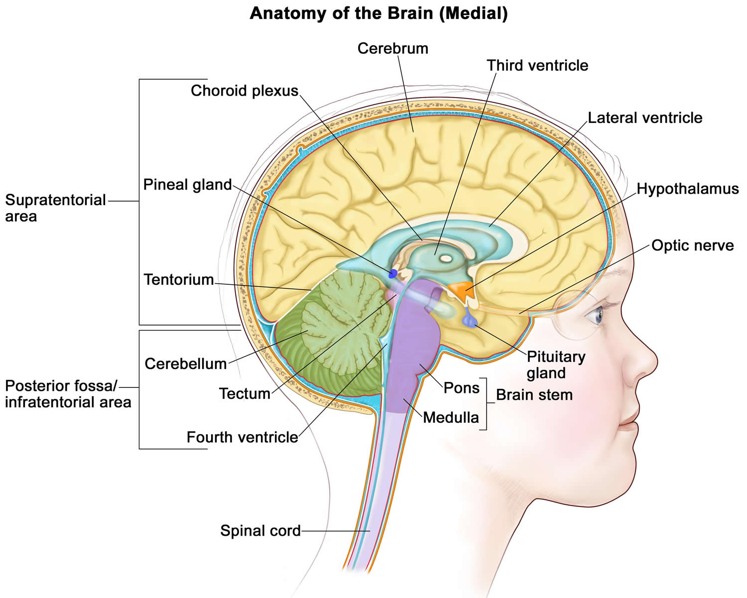
The colloid cyst is a benign (non-cancerous) growth usually located in the third ventricle and at or near the foramen of Monroe which is at the anterior aspect of the third ventricle of the brain 1. The colloid cyst is an epithelial lined cyst filled with gelatinous material. The gelatinous material commonly contains, mucin, old blood, cholesterol, and ions 2.
Colloid cyst accounts for less than 2% of all primary brain tumors. More than 99% of all colloid cysts are reported to occur at the frontend (rostral) of the third ventricle, at or near the foramen of Monro. The foramen of Monro is the conduit of cerebrospinal fluid (CSF) outflow from the lateral ventricles to the third ventricle. A colloid cyst can act as a ball valve, stopping CSF flow out of the lateral ventricles. If this occurs, CSF backs up into the lateral ventricles and causes ventriculomegaly and hydrocephalus. Some have posited that colloid cysts can cause intermittent obstructive hydrocephalus thus causing intermittent symptoms. Over time colloid cysts tend to growly very slowly. Some colloid cysts may never reach a size which will cause an issue and can be followed whereas others grow more quickly and become symptomatic with time.
Colloid cysts account for approximately one in five intraventricular primary brain tumors. Most patients diagnosed with a colloid cyst are in their third through the seventh decade of life, but cases have rarely been reported as early as the first year of life 1.
Colloid cysts can cause various symptoms including headaches, diplopia, memory issues and vertigo 1. Rarely colloid cysts have been cited as a cause for sudden death 1. When colloid cysts are symptomatic, they most commonly cause headaches, nausea and vomiting secondary to an obstructive hydrocephalus. The obstructive hydrocephalus is precipitated by blocking the egress of cerebrospinal fluid (CSF) from the lateral ventricles, at the foramen of Monro which connects the lateral and third ventricles 3.
When a colloid cyst is diagnosed, the biggest dilemma is how to manage it. The minimally invasive approaches have less morbidity but have a higher rate of recurrence and reoperation. Removing a colloid cyst via a craniotomy has the highest up-front surgical risk but may have the lowest recurrence and reoperation rate. The open craniotomy provides more degrees of freedom for access to the colloid cyst and may be more suitable for larger colloid cysts but does have limitations based on the approach chosen.
Figure 1. Brain anatomy
Colloid cyst causes
The precise cause of colloid cysts is unclear and still a topic of debate 1. In the early 20th century, the suggested cause was the colloid cyst was a remnant of the paraphysis element. The paraphysis element is an embryonic structure located at the anterior portion of the diencephalon between the two hemispheres of the telencephalon.
As colloid cysts have also been found in the cerebellum, frontal lobe and pontomesencephalon there have been other theorized origins of the colloid cysts. Other etiologies include remnants of respiratory epithelium, an ependymal cyst from the diencephalon and invagination of the neuroepithelium of the lateral ventricle causing a cyst to form 3.
Colloid cyst symptoms
Most colloid cysts identified are currently asymptomatic and identified incidentally on imaging. When a colloid cyst does cause issues, it most commonly causes obstructive hydrocephalus.
Colloid cysts can cause various symptoms including headaches, diplopia, memory issues and vertigo 1. Rarely colloid cysts have been cited as a cause for sudden death 1. When colloid cysts are symptomatic, they most commonly cause headaches, nausea and vomiting secondary to an obstructive hydrocephalus. The obstructive hydrocephalus is precipitated by blocking the egress of cerebrospinal fluid (CSF) from the lateral ventricles, at the foramen of Monro which connects the lateral and third ventricles 3.
Colloid cyst diagnosis
The majority of colloid cysts are found incidentally on imaging of the brain occurring for other reasons. When a colloid cyst is symptomatic, it most commonly causes non-communicating hydrocephalus. Symptoms of the hydrocephalus can include headaches, nausea, vomiting, lethargy, coma, and death. If the hydrocephalus is slowly progressive the patient can have more subtle findings including urinary incontinence, trouble with walking, falls, altered mentation and memory deficits.
For asymptomatic colloid cysts, the physical exam should be normal. If the patient has hydrocephalus from the colloid cyst physical exam findings may include lethargy, failure of upward gaze, unsteady gait, ataxia, increased reflexes, and, if the hydrocephalus is chronic, papilledema and/or frontal release signs.
Immediate evaluation of suspected colloid cyst includes the airway, breathing, and circulation (ABCs) of emergency medical management if the patient may be at risk for acute hydrocephalus and neurologic deterioration. A thorough neurologic exam is important to identify any neurologic deficits, but imaging remains the cornerstone of evaluation for patients with a colloid cyst.
Colloid cyst radiology
A colloid cyst is typically not visualized on plain radiographs of the head and thus computed tomography (CT), and magnetic resonance imaging (MRI) of the head are more important imaging studies.
CT Head
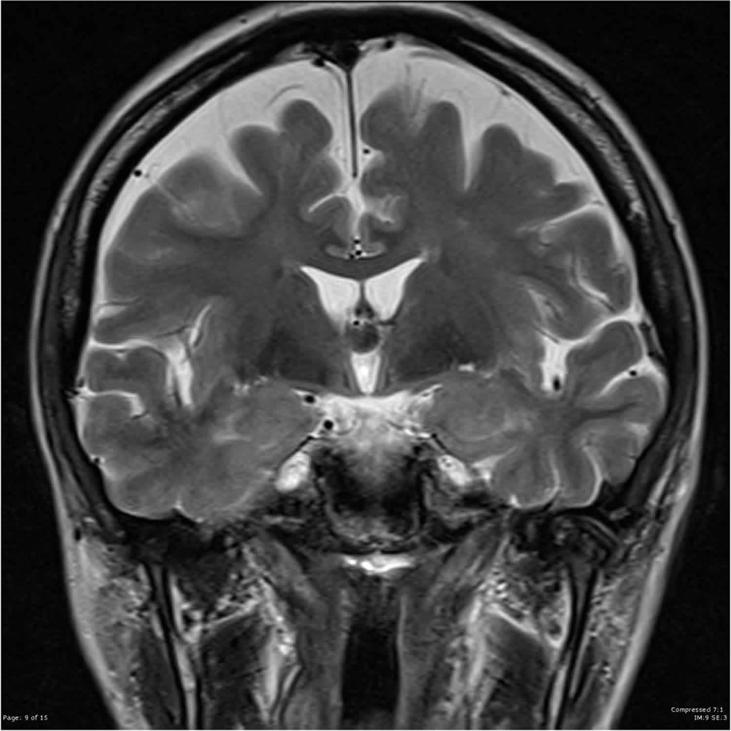
CT of the head can be quickly obtained to identify acute hydrocephalus. On CT imaging the colloid cyst is typically a circular, hyperdense mass at or near the foramen of Monro. Rarely are colloid cysts isodense, hypodense or calcified.
MRI Brain
MRI is the preferred method for imaging colloid cysts. On T1 sequencing a colloid cyst can have variable characteristics and be either hyperintense, isointense or hypointense. With gadolinium administration, the colloid cysts should not enhance. Rarely a peripheral enhancement will be noted around the colloid cyst which most likely represents a vessel stretched over the colloid cyst.
On T2 sequence imaging, most colloid cysts are hypointense. They may also have a heterogeneous T2 signal. A low signal intensity on T2 imaging may suggest the contents of the colloid cyst are more viscous and thus harder to aspirate. On FLAIR sequencing most colloid cysts have a similar intensity to the surrounding CSF. Most colloid cysts have decreased signal intensity on diffusion-weighted imaging.
Colloid cyst treatment
Treatment and management depend if the colloid cyst is found incidentally or it is symptomatic.
If the colloid cyst is symptomatic and causing hydrocephalus, the colloid cyst should be treated. For acute, life-threatening hydrocephalus, treatment of the hydrocephalus should be the next priority after ensuring adequate airway, breathing, and circulation. An external ventricular drain can be placed to relieve acute hydrocephalus and may be a life-saving procedure.
After acute, life-threatening hydrocephalus has either been treated or ruled out, the clinician can deal with the colloid cyst. Current treatment options include craniotomy with excision via a transcallosal or transcortical route, endoscopic removal, and stereotactic aspiration.
Craniotomy for Colloid Cyst Removal
A colloid cyst can be removed with a craniotomy. A craniotomy is a surgery where an incision is made in the scalp and part of the skull is removed for the duration of the surgery then the skull is put back in place. Two separate routes exist to remove the colloid cysts: transcallosal and transcortical. In the transcallosal approach, the two frontal hemispheres are split apart, and a surgical corridor is created through the rostral end of the genu of the corpus callosum to access the colloid cyst. For the transcortical route, a surgical corridor is developed directly through the brain cortex, most commonly through the right frontal, middle gyrus, to access the lateral ventricle. The colloid cyst can then be removed through the lateral ventricle.
Removing a colloid cyst via a craniotomy has the highest up-front surgical risk but may have the lowest recurrence and reoperation rate. The open craniotomy provides more degrees of freedom for access to the colloid cyst and may be more suitable for larger colloid cysts but does have limitations based on the approach chosen 4.
Endoscopic Removal of a Colloid Cyst
An endoscopic surgery consists of making a small incision in the scalp and a small hole in the bone. A small tube, typically called a sheath, is advanced through the brain to get access to the lateral ventricle. An endoscope can then be passed into the lateral ventricle to remove the colloid cyst. An endoscopic in its simplest form is a tube with a light, camera and working channel. The light provides illumination for the camera to see what is going on. The working channel provides the surgeon a way to get instruments and tools in front of the camera to perform surgery.
Endoscopic removal of a colloid cyst tends to have less up-front risk than an open surgery but also may have a slightly higher reoperation rate than open surgery. The endoscopic approach may not be suitable for all colloid cysts depending on the size and location of the cyst 4.
Stereotactic Aspiration of a Colloid Cyst
A third option to treat a colloid cyst is a stereotactic aspiration. This is performed by making a small incision in the scalp then a small hole in the bone. The surgeon then advances a needle through the brain and into the cyst using some variety of either frame-based or frameless neuronavigation. The contents of the colloid cyst may be able to be aspirated decreasing its size.
Aspiration of a colloid cyst may not be achievable if the contents of the colloid cyst are particularly thick or if there is no safe corridor to the colloid cyst. Stereotactic aspiration of a colloid cyst has less relative surgical risk than an endoscopic or open resection of the colloid cyst but has the highest reoperation rate compared to the other two treatment modalities. With the aspiration of the colloid cyst, the cyst is left in place and simple decompressed. The cyst may re-expand over time and become symptomatic again 4.
Asymptomatic Colloid Cyst
An asymptomatic colloid cyst does not necessarily warrant treatment. If there is hydrocephalus most, all surgeons would agree surgery is warranted, but if a colloid cyst is found incidentally, then surgery is not necessarily warranted. Colloid cysts which are smaller than 10 mm or more centrally located in the third ventricle are less likely to obstruct the near term. Such colloid cysts may be monitored over time with serial imaging looking for colloid cyst size and location as well as any evidence of hydrocephalus. There have been infrequent reported cases of colloid cysts which were followed clinically but caused acute hydrocephalus and death.
Colloid cyst survival rate
Some colloid cysts can be watched for years to decades without any issue. Others can slowly grow in size or cause subacute or acute hydrocephalus. With complete surgical resection, the prognosis is good, and colloid cysts are rare to recur after complete resection. Rare cases of sudden death have been reported with colloid cysts which is usually attributed to acute obstructive hydrocephalus.
References- Tenny S, Thorell W. Colloid Brain Cyst. [Updated 2019 Jun 4]. In: StatPearls [Internet]. Treasure Island (FL): StatPearls Publishing; 2019 Jan-. Available from: https://www.ncbi.nlm.nih.gov/books/NBK470314
- Ahmed SI, Javed G, Laghari AA, Bareeqa SB, Aziz K, Khan M, Samar SS, Humera RA, Khan AR, Farooqui MO, Shahbaz A. Third Ventricular Tumors: A Comprehensive Literature Review. Cureus. 2018 Oct 05;10(10):e3417
- Barbagallo GM, Raudino G, Visocchi M, Maione M, Certo F. Out-of-third ventricle colloid cysts: review of the literature on pathophysiology, diagnosis and treatment of an uncommon condition, with a focus on headache. J Neurosurg Sci. 2019 Jun;63(3):330-336
- Yadav YR, Yadav N, Parihar V, Kher Y, Ratre S. Management of colloid cyst of third ventricle. Turk Neurosurg. 2015;25(3):362-71


