What is congenital ptosis
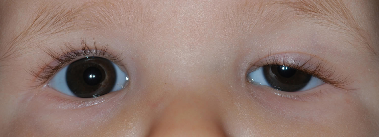
Congenital ptosis also called congenital blepharoptosis, is a drooping eyelid that is present at birth or within the first year of life. Congenital ptosis is usually present at birth but may manifest within the first years of life. In ptosis, the upper eyelid falls to a position that is lower than normal. In severe cases of ptosis, the drooping eyelid can cover part or all of the pupil and interfere with vision, resulting in amblyopia.
Amblyopia may result from obscuration of the vision directly or from development of astigmatism indirectly. Development of amblyopia is an indication for immediate surgical correction.
- Occlusion amblyopia
- Astigmatism from the compression of the droopy eyelid
- Ocular torticollis
In most cases of congenital ptosis, the problem is isolated and does not affect the vision. Any ptosis that develops over a period of days or weeks can signal a serious medical problem and needs further neurologic and physical evaluation.
Although not all patients with congenital ptosis need surgical intervention, patients need to be closely monitored for the possible development of deprivational amblyopia. Since amblyopia may not be reversed after age 7-10 years, appropriate and timely medical and surgical treatment of congenital ptosis is critical to preserve the child’s vision.
Uncorrected congenital ptosis can result in amblyopia secondary to deprivation or uncorrected astigmatism.
An abnormal eyelid position can have negative psychosocial effects.
Congenital ptosis occurs equally among the different races.
Congenital ptosis occurs equally between males and females.
In most cases of congenital ptosis, a droopy eyelid results from a localized myogenic dysgenesis. Rather than normal muscle fibers, fibrous and adipose tissues are present in the muscle belly, diminishing the ability of the levator to contract and relax. Therefore, the condition is commonly called congenital myogenic ptosis.
Congenital ptosis can also occur when the innervation to the levator is interrupted through neurologic or neuromuscular junction dysfunction.
What problems can occur as a result of congenital ptosis?
One or more of the following abnormalities may accompany congenital eyelid ptosis: astigmatism (refractive error), obstruction of the visual axis (the path that light takes into the eye), a chin-up head position, and amblyopia. The abnormal resting position of the eyelid on the cornea may result in astigmatism (a misshaping of the cornea) or other refractive errors, and is a risk factor for developing amblyopia (refractive amblyopia). Another risk factor for amblyopia is an eyelid drooping so low that it actually prevents light from entering the eye and creating an image on the retina at the back of the eye (deprivation amblyopia) Also, a chin-up head position may be present. This head position is adopted in order to be able to see beneath the edge of the drooping upper eyelid. Contraction of the frontalis muscle (in the forehead) to further elevate the upper eyelid is a very common compensatory mechanism.
Congenital ptosis causes
In most cases of congenital ptosis, the cause is idiopathic (unknown).
Histologically, the levator muscles of patients with congenital ptosis are dystrophic 1. The levator muscle and aponeurosis tissues appear to be infiltrated or replaced by fat and fibrous tissue. In severe cases, little or no striated muscle can be identified at the time of surgery. This suggests that congenital ptosis is secondary to local developmental defects in muscle structure.
Congenital ptosis may occur through autosomal dominant inheritance. Common familial occurrences suggest that genetic or chromosomal defects are likely.
Other potential causes of congenital ptosis include:
- Blepharophimosis syndrome: This condition consists of short palpebral fissures, congenital ptosis, epicanthus inversus, and telecanthus.
- Third cranial nerve palsy: Signs of aberrant regeneration are usually present. The pupil may be paradoxically small and nonreactive.
- Horner syndrome: Ipsilateral findings of mild ptosis, miosis, and anhidrosis characterize this syndrome. The ipsilateral lower eyelid may be elevated. Also, because of the lack of sympathetic innervation to the iris melanocyte development, a difference in the iris color between the eyes may result (called heterochromia).
- Marcus Gunn jaw-winking syndrome: The motor nerve to the external pterygoid muscle is misdirected to the ipsilateral levator muscle. Lid elevation occurs with mastication or with movement of the jaw to the opposite side.
- Birth trauma
- Duane syndrome: In this condition, the sixth cranial nerve fails to innervate a lateral rectus muscle. Then, the muscle acquires an innervation of the third cranial nerve. Although the synkinesis produced does not involve lid innervation, enophthalmos with apparent ptosis may result. In Esotropic Duane syndrome, the upper eyelid droops further and the lower lid elevates when the eye is adducted because of a co-contraction of the horizontal rectus muscles.
- Periorbital tumor: Neuroblastoma, plexiform neuromas, lymphomas, leukemias, rhabdomyosarcomas, neuromas, neurofibromas, or other deep orbital tumors may produce ptosis or proptosis.
- Kearns-Sayre syndrome: This mitochondrial deletion disorder is characterized by progressive external ophthalmoplegia, heart block, retinitis pigmentosa, and central nervous system manifestations. This condition begins in childhood but is rarely present at birth. The conditions are most likely to become symptomatic in the first or second decade of life. Bilateral ptosis is a prominent feature of this syndrome.
- Myotonic dystrophy: Patients with this condition may present with polychromatic cataracts, gonadal atrophy, or premature thinning and/or loss of hair. Myotonic dystrophy is an autosomal dominant disorder that is characterized clinically by myotonia and progressive muscular weakness. Frontal balding and temporalis muscle wasting are also clinically evident.
- Myasthenia gravis: A defect at the neuromuscular junction produces relative unresponsiveness to released acetylcholine, resulting in ptosis.
- Pseudotumor of the orbit: Patients with this condition may present with ptosis due to inflammation and edema of the eyelid.
- Pseudoptosis: Less tissue in the orbit (eg, unilateral smaller eye, fat atrophy, blowout fracture) produces the appearance of ptosis secondary to the decreased volume of orbital contents.
Congenital ptosis diagnosis
All babies presenting with either unilateral droopy eyelid or bilateral droopy eyelids need a thorough examination that includes a medical history, a family history, a history of drug or allergic reactions, and a review of systems.
- Family photographs can help determine onset or variability of the ptosis. Providing photographs also gives the surgeon a chance to examine the other family members. A patient with a strong family history of congenital ptosis may not need an extensive workup.
- In severe cases of congenital ptosis in which surgery is needed, historical emphasis should be placed on any anticoagulant use or bleeding disorder to avoid potential complications during surgery. The surgeon should also inquire about a family history of malignant hyperthermia and cardiac disorders. Patients with ptosis and Kearns-Sayre syndrome or chronic progressive external ophthalmoplegia may also have a cardiac conduction disorder.
- A history of fluctuating ptosis with strabismus may indicate myasthenia gravis.
- A careful medical history regarding cancer should be obtained. Metastatic or primary orbital tumors can result in malpositioning of the eyelid.
- A history of trauma with orbital wall fractures can result in pseudoptosis with enophthalmos. Additionally, third cranial nerve palsy from trauma may result in ptosis.
- A history of drug or allergic reactions may be helpful. Allergic reactions can result in eyelid edema and droopy eyelid.
- A history of difference in the size of the pupil may be helpful in diagnosing Horner syndrome. Patients with Horner syndrome have ptosis and miosis on the same side. Cervical or apical thoracic tumors can cause damage to the sympathetic chain and result in this condition. Neuroblastoma, which is one of the most common childhood cancers, should be ruled out.
- A history of dry eyes, intermittent epiphora, or chronic conjunctivitis can indicate a dry eye disorder or corneal surface disease.
Physical examination
All pediatric patients presenting with either unilateral droopy eyelid or bilateral droopy eyelids need a thorough physical evaluation.
- Visual acuity, refractive error, and cycloplegic refraction should be recorded. In infants, the surgeon should make sure that the baby can fixate and follow objects with each eye individually.
- The patient should be evaluated for strabismus (misalignment) and undergo a dilated fundus examination.
- Serial external photographs of the eyes and the face may be included in the patient’s record for documentation.
- Tear function should be evaluated if any doubt exists about the adequacy of tear production. This evaluation would include a slit-lamp examination with fluorescein stain to examine the cornea, tear meniscus, and tear break-up time. The Schirmer test can also be performed for dry eye syndrome; to do so, a filter paper is applied at the junction of the middle and lateral one third of the lower eyelid.
- Corneal sensitivity should be tested if possible. This may be a difficult test in young pediatric patients.
- An exophthalmometer can be used to assess relative proptosis or enophthalmos of each eye. In pseudoptosis, a proptosis of the contralateral eye gives the false impression that the normal upper eyelid is droopy.
- The pupillary size and the iris color differences between the eyes should be examined for Horner syndrome.
- The lid height (palpebral fissure distance) should be observed and measured in millimeters with each eye fixating on a distant target. The distance is the measurement of the greatest width of the palpebral fissure with the patient’s eyes in straight gaze. The lid position in downgaze should be noted. In congenital ptosis, the ptotic lid appears higher in downgaze.
- After the palpebral fissure distance is measured, the levator function should be evaluated. The patient looks downward as a ruler is positioned with a mark adjacent to the upper lid margin. With the examiner’s hand eliminating any brow action by the patient, the patient looks upward as far as possible without a change in head position. Lid elevation is measured directly from the ruler and is recorded in millimeters of levator function.
- The patient should be examined for Bell phenomenon. The patient closes both eyes tightly as the examiner holds the upper and lower lids apart. If the globe elevates during the forced lid closure, a normal Bell phenomenon is present. This evaluation can help the surgeon to determine the risk of exposure keratopathy following the eyelid surgery.
- Careful external examination along with palpation of the eyelids and the orbital rim should be performed. A lid mass can cause extra weight in the lid, resulting in ptosis. Plexiform neuromas, lymphoma, or leukemia can result in an eyelid mass. Rhabdomyosarcoma may present with a mass that is palpable through the lid.
Laboratory test
If myasthenia gravis is suspected, check serum acetylcholine receptor antibody levels 2.
If myasthenia gravis is suspected, the following tests are recommended:
- Single fiber electromyography (EMG)
- Tensilon or Ice test
- Serum Acetylcholine receptor antibody
Imaging Studies
- The following are indications to perform neuroimaging studies (eg, MRI or CT scans) of the orbit and brain:
- History not consistent and onset not clear
- Other neurologic findings along with ptosis
- Orbital wall fracture suspected with history of trauma
- Visible or palpable lid mass
- Orbital tumors (eg, lymphoma, leukemia, rhabdomyosarcoma) suspected
- New onset of Horner syndrome with or without other neurologic findings
- New onset of third cranial nerve palsy with or without other neurologic findings
- Globe displacement with either enophthalmos or proptosis
Other Tests
If a mitochondrial disorder is suspected, an ECG is recommended.
If a mitochondrial disorder is suspected, a muscle biopsy should be performed.
Congenital ptosis treatment
- Early consultation to avoid amblyopia
- Must be able to rule out and document other possible causes of ptosis (eg, Horner syndrome, third cranial nerve palsy)
- Although not all patients with congenital ptosis need surgical intervention, patients need to be closely monitored for the possible development of deprivational amblyopia. Since amblyopia may not be reversed after age 7-10 years, appropriate and timely medical and surgical treatment of congenital ptosis is critical to preserve the child’s vision.
- Uncorrected congenital ptosis can result in amblyopia secondary to deprivation or uncorrected astigmatism.
- An abnormal eyelid position can have negative psychosocial effects.
- Uncorrected acquired eyelid ptosis results in decreased field of vision and frontal headaches.
Medical therapy
Observation is only required in mild cases of congenital ptosis if no signs of amblyopia, strabismus, and abnormal head posture are present.
- Depending on the severity of the congenital ptosis, patients should be monitored every 3-12 months for signs of amblyopia due to congenital ptosis.
- External photographs can be helpful in monitoring patients.
- Head posture should be carefully examined. If the patient acquires a chin-up posture due to the worsening of ptosis, surgery may be indicated.
- The patient should be checked for astigmatism due to the compression of the droopy eyelid.
When amblyopia is present, appropriate treatment is initiated. When astigmatism is significant enough to potentially cause amblyopia, glasses are prescribed. Early eyelid surgery is usually indicated for a drooping eyelid that blocks vision (which may cause delayed vision development), or leads to a significant chin-up head position (which may cause neck problems and/or delay of developmental skills). Children are usually monitored regularly for vision abnormalities. Surgery may also be indicated during preschool years if the ptosis does not improve with normal growth and maturation of the face.
Medical follow up
Patients with congenital ptosis may have other conditions that need to be addressed. These conditions include amblyopia, strabismus, craniofacial abnormalities, and other neurologic findings. Appropriate consultation may be needed depending on the associated findings.
- Pediatric ophthalmologist
- Pediatric oculoplastic service
- Pediatric neurologist
- Cardiologist (if mitochondrial disorder suspected)
Congenital ptosis surgery
Congenital ptosis has physical, functional, and psychological consequences. The method of repair depends on treatment goals, the underlying diagnosis, and the degree of levator function. Although the primary reason for the repair is functional, the surgeon has an opportunity through this procedure to produce symmetry in lid height, contour, and eyelid crease for better cosmesis 3.
Surgical correction of congenital ptosis can be undertaken at any age depending on the severity of the disease. Earlier intervention may be required if significant amblyopia or ocular torticollis is present. Severe cases of ocular torticollis may delay mobility in infants and toddlers because of the balance problems from extreme chin-up head posture. If intervention is not urgent, surgery is often delayed until age 3-4 years. Waiting until this age allows for more accurate measurements preoperatively 4.
Surgery for ptosis in patients with a history of dry eyes, seventh cranial nerve palsy, or significant extraocular muscle abnormalities, such as severe Graves ophthalmopathy, double elevator palsy, or progressive external ophthalmoplegia, should be approached with great caution to avoid exposure keratopathy following the surgery.
Levator muscle resection
- This procedure is the shortening of the levator-aponeurosis complex through a lid-crease incision. The skin incision is hidden either in the existing lid fold or in a new lid fold created to match that of the contralateral eyelid.
- Indications: Moderate levator function must be present to offer a chance for correction with a levator resection. If the levator function is greater than 4 mm but less than 6 mm, a levator resection of greater than or equal to 22 mm is recommended. If the levator function is 6-8 mm, a levator resection of 16-18 mm is indicated. If the levator function is greater than 8 mm, a levator resection of 10-13 mm is indicated.
- Contraindications: An external levator resection is not indicated when the levator function is less than 4 mm. In such cases, a long-term surgical outcome may result in undercorrection. Poor Bell phenomenon (limited elevation of the eye), reduced corneal sensitivity, or poor tear production can produce exposure keratopathy.
Frontalis suspension procedure
- This procedure is designed to augment the patient’s lid elevation through brow elevation. Frontalis suspension procedures produce lagophthalmos in most cases. Some surgeons prefer to perform a bilateral suspension procedure for severe unilateral congenital ptosis to obtain symmetry.
- Indications: The procedure is indicated when the levator function is less than 4 mm.
- Relative contraindications: Poor Bell phenomenon (limited elevation of the eye), reduced corneal sensitivity, or poor tear production can produce exposure keratopathy. If surgery is still indicated, these patients need close postoperative follow-up care to avoid corneal exposure, infection, corneal ulcer and amblyopia.
- Surgical technique: Several materials are available to secure the lids to the frontalis muscles 5. These materials include:
- Autogenous fascia lata: Autogenous fascia lata can be obtained from the leg of patients older than 3 years.
- Preserved (tissue bank) fascia lata
- Nonabsorbable suture material (eg, 2-0 Prolene, Nylon (Supramid) or Mersilene)
- Silicone bands, silicone rods
- ePTFE (expanded Poly Tetra Fluoro Ethylene), Gore-Tex
- Autogenous materials used less frequently include palmaris longus tendon and temporalis fascia.
- Surgical outcome: Patients may not be able to close their eyelids during sleep from a few weeks to several months following surgery. Families must be warned of this outcome before the operation. The problem of open lids during sleep improves with time; however, aggressive lubrication is needed to avoid exposure keratopathy.
Fasanella-Servat procedure
- The upper lid is elevated by removing a block of tissue from the underside of the lid. This tissue includes the tarsus, conjunctiva, and Müller muscle.
- This procedure is not commonly performed for cases of congenital ptosis.
Müller muscle–conjunctival resection
- This surgery is chosen if the eyelid has had a good response to phenylephrine.
- The conjunctiva and the Müller muscle are marked off, clamped, and sutured. The tissues are resected. Then, the conjunctival layer is closed.
- This procedure is not commonly performed for cases of congenital ptosis, although its use has been well-documented and its utility has increased in recent literature.
Congenital ptosis surgical follow up
Close follow up is necessary in the first few weeks following surgery to make sure that exposure keratoconjunctivitis doesn’t develop and is controlled if it does develop. This also allows for evaluation of the wound itself and for signs of infection or inflammation of any foreign implanted material.
- Patients who underwent surgery for congenital ptosis are initially monitored every 2-4 weeks for signs of exposure keratopathy, infection, granuloma formation, and overcorrection and undercorrection. External photographic documentation can be helpful in monitoring patients.
- Following the surgery, visual acuity, head posture, and refractive error should be carefully monitored. Any residual amblyopia should be treated aggressively.
Congenital ptosis surgery complications
Complications associated with the frontalis suspension procedure for congenital ptosis repair include the following:
- Granuloma: If suspension materials are not placed well beneath the skin, granuloma formation may occur. Granulomas should be treated conservatively because they tend to eventually resolve.
- Lid asymmetry
- Overcorrection with exposure keratopathy and dry eyes
- Undercorrection: Suspension materials may dissolve or break. Suture material may tear through soft tissue. Undercorrected congenital ptosis repair may require repeat surgery.
- Infection
Congenital ptosis prognosis
- The repair of congenital ptosis can produce excellent functional and cosmetic results.
- With careful observation and treatment, amblyopia can be treated successfully.
- Of patients who require surgical intervention, 50% or more may require repeat surgery in 8-10 years following the initial surgery.
- Congenital ptosis. https://eyewiki.org/Ptosis,_Congenital
- Wabbels B, Schroeder JA, Voll B, Siegmund H, Lorenz B. Electron microscopic findings in levator muscle biopsies of patients with isolated congenital or acquired ptosis. Graefes Arch Clin Exp Ophthalmol. Oct 2007;245(10):1533-41.
- Chong KK, Fan DS, Lai CH, Rao SK, Lam PT, Lam DS. Unilateral ptosis correction with mersilene mesh frontalis sling in infants: thirteen-year follow-up report. Eye (Lond). Jan 2010;24(1):44-9.
- Bernardini FP, Devoto MH, Priolo E. Treatment of unilateral congenital ptosis. Ophthalmology. Mar 2007;114(3):622-3
- Lee MJ, Oh JY, Choung HK, Kim NJ, Sung MS, Khwarg SI. Frontalis sling operation using silicone rod compared with preserved fascia lata for congenital ptosis a three-year follow-up study. Ophthalmology. Jan 2009;116(1):123-9.


