
What is erythema toxicum neonatorum
Toxic erythema of the newborn is also known as erythema toxicum and erythema toxicum neonatorum, is a common and harmless rash seen in newborn infants. This newborn rash erythema toxicum goes away on its own and has no symptoms. Erythema toxicum is an extremely common rash that does not require any treatment, as it will spontaneously go away in 5–7 days. Do not pop the pus-filled bumps, as irritating the rash may lead to an infection. Erythema toxicum of newborn has been associated with a reaction to meconium (fetal poop) to the skin of the baby, and the name of the condition has changed several times over the years, from erythema populated to erythema dyspepsia and erythema neonatorum allergicum 1. In 1912, Dr. Karl Leiner, an Austrian pediatrician, named the condition erythema toxicum neonatorum and currently this is the term used for these skin findings 2.
Erythema toxicum of newborn affects approximately 48% to 72% of full-term newborn infants, but is less common in infants born prematurely. There is no known sex or racial predilection, although it has been suggested that it may be under-recognized in infants with darker skin types. In 356 Spanish newborns, 25.3% had erythema toxicum neonatorum (61.9% were males, and 38.1% were females). Recurrence is exceedingly rare, although there have been some reports of recrudescence within the first month of life 3. A 1986 study reported that 40.8% of 5387 Japanese neonates examined over a period of 10 years were identified as having erythema toxicum neonatorum 1. Preterm infants with birth weight less than 2500 grams are less likely to be affected 4.
Clinical features of erythema toxicum of the newborn
Most cases of erythema toxicum of the newborn begin in the first few days after birth, although onset can be as late as two weeks of age. Toxic erythema of the newborn is evident as various combinations of erythematous macules (flat red patches), papules (small bumps) and pustules. The eruption typically waxes and wanes over several days and it is unusual for an individual lesion to persist for more than a day.
Erythema toxicum of the newborn often begins on the face and spreads to affect the trunk and limbs. Palms and soles are not usually affected.
The infant is otherwise well.
The lesions are firm, yellow-white, 1 to 3-mm papules or pustules with a surrounding irregular erythematous base 1. Erythema toxicum of the newborn have been described as having a “flea-bitten appearance” or a papule surrounded by a sea of erythema. At times, splotchy erythema is the only manifestation. Lesions may be sparse or numerous and they can be either clustered in several sites or widely dispersed over much of the body surface. Erythema toxicum neonatorum has been described as consisting of two likely variations: an erythematous papular or a pustular variant. During the first day of presentation, macules tend to appear within and outside the erythematous lesions. It usually begins on the cheeks and rapidly spreads to the forehead, the rest of the face, chest, trunk, and extremities but there are several case reports of patients with erythema toxicum neonatorum lesions over the skin of the scrotum. The blotchy appearance ensues as the macules become confluent, sometimes resembling urticarial lesions on the trunk. The palms and soles are usually spared, this pattern of involvement may be correlated to the distribution of hair follicles. Clinicians have a harder time seeing erythema toxicum neonatorum in babies with darker skin. This makes the identification of the problem somewhat challenging. Erythematous macules often are evanescent, but when they persist, papules then tend to appear, papules also may arise as de novo. Some papules may become superficial, particularly on the skin of the back and abdomen. It is very uncommon for the pustules to become secondarily infected. The peak incidence occurs on the second day of life, but new lesions may erupt during the first few days as the rash waxes and wanes, although recurrences may occur in up to 11% of neonates. The onset may occasionally be delayed for a few days to weeks in premature infants. There is no systemic manifestation associated with the condition, and one might rarely see some eosinophilia in blood studies and counts as high as 18% have been reported 5.
Figure 1. Erythema toxicum neonatorum
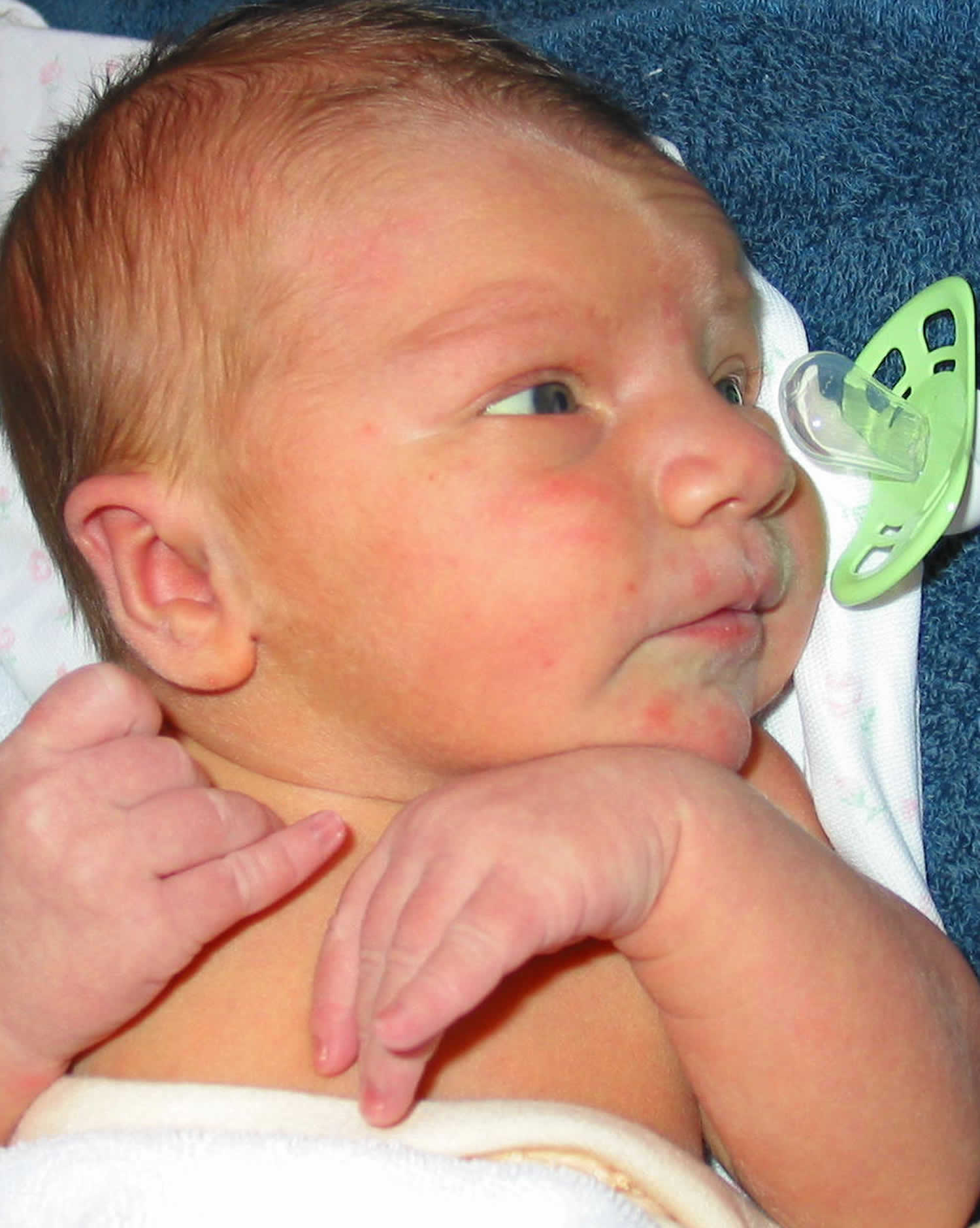
Footnote: 2 days old newborn infant with pink macules and papules.
Figure 2. Newborn rash erythema toxicum
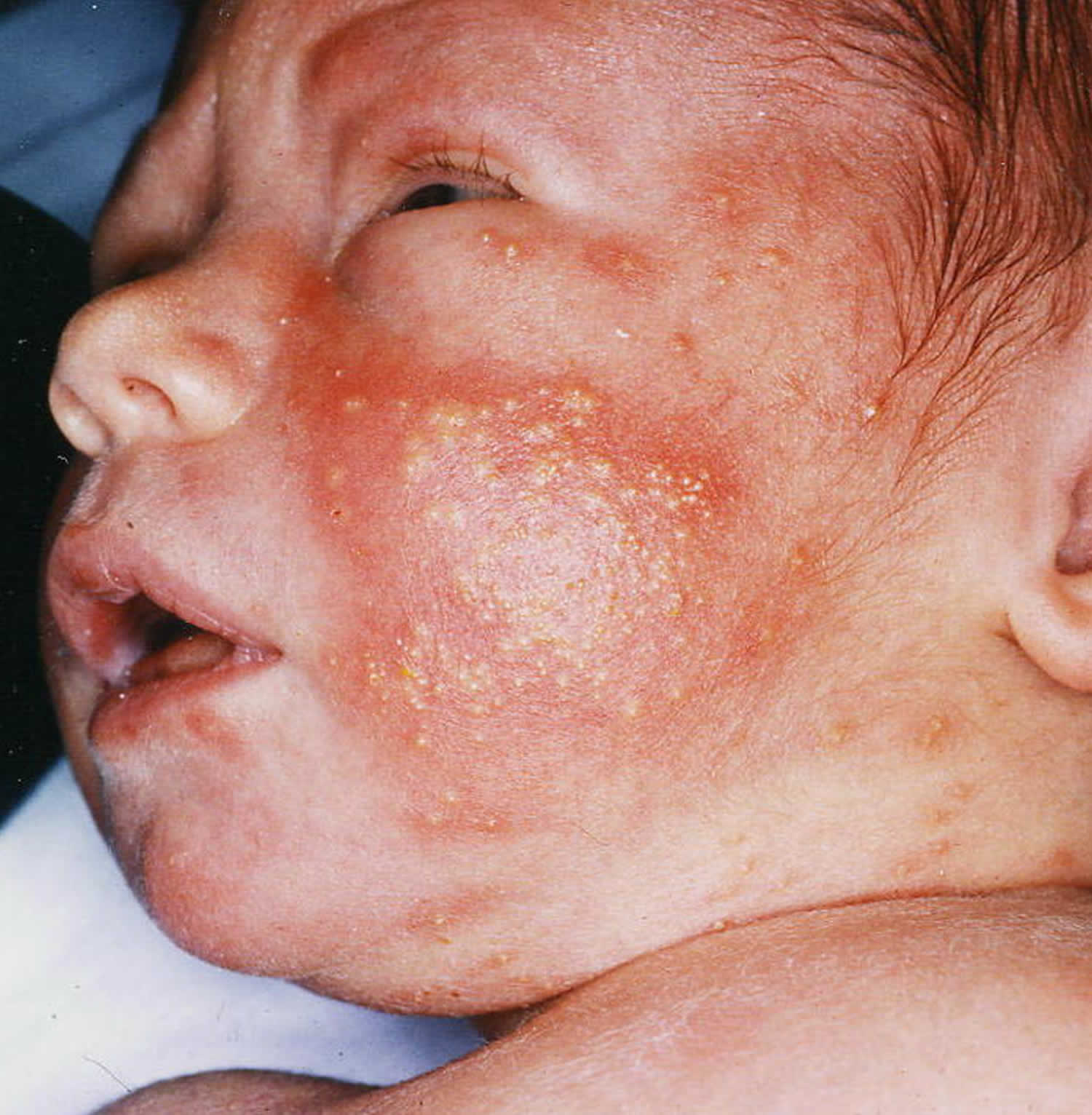
Footnote: 2 days old term infant with a new rash characterized by yellow pustules on an erythematous base involving the trunk and extremities but sparing the palms and soles.
Erythema toxicum causes
The etiology of erythema toxicum is completely unknown: a graft-versus-host reaction against maternal lymphocytes has been postulated as a possible mechanism, but recent studies failed to show the presence of maternal cells in these lesions. Another theory proposes an immune response to microbial colonization through the hair follicles as early as one day of age. Immunohistochemical study of lesions from 1-day-old infants supports the accumulation and activation of immune cells in erythema toxicum neonatorum lesions. A variety of inflammatory mediators such as IL-1alpha, IL-1beta, IL-8, eotaxin, aquaporins 1 and 3, psoriasin, nitric oxide synthetases 1, 2 and 3, and HMGB-1 have been associated with erythema toxicum neonatorum immunohistochemically 1. Tryptase-expressing mast cells, however not the cathelicidin antimicrobial peptide LL-37, have been located in erythema toxicum neonatorum biopsy specimens 1.
Erythema toxicum signs and symptoms
Erythema toxicum appears as small (1–3 mm), firm, yellow or white raised bumps filled with pus on top of a red area of skin. There may be a few to many lesions, and they may be found on any area of the body, with the exception of palms and soles. Erythema toxicum often begins on the face and spreads, and it may be clustered in areas where there is pressure on the skin. Although it most frequently appears during the first 3–4 days of life, erythema toxicum can be seen at birth and may not be present until 10 days of life. New lesions may appear as older ones resolve.
Erythema toxicum neonatorum diagnosis
The diagnosis of erythema toxicum of the newborn is usually made on clinical grounds. No laboratory work up is required. Histology from skin biopsy samples taken from flat patches shows a diffuse infiltrate of eosinophils and neutrophils (inflammatory cells) in the upper dermis; from papules shows an eosinophilic infiltration around the hair follicle; and from pustular lesions shows perifollicular subcorneal pustules filled predominantly with eosinophils.
How erythema toxicum of the newborn arises is unknown. A number of theories have been proposed including that it is a reaction to mechanical and thermal (heat) stimuli, an ‘allergic’ reaction or a mild form of graft vs host disease.
Although erythema toxicum of the newborn is benign and requires no treatment, a number of differential diagnoses should be considered. These include:
- infections (folliculitis, impetigo, listeriosis, congenital cutaneous candidiasis, herpes simplex, varicella and cytomegalovirus)
- transient neonatal pustular melanosis
- infantile acropustulosis
- miliaria rubra
- eosinophilic pustulosis (infantile Ofuji syndrome)
- incontinentia pigmenti
- Omenn syndrome
- self-healing histiocytosis.
These usually be recognized by clinical features, although in some cases diagnosis may be helped by Tzanck smears or skin biopsy. Neonatal herpes presents with blisters or erosions and an ill-appearing child. Neonatal herpes diagnosis is made via a Tzanck smear demonstrating multinucleated giant cells, viral culture, or direct fluorescent antibody for HSV 1 or 2. Commonly seen in warm climates. Transient neonatal pustular melanosis and incontinentia pigmenti can be distinguished without any difficulty from erythema toxicum neonatorum. Transient neonatal pustular melanosis present at birth as does erythema toxicum neonatorum, but transient neonatal pustular melanosis involves the palms and soles, there is no erythematous component, and it shows only neutrophils with cytologic debris. However, both condition can present at the same time. Incontinentia pigment presents with vesicles filled with eosinophils, and it is more common in boys but is rarely pustular, and the appearance is in a linear pattern different from the pattern of erythema toxicum neonatorum. The distribution of the rash can also be useful to differentiate erythema toxicum neonatorum from infantile acropustulosis, while erythema toxicum neonatorum spares the palm and soles, the hallmark of infantile acropustulosis characteristically include the acral surfaces hence its name. Erythema toxicum neonatorum can be differentiated from miliaria rubra, a condition in which the vesicles are related to the sweat ducts rather than hair follicles and the lesions containing mononuclear cells rather than eosinophils. Congenital cutaneous candidiasis can also present at birth or in the first days of life, with a diffuse erythematous rash with overlying papules, pustules and vesicles, but the palms and soles are not spared.
Erythema toxicum treatment
Treatment starts with reassuring the parents about the natural course of erythema toxicum neonatorum: it is benign (harmless) and will resolve without any complications, the rash will spontaneously go away in 5–7 days. Erythema toxicum is a benign self-limited skin disorder that is only seen in newborn infants. Because of the appearance of the rash creates great parental concerns, reassuring the parents has paramount significance.
References- Roques E, Mendez MD. Erythema Toxicum. [Updated 2018 Nov 24]. In: StatPearls [Internet]. Treasure Island (FL): StatPearls Publishing; 2018 Jan-. Available from: https://www.ncbi.nlm.nih.gov/books/NBK470222
- Reginatto FP, Muller FM, Peruzzo J, Cestari TF. Epidemiology and Predisposing Factors for Erythema Toxicum Neonatorum and Transient Neonatal Pustular: A Multicenter Study. Pediatr Dermatol. 2017 Jul;34(4):422-426.
- Ghosh S. Neonatal pustular dermatosis: an overview. Indian J Dermatol. 2015 Mar-Apr;60(2):211.
- Reddy HB, Gandra NR, Katta TP. Cutaneous Changes in Neonates in the First 72 Hours of Birth: An Observational Study. Curr Pediatr Rev. 2017;13(2):136-143.
- Reginatto FP, DeVilla D, Muller FM, Peruzzo J, Peres LP, Steglich RB, Cestari TF. Prevalence and characterization of neonatal skin disorders in the first 72h of life. J Pediatr (Rio J). 2017 May – Jun;93(3):238-245.


