What is nabothian cyst
A nabothian cyst is also known as a retention cysts of the cervix or mucinous retention cyst or epithelial cysts, is a harmless (benign) lump filled with mucus on the surface of the cervix below the squamous epithelium 1, 2. Nabothian cysts are always situated in the transformation zone. Nabothian cysts are multiple-opaque nodules (whitish to yellow) on the cervix 3. However, nabothian cyst is often single. The shape is circular and there is a central elevation of the mucosal surface. The blood vessels seem to be pushed aside or may cross over the surface 1.
Nabothian cysts are a common gynecological finding in women of reproductive age without clinical significance 3. Nabothian cyst is caused by chronic inflammation of the uterine cervix and are harmless and usually disappear on their own. Nabothian cysts usually are small in size, it is quite rare to reach a size above 4 cm 4. The presence of many nabothian cysts or nabothian cysts that are large and blocked can make it hard for the provider to do a Pap test. This is rare. Intraabdominal giant nabothian cyst has been reported 5; a large nabothian cyst causing genital prolapse due to the weight of the large nabothian cyst on the cervix which could have caused the elongation of the vaginal portion of the cervix 6.
The cervix is located at the lower end of the womb (uterus) at the top of the vagina. It is about 1 inch (2.5 centimeters) long. The cervix is lined with glands that normally secrete mucus. These endocervical glands can become filled with secretions that accumulate as a pimple-like elevation called Nabothian cysts. These cysts are not a threat to health and no treatment is necessary.
Nabothian cysts do not cause any harm. They are a benign (harmless) condition.
Figure 1. Cervix location
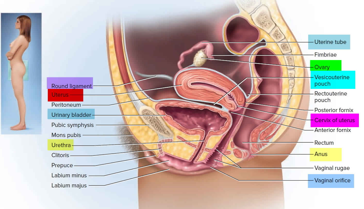
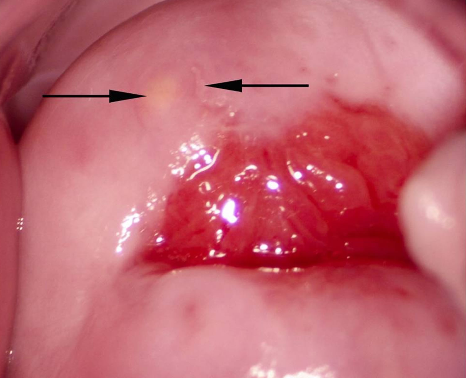
Figure 2. Nabothian cyst

Footnote: Normal cervical surface with a small yellow Nabothian cyst (arrow) 11 o’clock in the anterior lip of the transformation zone. These may be confused with rubbery papules but the Nabothian cysts are often bigger, do not protrude so acutely, and they are only found in the transformation zone. Rubbery papules, however, may be situated anywhere on the vaginal and cervical surface. Also note, next to the Nabothian cyst (left arrow) a small irregular-shaped leukoplakia area (right arrow) that could be a herpes simplex viral infection.
[Source 1 ]Figure 3. Nabothian cyst
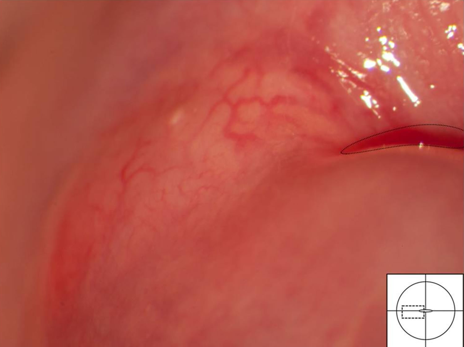
Footnote: Typical blood vessels across a Nabothian cyst. The underlying cyst is pale yellow adjacent to the squamo-columnar junction (dashed line) and the vascular network shows regular branching.
[Source 1 ]Nabothian cyst and pregnancy
Few cases of large nabothian cyst have been reported in the literature 7. Vural and colleagues 3 reported a case of a woman with 38 weeks of gestation presented to the maternity unit with labor pain and protruding cystic mass (60×70 mm) out of the vagina. According to Vural et al. 3, their patient was the first reported of nabothian cysts obstructing labor passage. Simple drainage was performed to allow the vaginal delivery. She delivered 4130 grams, 9-10 Apgar, male baby by spontaneous vaginal delivery. The patient’s and the newborn postpartum course was uneventful. The postpartum course is uneventful without hemorrhagic or infectious complication 3.
Nabothian cyst causes
Nabothian cysts are thought to occur secondarily to healing process of chronic cervicitis 3. The cervix is lined with glands and cells that release mucus. The glandular or columnar epithelium covers the endocervical canal and variably extend the ectocervix. This epithelium contains the single layer of mucin-secreting cells and invaginations of this epithelium make up endocervical glands. The border between the ectocervix (stratified squamous epithelium) and the endocervical canal (columnar epithelium) is called the squamo columnar junction 8. There is a continuous process of repair in the squamo columnar junction. The inflammatory process may block an endocervical gland orifice and retention cysts occur 8. The glands can become covered by a type of skin cells called squamous epithelium. When this happens, the secretions build up in the plugged glands. They form a smooth, rounded bump on the cervix. The bump is called a nabothian cyst.
Symptomatic Nabothian cysts can also occur as a late complication of subtotal hysterectomy, in which internalisation of the transformation zone and partial obliteration of the canal are postulated as predisposing factors.
Histopathologically, nabothian cysts are lined by single layer of columnar epithelium or flattened epithelium without cellular mitosis or atypia. Huge nabothian cysts need a pathologic diagnosis to exclude other tumors of cervix and adenoma malignum that contains well-differentiated glandular cells with atypia and mitosis 9. Adenoma malignum is also called minimal deviation adenocarcinoma and is quite a rare phenomenon. Adenoma malignum mimics an appearance of deep and larger sized nabothian cysts. Histopathologically, adenoma malignum lesion shows a benign histological appearance and it contains well-differentiated mucinous glands, deeply invading the cervical stroma. Despite benign histopathologic findings, the clinical behavior of adenoma malignum is aggressive. Thus, adenoma malignum is a diagnostic challenge and accurate diagnosis is important and depends on high suspicion of gynaecologist 10.
Nabothian cyst symptoms
Each nabothian cyst appears as a small, white raised bump. There can be more than one.
Nabothian cyst diagnosis
During a pelvic exam, your health care provider will see a small, smooth, rounded lump (or collection of lumps) on the surface of the cervix. Rarely, magnifying the area (colposcopy) may be needed to tell these cysts from other bumps that can occur.
Most women have small nabothian cysts. These are detected by vaginal ultrasound. If you’re told you have a nabothian cyst during a vaginal ultrasound exam, don’t be concerned, as their presence is normal.
Sometimes the nabothian cyst is opened to confirm the diagnosis.
Figure 4. Nabothian cyst ultrasound
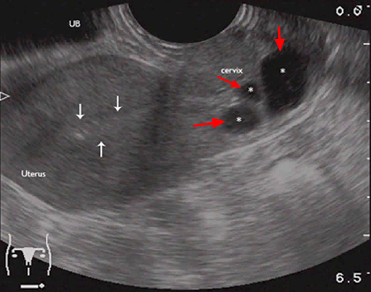
Footnote: Sagittal view. Nabothian cysts (red arrows), located in the cervix. Normal appearing uterus with thin endometrial stripe (white arrows).
Nabothian cyst treatment
No treatment is necessary. Nabothian cysts do not cause any problems.
Most Nabothian cysts are clinically insignificant. The majority of cases are not treated because the cysts are entirely benign; however, an excision biopsy is sometimes indicated in large, complex, cystic lesions to rule out rare forms of mucus-producing neoplasia, including adenoma malignum 11.
References- Norseth HM, Ndhlovu PD, Kleppa E, et al. The colposcopic atlas of schistosomiasis in the lower female genital tract based on studies in Malawi, Zimbabwe, Madagascar and South Africa. PLoS Negl Trop Dis. 2014;8(11):e3229. Published 2014 Nov 20. doi:10.1371/journal.pntd.0003229 https://www.ncbi.nlm.nih.gov/pmc/articles/PMC4238986/
- Fogel SR, Slasky BS. Sonography of Nabothian cysts. AJR Am J Roentgenol. 1982;138 (5): 927-30. https://www.ajronline.org/doi/pdf/10.2214/ajr.138.5.927
- Vural F, Sanverdi I, Coskun AD, Kusgöz A, Temel O. Large Nabothian Cyst Obstructing Labour Passage. J Clin Diagn Res. 2015;9(10):QD06-7. https://www.ncbi.nlm.nih.gov/pmc/articles/PMC4625292/
- Casey PM, Long ME, Marnach ML. Abnormal cervical appearance: What to do, when to worry? Mayo Clin Proc. 2011;86:147–51
- An intra-abdominal giant Nabothian cyst. SOLOMKIN M, TAUBER W. Conn Med. 1959 Jan; 23(1):27.
- Nigam A, Choudhary D, Raghunandan C. Large nabothian cyst: a rare cause of nulliparous prolapse. Case Rep Obstet Gynecol. 2012;2012:192526. https://www.ncbi.nlm.nih.gov/pmc/articles/PMC3384920/
- Nigam A, Choudhary D, Raghunandan C. Large nabothian cyst: a rare cause of nulliparous prolapse. Case Rep Obstet Gynecol. 2012;2012:192526.
- Malpica A, Robboy SJ. Cervical Benign and Nonneoplastic conditions in: Robboy Pathology of Female Reproductive Tract. 2nd Ed. Elsevier; 2009. pp. 141–166.
- Complex Nabothian cysts: a diagnostic dilemma. Sosnovski V, Barenboim R, Cohen HI, Bornstein J. Arch Gynecol Obstet. 2009 May; 279(5):759-61.
- Adenoma malignum of the uterine cervix: report of four cases. Ki EY, Byun SW, Park JS, Lee SJ, Hur SY. World J Surg Oncol. 2013 Jul 26; 11():168.
- Kuligowska E, Deeds L, Lu K. Pelvic pain: overlooked and underdiagnosed gynecologic conditions. Radiographics. 2005;25 (1): 3-20. Radiographics (full text) – doi:10.1148/rg.251045511 https://pubs.rsna.org/doi/pdf/10.1148/rg.251045511


